Setting the Standard: A Comprehensive Guide to Benchmarking Computational Neuroscience Models
This article provides a rigorous framework for benchmarking computational neuroscience models, addressing a critical need for standardization in the field.
Setting the Standard: A Comprehensive Guide to Benchmarking Computational Neuroscience Models
Abstract
This article provides a rigorous framework for benchmarking computational neuroscience models, addressing a critical need for standardization in the field. Aimed at researchers, scientists, and drug development professionals, it explores the foundational principles of model validation, from canonical neuron models to large-scale network simulations. It details methodological approaches for creating and applying benchmarks, including the use of synthetic and experimental data. The guide further offers practical strategies for troubleshooting and optimizing model performance, and finally, establishes robust protocols for the comparative validation of models against empirical data and other models. By synthesizing insights from recent literature and community initiatives, this article serves as a vital resource for advancing reproducible and clinically relevant computational neuroscience.
The Why and What: Core Principles and Canonical Models in Computational Neuroscience
In computational neuroscience, the journey from simulating a single ion channel to modeling an entire brain represents a spectrum of immense methodological complexity. Benchmarking serves as the essential practice that grounds this endeavor, enabling researchers to validate model correctness, assess computational efficiency, and facilitate meaningful comparisons across diverse simulation technologies. As the field progresses toward more integrated multi-scale models, establishing robust and standardized benchmarking practices becomes paramount for accelerating scientific discovery and ensuring the reliability of computational findings. This guide provides a comprehensive framework for defining benchmarking scope, offering practical methodologies and tools tailored for researchers and drug development professionals operating across biological scales.
Foundational Concepts and Definitions
Benchmarking in computational neuroscience systematically evaluates models and simulation technologies against standardized criteria, metrics, and datasets. This process transcends simple performance measurement; it provides the fundamental infrastructure for validating biological plausibility, quantifying computational efficiency, and ensuring reproducible results across different research environments. For drug development applications, rigorous benchmarking directly impacts risk assessment by providing data-driven estimates of a model's predictive power for clinical outcomes [1].
The scope of a benchmarking initiative must explicitly define the biological scale, specific research questions, and evaluation criteria. Clear scoping prevents mission creep—the tendency for a project's objectives to expand uncontrollably—by maintaining focus on distinguishing features of the phenomena and intuition about which factors require inclusion in an explanation [2]. Properly delineated scope acts as a natural Occam's razor, ensuring models address core knowledge gaps while minimizing unnecessary complexity.
Benchmarking Across Biological Scales
Computational neuroscience encompasses multiple biological scales, each requiring specialized benchmarking approaches. The following table summarizes key characteristics, challenges, and primary benchmarking metrics relevant to each scale.
Table 1: Benchmarking Considerations Across Biological Scales in Computational Neuroscience
| Biological Scale | Core Modeling Focus | Primary Benchmarking Metrics | Unique Challenges |
|---|---|---|---|
| Ion Channels [3] | Permeation, selectivity, and gating mechanisms | Conductance rates, selectivity ratios, gating kinetics, energy profiles | Reconciling high selectivity with high permeability; simulating rare events |
| Single Neurons | Electrical activity and signal integration | Spike timing accuracy, input resistance, firing patterns, computational cost | Balancing biophysical detail with simulation speed |
| Microcircuits & Networks [4] | Emergent dynamics from neuronal populations | Firing rate distributions, oscillation patterns, synchronization measures, scaling efficiency | Managing combinatorial complexity; interpreting population-level dynamics |
| Whole-Brain Systems [5] [6] | Large-scale functional connectivity and dynamics | Structure-function coupling, individual fingerprinting, brain-behavior prediction | Data integration across modalities; massive computational resources required |
Cross-Scale Integration and Validation
A critical challenge in multi-scale benchmarking involves validating how phenomena emerging at one level (e.g., network oscillations) relate to mechanisms at lower levels (e.g., channel kinetics). Effective benchmarking protocols must establish quantitative bridges between scales, ensuring that simplifications at lower levels do not invalidate emergent properties at higher levels. This often requires designing specific experiments that probe the sensitivity of macro-scale outputs to micro-scale parameters, creating a cohesive framework for integrated model validation across biological hierarchies.
Quantitative Benchmarking Data and Metrics
Establishing comprehensive quantitative profiles is fundamental to effective benchmarking. The table below synthesizes key benchmarking data from recent studies, providing a reference for expected performance across different metrics and methodologies.
Table 2: Quantitative Benchmarking Data for Neuroscience Models
| Benchmark Category | Specific Metric | Reported Values / Range | Context & Implications |
|---|---|---|---|
| Functional Connectivity (FC) Mapping [5] | Structure-Function Coupling (R²) | 0 to 0.25 | Precision-based statistics showed strongest correspondence with structural connectivity |
| FC Mapping [5] | Weight-Distance Correlation (∣r∣) | < 0.1 to > 0.3 | Fundamental network property varies significantly with FC estimation method |
| Ion Channel Properties [3] | K⁺ Conductance (KcsA) | 10⁷ to 10⁸ ions/second | Approaches theoretical diffusion limit; creates selectivity-permeability paradox |
| Ion Channel Selectivity [3] | K⁺/Na⁺ Selectivity (KcsA) | ~150:1 | Challenges simple size-exclusion models; highlights role of dehydration energy |
| Drug Development [1] | Probability of Success (POS) | Varies by phase/therapeutic area | Traditional benchmarking often overestimates POS; dynamic benchmarks improve accuracy |
Interpreting Quantitative Benchmarks
When applying these quantitative benchmarks, researchers must consider contextual factors that significantly influence results. For example, functional connectivity benchmarks depend heavily on the specific pairwise statistic used (e.g., covariance versus precision), with different methods revealing distinct aspects of network organization [5]. Similarly, ion channel conductance measurements are sensitive to simulation parameters such as membrane potential, ion concentrations, and specific force fields used in molecular dynamics simulations. Effective benchmarking requires transparent reporting of these contextual parameters to enable meaningful cross-study comparisons and scientific replication.
Experimental Protocols and Methodologies
Protocol for Benchmarking Functional Connectivity Methods
Based on large-scale comparisons of 239 pairwise statistics, the following protocol provides a standardized approach for evaluating functional connectivity methods [5]:
- Data Preparation: Utilize resting-state fMRI data from established public repositories (e.g., Human Connectome Project). Preprocess data using standardized pipelines for motion correction, normalization, and denoising. Parcellate brains using consistent atlases (e.g., Schaefer 100 × 7).
- FC Matrix Calculation: Apply multiple pairwise interaction statistics across different families (covariance, precision, information-theoretic, spectral, distance, linear model fits). The
pyspiPython package provides a standardized implementation of these measures. - Topological Analysis: Calculate probability density of edge weights and weighted degree (strength) for each brain region across all FC matrices. Identify network hubs and compare spatial distributions across methods.
- Geometric and Structural Validation: Quantify the relationship between functional connectivity and Euclidean distance between regions. Evaluate structure-function coupling by correlating FC matrices with diffusion MRI-based structural connectivity.
- Biological Alignment Assessment: Compute correlation between FC matrices and multimodal neurophysiological data, including gene expression, laminar similarity, neurotransmitter receptor similarity, electrophysiological connectivity, and metabolic connectivity.
- Individual Differences Quantification: Assess fingerprinting capability by measuring how well FC matrices can identify individuals across scanning sessions. Evaluate brain-behavior prediction performance using cross-validated models.
Protocol for Benchmarking Spiking Neural Network Simulators
For large-scale network simulations, the following modular workflow ensures comprehensive benchmarking [4]:
- Benchmark Specification: Define the benchmarking goal (e.g., weak scaling, strong scaling, energy efficiency). Select appropriate network models with different complexity levels (e.g., balanced random networks, multi-area models with natural density).
- Hardware Configuration: Document complete hardware specifications including compute nodes, processors, memory, interconnect, and GPU accelerators if applicable. For energy measurements, specify whether consumption includes only compute nodes or entire support infrastructure.
- Software Configuration: Record simulator versions, compiler versions, math libraries, and all relevant configuration options. Maintain consistent software environments across benchmarking runs.
- Model Implementation: Implement standardized network models (e.g., Brunel-type balanced networks with leaky integrate-and-fire neurons). For weak scaling, increase network size proportionally with computational resources. For strong scaling, maintain fixed network size while increasing resources.
- Execution and Monitoring: Execute simulations with detailed timing measurements separated into setup phase and simulation phase. Monitor memory usage, power consumption, and network activity dynamics throughout execution.
- Data Collection and Analysis: Record benchmarking data and metadata in standardized formats. Calculate key metrics including time-to-solution, energy-to-solution, memory consumption, and scaling efficiency. Compare activity statistics (firing rates, distributions) across simulators.
A Generalized 10-Step Modeling Process
For developing new models across biological scales, the following systematic process ensures rigorous benchmarking [2]:
- Frame the Question: Identify a specific phenomenon and formulate a precise question. Define evaluation criteria and stopping conditions.
- Define the Core assumptions: Make simplifying assumptions explicit and determine the model's intended scope.
- Formalize the Model: Translate concepts into mathematical frameworks and computational structures.
- Implement the Model: Develop code using appropriate programming languages and simulation technologies.
- Check the Implementation: Verify code correctness through unit tests and sanity checks.
- Test the Model: Assess basic operation and qualitative behavior against expected outcomes.
- Perform Preliminary Fitting: Compare initial model outputs with target data using simple parameter adjustments.
- Formally Fit the Model: Employ rigorous estimation procedures to optimize model parameters.
- Validate the Model: Evaluate performance on held-out data not used during fitting.
- Interpret the Model: Draw scientific conclusions based on model behavior and performance.
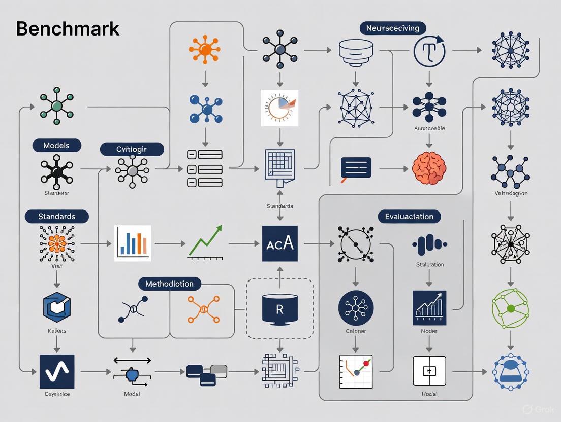
The diagram below illustrates this iterative modeling and benchmarking workflow:
Visualization and Workflow Diagrams
Functional Connectivity Benchmarking Workflow
The following diagram outlines the key stages in benchmarking functional connectivity methods, from data preparation to quantitative evaluation:
Ion Channel Simulation and Analysis Pipeline
For ion channel research, computational studies typically follow this pathway to characterize key biophysical properties:
The Scientist's Toolkit: Essential Research Reagents and Materials
Successful benchmarking requires both computational tools and conceptual frameworks. The following table details essential components for designing and executing rigorous benchmarking studies.
Table 3: Essential Research Reagents and Tools for Computational Neuroscience Benchmarking
| Tool Category | Specific Tool/Resource | Function/Purpose | Example Applications |
|---|---|---|---|
| Simulation Engines [4] | NEST, Brian, NEURON, Arbor, GeNN | Simulate spiking neuronal networks at different scales | Large-scale network models, detailed cellular simulations |
| FC Analysis Packages [5] | pyspi (Python) |
Implements 239 pairwise interaction statistics | Benchmarking functional connectivity methods |
| Benchmarking Frameworks [4] | beNNch |
Configures, executes, and analyzes simulation benchmarks | Standardized performance comparisons across HPC systems |
| Data Resources [5] [6] | Human Connectome Project, ZAPBench Dataset | Provides standardized neuroimaging data for benchmarking | Testing FC methods, whole-brain activity prediction |
| Modeling Guidance [2] | 10-Step Modeling Process | Provides systematic framework for model development | Structuring modeling projects across biological scales |
| Visualization Tools [7] [8] | Sigma, ColorBrewer, specialized neuro tools | Creates clear, honest data visualizations | Presenting benchmarking results, uncertainty visualization |
Defining the scope for benchmarking computational neuroscience models requires careful consideration of biological scale, research questions, and appropriate evaluation metrics. By adopting the standardized protocols, quantitative benchmarks, and systematic workflows outlined in this guide, researchers can establish rigorous benchmarking practices that enhance model reliability, facilitate cross-study comparisons, and accelerate scientific discovery. The ongoing development of specialized benchmarking tools and shared datasets will further strengthen these efforts, ultimately contributing to more robust, reproducible, and biologically-grounded computational models across all scales of neuroscience inquiry.
Computational neuroscience builds quantitative models of neural systems across scales, from single ion channels to entire networks and behavior [9]. Canonical models provide a shared vocabulary for researchers, enabling effective communication, collaboration, and benchmarking across the discipline [9]. These families of models capture fundamental neural phenomena including excitability, rhythms, and circuit-level dynamics, forming the foundation for both theoretical exploration and experimental validation.
This technical guide examines three foundational canonical models that operate at different biological scales: the Hodgkin-Huxley model (single neuron biophysics), the Izhikevich model (single neuron phenomenology), and the Wilson-Cowan model (population dynamics). For each model, we provide the mathematical formalisms, experimental protocols for validation, and their specific roles in establishing standards for computational neuroscience research.
The Hodgkin-Huxley Model: Cellular-Level Biophysical Foundation
Model Definition and Mathematical Formalisms
The Hodgkin-Huxley (HH) model, developed in 1952, represents the biophysical foundation of neuroscience and describes how action potentials in neurons are initiated and propagated [10] [11]. It approximates the electrical characteristics of excitable cells through nonlinear differential equations that model the neuron as an electrical circuit [12] [13].
The lipid bilayer is represented as a capacitance ((Cm)), voltage-gated ion channels as electrical conductances ((gn)), leak channels as linear conductances ((gL)), and electrochemical gradients as voltage sources ((En)) [10]. The model describes three types of ion currents: sodium (Na⁺), potassium (K⁺), and a leak current that consists mainly of Cl⁻ ions [12].
The core equation describes the current balance across the membrane:
[I(t) = Cm\frac{dVm}{dt} + \bar{g}\text{K}n^4(Vm - VK) + \bar{g}\text{Na}m^3h(Vm - V{Na}) + \bar{g}l(Vm - V_l)]
where (I(t)) is the total membrane current per unit area, (Vm) is the membrane potential, (Cm) is the membrane capacitance per unit area, (\bar{g}i) are the maximum conductances, and (Vi) are the reversal potentials for each ion species [10].
The gating variables (m), (n), and (h) (representing sodium activation, potassium activation, and sodium inactivation respectively) evolve according to:
[\frac{dx}{dt} = \alphax(Vm)(1 - x) - \betax(Vm)x]
where (x) represents (m), (n), or (h), and (\alphax) and (\betax) are voltage-dependent rate functions that describe the transition rates between open and closed states of ion channels [12] [10].
Figure 1: Hodgkin-Huxley Model Conceptual Workflow. The diagram shows the mathematical and experimental relationships between core components in the Hodgkin-Huxley framework, highlighting how voltage clamp data informs gating variables that ultimately generate action potentials.
Experimental Protocol and Parameterization
The original HH model was parameterized using voltage-clamp experiments on the giant axon of the squid [12] [10]. This experimental approach holds the membrane potential at a constant value while measuring ionic currents, allowing researchers to characterize the nonlinear conductance properties of voltage-gated ion channels.
Voltage-Clamp Experimental Protocol:
- Setup Preparation: Insert microelectrodes into squid giant axon (maintained at appropriate physiological temperature)
- Voltage Control: Clamp membrane potential at predetermined values (-100 mV to +100 mV range)
- Current Measurement: Record ionic currents flowing through membrane at each voltage level
- Ionic Separation: Use specific channel blockers (TTX for Na⁺ channels, TEA for K⁺ channels) to isolate individual current components
- Kinetic Analysis: Fit α and β rate functions to current traces at different voltage levels
The standard parameters derived from these experiments are summarized in Table 1 [12].
Table 1: Standard Parameters for the Hodgkin-Huxley Model
| Parameter | Symbol | Value | Units |
|---|---|---|---|
| Sodium Reversal Potential | (E_{Na}) | 55 | mV |
| Potassium Reversal Potential | (E_K) | -77 | mV |
| Leak Reversal Potential | (E_L) | -65 | mV |
| Maximum Sodium Conductance | (\bar{g}_{Na}) | 40 | mS/cm² |
| Maximum Potassium Conductance | (\bar{g}_K) | 35 | mS/cm² |
| Leak Conductance | (\bar{g}_L) | 0.3 | mS/cm² |
| Membrane Capacitance | (C_m) | 1 | μF/cm² |
The voltage-dependent rate functions are typically parameterized as [10]:
[ \alphan(Vm) = \frac{0.01(10 - V)}{\exp((10 - V)/10) - 1} \quad \betan(Vm) = 0.125\exp(-V/80) ] [ \alpham(Vm) = \frac{0.1(25 - V)}{\exp((25 - V)/10) - 1} \quad \betam(Vm) = 4\exp(-V/18) ] [ \alphah(Vm) = 0.07\exp(-V/20) \quad \betah(Vm) = \frac{1}{\exp((30 - V)/10) + 1} ]
where (V = V{rest} - Vm) [10].
Benchmarking Applications
The HH model serves as a gold standard for biophysically detailed single-neuron simulations. It accurately reproduces the shape and propagation velocity of action potentials, refractory periods, and anode-break excitation [12] [11]. Modern implementations can incorporate additional channel types, making it suitable for studying channelopathies and pharmacological interventions relevant to drug development [11].
The Izhikevich Model: Efficient Single-Neuron Phenomenology
Model Definition and Mathematical Formalisms
The Izhikevich model represents a compromise between biophysical realism and computational efficiency, capable of reproducing the firing patterns of various cortical neuron types with minimal computational overhead [9] [14]. The model combines continuous spike-generation mechanisms with a discrete reset condition, capturing the essential dynamics of neural excitability with just two variables.
The model is described by the following equations:
[ \frac{dv}{dt} = 0.04v^2 + 5v + 140 - u + I ] [ \frac{du}{dt} = a(bv - u) ]
with the reset condition:
If (v \geq 30) mV, then (v \leftarrow c) and (u \leftarrow u + d)
where (v) represents the membrane potential, (u) represents a membrane recovery variable, (I) is the input current, and (a), (b), (c), (d) are dimensionless parameters that determine the firing pattern of the neuron [14].
Parameterization for Different Neuron Types
The Izhikevich model can reproduce various firing patterns by adjusting just four parameters ((a), (b), (c), (d)), as summarized in Table 2.
Table 2: Izhikevich Model Parameters for Different Neural Firing Patterns
| Neuron Type | Parameter a | Parameter b | Parameter c | Parameter d |
|---|---|---|---|---|
| Regular Spiking (RS) | 0.02 | 0.2 | -65 | 8 |
| Intrinsically Bursting (IB) | 0.02 | 0.2 | -55 | 4 |
| Chattering (CH) | 0.02 | 0.2 | -50 | 2 |
| Fast Spiking (FS) | 0.1 | 0.2 | -65 | 2 |
| Thalamo-Cortical (TC) | 0.02 | 0.25 | -65 | 0.05 |
| Resonator (RZ) | 0.1 | 0.26 | -65 | 2 |
Benchmarking Applications
The computational efficiency of the Izhikevich model (approximately 13 FLOPs per integration step) makes it particularly suitable for large-scale network simulations involving thousands to millions of neurons [14] [15]. This enables researchers to investigate emergent phenomena in neural networks while maintaining biological plausibility of individual unit dynamics. The model has been extensively used in studies of network synchronization, pattern generation, and memory formation.
The Wilson-Cowan Model: Population-Level Dynamics
Model Definition and Mathematical Formalisms
The Wilson-Cowan model describes the population dynamics of synaptically coupled excitatory and inhibitory neurons in the neocortex [16] [17]. Introduced in 1972-1973, it tracks the mean numbers of activated and quiescent excitatory and inhibitory neurons, providing a mean-field description of large-scale neuronal network activity [16].
The model consists of two coupled differential equations representing excitatory ((E)) and inhibitory ((I)) neuronal populations:
[ \tauE\frac{dE}{dt} = -E + (1 - E)SE(c{EE}E - c{IE}I + P) ] [ \tauI\frac{dI}{dt} = -I + (1 - I)SI(c{EI}E - c{II}I + Q) ]
where (E(t)) and (I(t)) represent the proportion of excitatory and inhibitory cells firing per unit time, (\tauE) and (\tauI) are time constants, (c{ij}) are connection weights between populations, (P) and (Q) represent external inputs, and (SE) and (S_I) are sigmoidal response functions [16] [17].
The sigmoidal functions are typically of the form:
[ S(x) = \frac{1}{1 + \exp(-a(x - \theta))} - \frac{1}{1 + \exp(a\theta)} ]
where (a) determines the steepness and (\theta) the threshold of the sigmoid.
Figure 2: Wilson-Cowan Population Model Structure. The diagram illustrates the connectivity between excitatory and inhibitory populations in the Wilson-Cowan model, showing both recurrent connections and external inputs that drive the system dynamics.
Experimental Validation and Neural Mass Effects
The Wilson-Cowan equations successfully explain several phenomena observed in large-scale neural recordings [16]:
Stimulus Response Patterns:
- Weak stimuli trigger waves propagating at ~0.3 m/s with exponential amplitude decay
- Strong stimuli produce larger responses that remain localized
Pair Correlation Measurements:
- Resting cortex shows slowly decaying pair correlations between neighboring populations
- Stimulated cortex exhibits rapidly decaying pair correlations
Spontaneous Activity:
- Isolated cortical slabs generate spontaneous bursts of propagating activity
- Activity follows power-law avalanche size distributions with exponent ≈ -1.5
Recent extensions incorporate synaptic resources through combined effects of synaptic depression and recovery, following the Tsodyks-Markram scheme [17]:
[ \dot{R}E = \frac{(\xiE - RE)}{\tau{RE}} - \frac{ERE}{\tau{DE}} ]
where (RE) represents the fraction of available excitatory synaptic resources, (\tau{RE}) and (\tau{DE}) are recovery and depletion time constants, and (\xiE) is the baseline resource level [17].
Benchmarking Applications
The Wilson-Cowan model provides a framework for studying population-level phenomena including oscillations, multistability, traveling waves, and self-organized patterns [16] [17]. Its simplicity enables analytical treatment while capturing essential features of large-scale brain activity, making it invaluable for interpreting EEG, fMRI, and LFP recordings.
Integrated Benchmarking Framework
Multi-Scale Model Integration
A unified benchmarking framework for computational neuroscience requires integration across biological scales, from subcellular mechanisms to population dynamics [13]. Recent work has focused on developing models that unify temporal and spatial excitability mechanisms, bridging the gap between HH-type cellular models and Wilson-Cowan-type population models [13].
The memristive modeling approach provides one promising framework, representing each neuron as the parallel connection of a capacitive element with voltage-gated current sources, where the conductances have memory (memductances) [13]. This principle operates effectively at both cellular and population scales.
Standardized Research Toolkit
Table 3: Essential Research Reagents and Computational Tools for Neuroscience Benchmarking
| Tool/Reagent | Function | Application Context |
|---|---|---|
| Voltage-Clamp Apparatus | Measures ionic currents across membrane | Hodgkin-Huxley parameterization |
| Tetrodotoxin (TTX) | Blocks voltage-gated sodium channels | Isolating potassium currents |
| Tetraethylammonium (TEA) | Blocks potassium channels | Isolating sodium currents |
| Microelectrode Arrays | Records extracellular potentials | Wilson-Cowan model validation |
| NEURON Simulation Environment | Simulates biophysically detailed neurons | Hodgkin-Huxley implementation |
| NEST (Neural Simulation Tool) | Simulates large neural networks | Izhikevich model deployment |
| Local Field Potential (LFP) Recording | Measures population activity | Wilson-Cowan model validation |
Validation Metrics and Standards
Rigorous benchmarking requires standardized metrics across spatial and temporal scales:
Single-Neuron Level:
- Action potential shape characteristics (width, amplitude, after-hyperpolarization)
- Firing frequency versus input current (F-I) curves
- Phase response curves
Network Level:
- Power spectral density of population activity
- Pair correlation functions between neural units
- Avalanche size and duration distributions
Computational Performance:
- Simulation time per unit biological time
- Memory requirements for network simulations
- Scalability with network size
Canonical models provide an essential shared vocabulary that enables productive collaboration and cumulative progress in computational neuroscience. The Hodgkin-Huxley model offers biophysical precision at the cellular level, the Izhikevich model balances efficiency and biological plausibility for network simulations, and the Wilson-Cowan model captures population-level dynamics essential for understanding large-scale brain activity.
As computational power continues to grow exponentially (from ~10 TeraFLOPS in the early 2000s to above 1 ExaFLOPS in 2022) [15], the integration of these canonical models across scales becomes increasingly feasible. Future benchmarking standards should focus on cross-validation between models operating at different biological scales, ensuring that insights gained at one level of analysis can inform and constrain models at adjacent levels.
The development of unified frameworks that incorporate both temporal and spatial excitability mechanisms [13], along with standardized validation metrics across biological scales, will accelerate progress toward more accurate, efficient, and biologically grounded models of neural function with significant implications for basic research and therapeutic development.
The field of computational neuroscience is experiencing a data deluge, fueled by rapid developments in experimental methods for acquiring neural data. However, this abundance has revealed a critical bottleneck: the lack of standardized datasets and benchmarks for comparing the proliferation of models that aim to preprocess this data or explain brain function. Without these standards, researchers struggle to answer fundamental questions about model accuracy, performance dependencies on specific datasets, and which model is appropriate for a given neuroscientific question [18]. This benchmarking crisis stands in stark contrast to the field of computer vision, where the establishment of the ImageNet Large Scale Visual Recognition Challenge in 2010 catalyzed explosive progress, catapulting model accuracy from just over 50% to more than 90% within a decade [18]. Many experts trace this rapid growth to two key elements: widespread adoption of a standardized dataset for tracking progress, and a structured framework for conducting this tracking [18]. This whitepaper examines how neuroscience can adapt the lessons from ImageNet's success to address its own benchmarking challenges, with a focus on establishing rigorous standards for evaluating computational neuroscience models.
Deconstructing the ImageNet Success Story
The ImageNet Challenge demonstrated how carefully designed benchmarks can accelerate progress across an entire field. Its success was not accidental but resulted from specific, replicable elements that neuroscience can adapt.
Core Success Factors
- Standardized Datasets: ImageNet provided a massive, curated dataset with clear ground truth labels, enabling direct comparison between different approaches [18].
- Unified Evaluation Framework: The challenge established consistent evaluation metrics and procedures, ensuring results were comparable across research groups [18].
- Competitive yet Collaborative Structure: The annual challenge fostered healthy competition while building a community around shared goals [18].
- Progressive Difficulty: As performance improved, the challenge evolved to address more difficult aspects of visual recognition, including robustness to real-world variations [19].
The ImageNet Legacy in AI Robustness
The ImageNet legacy continues through next-generation benchmarks like ImageNet-D, which uses diffusion models to create challenging test images with diversified backgrounds, textures, and materials. This benchmark has revealed significant accuracy drops (up to 60%) in state-of-the-art vision models, including foundation models like CLIP and MiniGPT-4 [19]. The methodology of creating "hard image mining with shared perception failures"—selectively retaining images that cause failures in multiple models—demonstrates how benchmarks can evolve to probe specific weaknesses in computational models [19].
Table 1: Evolution of ImageNet-style Benchmarks
| Benchmark | Focus | Key Innovation | Impact |
|---|---|---|---|
| Original ImageNet Challenge | Object recognition | Large-scale standardized dataset | Increased accuracy from ~50% to >90% |
| ImageNet-C | Robustness to corruptions | Synthetic corruptions (noise, blur) | Tested resilience to low-level distortions |
| ImageNet-9 | Background independence | Foreground-background separation | Evaluated reliance on contextual information |
| ImageNet-D | Real-world variations | Diffusion-generated hard examples | Revealed vulnerabilities in foundation models |
Current Benchmarking Initiatives in Neuroscience
A patchwork collective of investigators is spearheading benchmarking initiatives across different areas of neuroscience research, employing community challenges, standardized datasets, publicly available code, and accessible websites [18].
Standardizing Foundational Analysis
The critical first steps in neural data analysis have become major foci for benchmarking efforts:
Spike Sorting: SpikeForest, an initiative from the Flatiron Institute, standardizes and benchmarks spike-sorting algorithms through curated benchmark datasets (including gold-standard, synthetic, and hybrid-synthetic data), maintains up-to-date performance results, and lowers technical barriers to using spike-sorting software [18]. The platform runs algorithms on benchmark datasets and publishes accuracy metrics on an interactive website, giving users current information on algorithm performance [18]. Worryingly, these efforts have revealed low concordance between different sorters for challenging cases where spike size is small compared to background noise [18].
Functional Microscopy: For optical microscopy data, tools like the NAOMi simulator generate detailed, end-to-end simulations of brain activity with natural adulterations that imaging methods introduce [18]. Because the ground truth is known, researchers can precisely determine how effective their preprocessing models are and where they fall short [18].
Modeling Brain Function
Beyond preprocessing, benchmarks are emerging for models of brain function:
Brain-Score: This initiative compares models of the ventral visual stream by ranking them based on how well they account for benchmark datasets, with composite "brain scores" based on multiple criteria: how well models capture real neural responses in multiple brain regions during object identification tasks, and how well they predict behavioral choices [18]. This multi-faceted approach allows researchers to examine trade-offs—whether improvements in explaining one aspect enhance or diminish performance on others [18].
Functional Connectivity Mapping: A 2025 comprehensive benchmarking study evaluated 239 pairwise interaction statistics for mapping functional connectivity from fMRI data, examining how network features varied with the choice of statistical method [5]. The study found substantial quantitative and qualitative variation across methods, with measures like covariance, precision, and distance displaying multiple desirable properties including correspondence with structural connectivity and capacity to differentiate individuals [5].
Table 2: Key Neuroscience Benchmarking Initiatives
| Initiative | Neuroscience Domain | Benchmarking Approach | Key Finding |
|---|---|---|---|
| SpikeForest | Electrophysiology | Standardized datasets + consistent evaluation | Low concordance between sorters for challenging cases |
| NAOMi | Optical microscopy | Synthetic ground-truth data | Enables precise quantification of preprocessing effectiveness |
| Brain-Score | Visual processing | Multi-region neural + behavioral prediction | Reveals trade-offs between explaining different neural responses |
| Functional Connectivity Benchmark | Network neuroscience | Comparison of 239 interaction statistics | Precision-based statistics optimize structure-function coupling |
Essential Methodologies for Rigorous Benchmarking
Based on lessons from successful benchmarking efforts across computational fields, several essential methodologies emerge for rigorous benchmark design and implementation.
Benchmark Design Principles
Well-designed benchmarks must balance comprehensiveness with practical constraints while avoiding inherent biases:
- Clear Purpose and Scope: The benchmark's purpose should be clearly defined at the study's outset, whether it's demonstrating a new method's merits, neutrally comparing existing methods, or organized as a community challenge [20].
- Comprehensive Method Selection: Neutral benchmarks should include all available methods for a specific analysis type, or define explicit, unbiased inclusion criteria [20].
- Diverse Dataset Selection: Including a variety of datasets ensures methods are evaluated under a wide range of conditions, using either simulated data (with known ground truth) or real experimental data [20].
- Balanced Parameter Tuning: To avoid bias, all methods should receive equivalent attention to parameter tuning rather than extensively tuning some while using defaults for others [20].
Evaluation Criteria and Metrics
Choosing appropriate evaluation criteria is fundamental to meaningful benchmarking:
- Multiple Performance Metrics: Benchmarks should employ multiple quantitative metrics to capture different aspects of performance, as single metrics can provide over-optimistic estimates [20].
- Scientifically Relevant Dimensions: Beyond pure performance metrics, evaluation should consider explainability, robustness, uncertainty, computational efficiency, and code quality [21].
- Interpretable Rankings and Guidelines: Results should be summarized to provide clear guidelines for method users while highlighting weaknesses for developers to address [20].
Diagram 1: Benchmarking Workflow
Implementation Framework for Neuroscience Benchmarks
Translating benchmarking principles into practical neuroscience applications requires specialized frameworks addressing the field's unique challenges.
Addressing Neuroscience-Specific Challenges
Neuroscience presents distinct benchmarking challenges that require tailored solutions:
Ground Truth Data Acquisition: Unlike computer vision with its clear labels, obtaining ground truth in neuroscience is particularly challenging. For spike sorting, creating "gold standard" datasets requires simultaneously recording from neurons using extracellular methods and from within the cell itself—a time-consuming approach feasible only for small neuron numbers [18]. Synthetic data generation through tools like MEArec provides a viable alternative by simulating physical processes underlying data generation [18].
System-Level Brain Modeling: System-level brain models occupy an intermediate position between detailed neuronal circuit models and abstract cognitive models, distinguished by their structural and functional resemblance to the brain while allowing thorough testing and evaluation [22]. Effective benchmarking requires clear specification of model components, their modeling level, connection structures, component functions, information flow between components, and coding schemes [22].
Integrating Behavioral and Neural Data: The Visual Accumulator Model (VAM) exemplifies how convolutional neural network models of visual processing can be integrated with traditional evidence accumulation models of decision-making in a unified Bayesian framework [23]. This approach jointly fits CNN and EAM parameters to trial-level response times and raw visual stimuli from individual subjects, constraining both visual representations and decision parameters with behavioral data [23].
Computational Infrastructure and Sustainability
Advanced benchmarking requires sophisticated computational infrastructure and long-term sustainability planning:
- High-Performance Computing: Supercomputer performance has increased from ~10 TeraFLOPS in the early 2000s to above 1 ExaFLOPS in 2022—a 100,000-fold increase enabling complex simulations previously impossible [24].
- Sustainable Software Development: Neuroscience must acknowledge that scientific software can have lifespans of 40+ years, making sustainability and portability crucial for community-serving tools [24].
- Emerging Computing Architectures: Future benchmarking platforms may leverage specialized components like neuromorphic hardware (SpiNNaker, BrainScales, Loihi) that are proving suitable for conventional computing applications [24].
Table 3: The Scientist's Toolkit: Essential Benchmarking Resources
| Resource Category | Specific Tools | Function | Application Context |
|---|---|---|---|
| Synthetic Data Generators | NAOMi, MEArec | Generate ground-truth data with known properties | Testing analysis methods for optical microscopy and electrophysiology |
| Standardized Evaluation Platforms | SpikeForest, Brain-Score | Provide consistent evaluation metrics and datasets | Comparing spike sorting algorithms and visual processing models |
| Simulation Environments | NEURON, NEST | Simulate neuronal networks at various scales | Testing hypotheses about neural computation and dynamics |
| Data Analysis Packages | PySPI | Calculate multiple pairwise interaction statistics | Functional connectivity mapping and method comparison |
| High-Performance Computing | GPU clusters, Neuromorphic hardware | Enable large-scale simulations and analyses | Processing complex models and massive datasets |
Future Directions and Implementation Roadmap
As neuroscience continues its benchmarking journey, several critical pathways emerge for advancing the field.
Developing a Collaborative Neuroimaging Benchmarking Platform
Future progress may hinge on establishing a collaborative neuroimaging benchmarking platform that combines multiple evaluation aspects in an agile framework, allowing researchers across disciplines to work together on key predictive problems in neuroimaging and psychiatry [21]. Such platforms should incorporate extended evaluation procedures that focus on scientifically relevant aspects including explainability, robustness, uncertainty, computational efficiency, and code quality [21].
Integrated Cognitive Computational Neuroscience
The future lies in integrative approaches that build bridges between theory (instantiated in task-performing computational models) and experiment (providing brain and behavioral data) [25]. This cognitive computational neuroscience would combine the strengths of cognitive science (task-performing models that explain behavior), computational neuroscience (neurobiologically plausible mechanisms explaining brain activity), and artificial intelligence (scalable algorithms for complex tasks) [25].
Diagram 2: VAM Architecture
Implementation Roadmap
Successfully implementing comprehensive benchmarking in neuroscience requires coordinated effort across multiple fronts:
- Community Consensus Building: Establish working groups to define priority areas and develop standards for data formats, evaluation metrics, and reporting [18] [20].
- Infrastructure Development: Create shared resources for dataset hosting, code distribution, and results dissemination following models like SpikeForest's interactive website [18].
- Incentive Structures: Develop recognition systems that reward creation of high-quality benchmarks and participation in community challenges [20].
- Iterative Refinement: Implement processes for regular benchmark updates to address new challenges and methodological advances [20].
The critical need for benchmarks in neuroscience represents both a challenge and opportunity. By learning from the ImageNet success story while adapting to neuroscience's unique complexities, the field can accelerate progress toward understanding brain function and disorders. The standardized datasets, evaluation frameworks, and community engagement that powered computer vision's revolution now stand ready to catalyze similar advances in computational neuroscience, potentially transforming our understanding of the brain and accelerating the development of treatments for neurological and psychiatric disorders.
The fundamental challenge in modern computational neuroscience is one of integration and scale. For years, research has produced models that successfully capture experimental results in individual behavioral tasks or account for neural activity in specific brain regions. However, this piecemeal approach has limited our ability to develop comprehensive theories of intelligence. Integrative benchmarking represents a paradigm shift that addresses this fragmentation by assembling suites of benchmarks from many laboratories, creating evaluation frameworks that push mechanistic models toward explaining entire domains of intelligence, such as vision, language, and motor control [26] [27]. This approach moves beyond isolated experimental validation to assess how well models generalize across diverse cognitive challenges, ultimately driving the field toward more unified, neurally mechanistic explanations of intelligence.
The timing for this approach is increasingly favorable. Recent years have witnessed both the rising success of neurally mechanistic models and an unprecedented surge in the availability of neural, anatomical, and behavioral data [26]. These developments create an ideal environment for implementing integrative benchmarking platforms that can incentivize the development of more ambitious, unified models. By establishing clear standards for model evaluation across multiple dimensions of intelligence, the field can accelerate progress toward its organizing goal: accurately explaining domains of human intelligence as executable, neurally mechanistic models [26].
Core Principles and Methodological Framework
Defining Integrative Benchmarking
Integrative benchmarking constitutes a systematic approach to model evaluation that differs fundamentally from traditional single-measure validation. At its core, it involves:
- Aggregated Benchmark Suites: Curating diverse benchmarks from multiple laboratories and experimental paradigms into unified evaluation platforms [26]
- Multi-Dimensional Assessment: Evaluating models against neural, behavioral, and anatomical data simultaneously rather than optimizing for single metrics
- Domain-Wide Explanatory Scope: Pushing models beyond task-specific performance toward explaining entire domains of intelligence [26]
- Mechanistic Grounding: Ensuring models implement biologically plausible computations rather than merely fitting input-output relationships
This approach stands in stark contrast to traditional modeling practices in computational neuroscience, which have often focused on reproducing results from individual publications or explaining activity in isolated brain regions.
The Brain-Score Case Study
The development of Brain-Score provides a concrete example of how integrative benchmarking operates in practice. This platform for visual intelligence implements several key principles:
- Hierarchical Neural Alignment: Evaluating how well model activity patterns match neural recordings along the ventral visual stream across multiple brain regions
- Behavioral Correspondence: Assessing whether model predictions align with behavioral measures such as psychophysical thresholds and categorization performance
- Unified Metric Generation: Combining neural and behavioral benchmarks into composite scores that reflect overall biological fidelity [26]
The power of this approach lies in its ability to objectively compare diverse models against the same set of benchmarks, creating a competitive environment that drives rapid improvement in biological plausibility.
Experimental Protocols and Implementation
Benchmark Assembly and Curation
Implementing integrative benchmarking begins with systematic benchmark assembly. The process requires:
- Data Aggregation: Collecting neural recordings, behavioral measurements, and anatomical constraints from published literature and shared datasets
- Standardization: Establishing common formats, pre-processing pipelines, and evaluation metrics for disparate data types
- Metric Definition: Creating quantitative measures that capture essential aspects of neural mechanisms and cognitive functions
- Platform Development: Building technical infrastructure for transparent, reproducible model evaluation
This process demands careful attention to experimental consistency while respecting the natural variability present in biological data. Effective benchmarks must be comprehensive enough to constrain theories yet flexible enough to accommodate legitimate biological variation.
Model Evaluation Procedures
The evaluation phase follows rigorous protocols to ensure fair comparison across modeling approaches:
Table: Core Components of Integrative Model Evaluation
| Evaluation Dimension | Data Sources | Key Metrics | Validation Approach |
|---|---|---|---|
| Neural Predictivity | fMRI, electrophysiology, ECoG, MEG | Pearson correlation, noise-normalized R² | Cross-validation across stimuli, subjects, and recording sites |
| Behavioral Alignment | Psychophysics, task performance, reaction times | Accuracy matching, choice probability, behavioral transfer | Out-of-distribution generalization testing |
| Architectural Constraints | Anatomical tracing, connectivity maps | Graph similarity, connection specificity | Ablation studies and lesion comparisons |
| Computational Principles | Theoretical neuroscience, circuit mechanisms | Dynamical regime analysis, robustness testing | Perturbation responses and stability assessment |
For each benchmark, models are evaluated using cross-validation procedures that test generalization to novel stimuli, conditions, and subjects. The evaluation must carefully separate training data from testing data to prevent overfitting and ensure genuine predictive power [26].
Current Applications and Implementation
The Algonauts Project: A Contemporary Example
The Algonauts Challenge represents a cutting-edge implementation of integrative benchmarking principles. The 2025 edition specifically focused on predicting human brain activity in response to long, multimodal movies, requiring models to account for temporal dynamics and cross-modal integration [28]. Key aspects included:
- Stimulus Complexity: Using nearly 80 hours of naturalistic movie stimuli from the CNeuroMod project, including both television series and feature films
- Comprehensive Neural Measurement: Predicting fMRI responses across 1,000 whole-brain parcels in four participants
- Generalization Testing: Evaluating models on both in-distribution (season 7 of Friends) and out-of-distribution (six held-out films) stimuli [28]
This approach moves significantly beyond static image presentation to capture how brains process complex, dynamic, real-world stimuli.
Methodological Insights from Leading Approaches
Analysis of top-performing models in the Algonauts 2025 Challenge reveals several critical success factors for modern integrative benchmarking:
Table: Comparative Analysis of Top Algonauts 2025 Approaches
| Team/Model | Architecture | Feature Sources | Ensembling Strategy | Key Innovation |
|---|---|---|---|---|
| TRIBE (1st) | Transformer | Unimodal vision, audio, text models | Parcel-specific softmax weighting | Modality dropout during training |
| VIBE (2nd) | Dual transformers | Multimodal (Qwen2.5, BEATs, SlowFast) | 20-model ensemble | Separate fusion and prediction transformers |
| Multimodal Recurrent (3rd) | Hierarchical RNNs | Mixed unimodal and multimodal extractors | 100-model diverse ensemble | Brain-inspired curriculum learning |
| MedARC (4th) | Convolution + linear | Multimodal (InternVL3, Qwen2.5-Omni) | Parcel-specific ensembles | Architectural simplicity (no nonlinearities) |
A striking observation across these approaches is that while multimodality was essential—all top teams used pre-trained models spanning vision, audio, and language—architectural choices mattered less than expected. First and second place used transformers, third place used RNNs, and fourth place used simple convolutions and linear layers without nonlinearities, yet all achieved remarkably similar performance [28]. This suggests that current benchmarking approaches effectively reward general computational principles rather than specific implementation details.
Essential Research Reagents and Computational Tools
The Scientist's Toolkit
Implementing integrative benchmarking requires leveraging a sophisticated ecosystem of research reagents and computational tools. The following table details essential components used in modern approaches:
Table: Essential Research Reagents for Integrative Benchmarking
| Tool Category | Specific Examples | Function in Research |
|---|---|---|
| Neural Recording Technologies | fMRI, ECoG, MEG, high-density electrophysiology | Provides ground truth neural data for benchmark development and model validation |
| Pre-trained Feature Extractors | V-JEPA2 (vision), Whisper (speech), Llama 3.2 (language), BEATs (audio) | Converts complex stimuli into feature representations for brain activity prediction [28] |
| Multimodal Fusion Models | Qwen2.5-Omni, InternVL3, CLIP | Integrates information across sensory modalities to predict activity in higher-order associative cortices [28] |
| Benchmarking Platforms | Brain-Score, Algonauts Challenge infrastructure | Provides standardized evaluation frameworks and comparative leaderboards [26] [28] |
| Simulation Technologies | NEST, PyNN, neuromorphic hardware | Enables large-scale simulation of neural circuit models for mechanistic testing [29] |
These tools collectively enable researchers to move from isolated model development to integrated evaluation against biological benchmarks. The predominance of pre-trained feature extractors in top Algonauts approaches is particularly notable—no leading team trained their own feature extractors from scratch, instead leveraging foundation models to convert stimuli into high-quality representations [28].
Visualizing Integrative Benchmarking Frameworks
Conceptual Workflow Diagram
The following diagram illustrates the comprehensive workflow for integrative benchmarking, from data aggregation through model evaluation and refinement:
Multimodal Integration Architecture
Modern approaches to integrative benchmarking increasingly require handling multiple data modalities, as demonstrated by leading Algonauts Challenge entries:
Future Directions and Implementation Challenges
Scaling and Methodological Refinements
As integrative benchmarking matures, several critical challenges must be addressed to advance its effectiveness:
- Neural Data Resolution: Current benchmarks rely heavily on fMRI data with limited temporal resolution. Future platforms must incorporate higher-resolution neural recording techniques, including ECoG, MEG, and large-scale electrophysiology, to constrain models with finer temporal dynamics and spatial specificity.
- Behavioral Complexity: Moving beyond simple perceptual tasks to incorporate complex behaviors, including naturalistic movement, social interactions, and multi-step decision making, will be essential for capturing the full scope of intelligence.
- Lifelong Learning and Adaptation: Developing benchmarks that assess how models adapt to experience over time, rather than just static performance, will better reflect the dynamic nature of biological intelligence.
- Cross-Species Validation: Creating parallel benchmarks for non-human species will enable testing of evolutionary theories and facilitate integration with rich neurophysiological datasets from animal models.
The Algonauts 2025 Challenge already points toward one important scaling relationship: encoding performance appears to increase with more training sessions (up to 80 hours per subject), though the relationship appears sub-linear and plateauing rather than following the clean power laws observed in large language models [28].
Institutional and Collaborative Frameworks
Successfully implementing integrative benchmarking requires more than technical solutions—it demands new collaborative structures and incentive systems:
- Data Sharing Standards: Establishing common formats, metadata requirements, and quality controls for neural and behavioral data
- Model Reproducibility: Developing containerization and workflow management tools to ensure model evaluations are exactly reproducible across research groups
- Academic Credit Systems: Creating citation and contribution frameworks that properly reward researchers for benchmark development and model contributions
- Industry-Academic Partnerships: Facilitating knowledge transfer between academic neuroscience and industry AI research while protecting scientific values
The experience with the Potjans-Diesmann model highlights both the challenges and opportunities of model sharing in computational neuroscience. Despite urgent calls for more systematic model sharing, research practice still shows limited re-use of circuit models, with the PD14 model being a rare exception [29]. Integrative benchmarking platforms can help address this by creating structured incentives for model reuse and extension.
Integrative benchmarking represents more than just a methodological refinement—it offers a pathway to transform how we build and evaluate theories of intelligence. By creating comprehensive evaluation frameworks that push models to explain entire domains of intelligence, the field can move beyond isolated findings toward cumulative, integrated knowledge. The initial successes of platforms like Brain-Score and the Algonauts Project demonstrate the feasibility and power of this approach, while current challenges highlight areas for continued development and refinement.
As the volume and quality of neural data continue to grow, and as computational models become increasingly sophisticated, integrative benchmarking provides the essential glue to connect these advancements. Through continued development of these approaches, the field can work toward its most ambitious goal: executable, neurally mechanistic models that genuinely explain, rather than merely describe, the foundations of intelligence.
The expansion of computational neuroscience necessitates robust frameworks for model validation and collaboration. This whitepaper examines how infrastructure initiatives like the International Neuroinformatics Coordinating Facility (INCF) and benchmarking platforms such as Brain-Score establish critical standards for the field. We detail how INCF's community-driven programs foster open, FAIR (Findable, Accessible, Interoperable, and Reusable) neuroscience, while Brain-Score provides quantitative, empirical benchmarks for evaluating model performance against neural data. Within the context of a broader thesis on benchmarking standards, this analysis explores their integrated role in advancing neurally mechanistic models of intelligence, supported by detailed methodologies, quantitative data summaries, and visual workflows.
Computational neuroscience builds quantitative models of neural systems across scales, from single ion channels to entire networks governing behavior [9]. The field employs canonical models like Hodgkin-Huxley for biophysical detail, Izhikevich for efficient spiking units, and Wilson-Cowan for population dynamics [9]. However, the proliferation of models and extensive datasets creates a critical challenge: the lack of standardized benchmarks to evaluate model accuracy and biological plausibility across different datasets and research questions. Without standardized datasets and benchmarks, researchers struggle to determine model accuracy and comparative performance [18].
This challenge mirrors one previously faced in machine learning, where the establishment of the ImageNet benchmark catalyzed rapid progress by allowing direct model comparisons [18]. Neuroscience initiatives are now adopting this successful paradigm. This whitepaper examines how the International Neuroinformatics Coordinating Facility (INCF) and the Brain-Score platform collaboratively establish and maintain the standards and infrastructure necessary for rigorous, reproducible, and cumulative progress in computational neuroscience.
INCF: Building the Community and Infrastructure for Standards
The International Neuroinformatics Coordinating Facility (INCF) is a pivotal organization that builds collaborative infrastructure and establishes standards for neuroscience. Its mission is to create an open, FAIR, and collaborative global neuroscience ecosystem.
Strategic Activities and Governance
INCF's work is executed through a network of specialized councils and committees, each focusing on a key area of neuroscience infrastructure [30]. Their 2025 calendar is populated with coordinated activities, including town halls, committee meetings, and major events, demonstrating a strategic, year-round approach to community building.
Table: INCF Committees and Core Functions
| Committee | Full Name | Core Function |
|---|---|---|
| GB | Governing Board | Strategic oversight and governance |
| CTSI | Council for Training, Science, & Infrastructure | Aligning training, scientific research, and infrastructure development |
| SBP | Standards & Best Practices committee | Developing and promoting community standards |
| TEC | Training & Education Committee | Fostering training and educational resources |
| IC | Infrastructure Committee | Overseeing technical infrastructure development |
| IAC | Industry Advisory Committee | Facilitating industry-academia collaboration |
A central theme of INCF's 2025 agenda is strengthening collaboration between large-scale initiatives. A dedicated session at the joint EBRAINS Summit 2025 – INCF Assembly 2025 will bring together leaders from major international research infrastructures to identify common gaps and explore opportunities for partnership and interoperability [31]. This reflects a mature understanding that overcoming fragmentation is essential for advancing the field.
Brain-Score: A Platform for Empirical Model Benchmarking
Brain-Score is an open-source, community-driven platform that quantitatively evaluates computational models of brain function by testing them against a wide array of biological measurements [32] [33]. Its core mission is to "democratize the search for scientific models of natural intelligence" by providing a standardized suite of benchmarks.
Platform Architecture and Core Principles
The platform operates on several key principles. It is integrative, demanding that a model predict multiple aspects of neural data and behavior, not just a single dataset [18]. Its collaborative and open-source nature ensures all code is freely available, and many community members make their data and model weights public [33]. The platform is also domain-agnostic, beginning with primate vision and expanding to include human language processing, enabling the evaluation of large language models (LLMs) [32].
At a technical level, Brain-Score operationalizes experimental data into quantitative, machine-executable benchmarks. Models adhering to the defined BrainModel interface can be automatically scored on dozens of neural and behavioral benchmarks [34]. A modular plugin system simplifies the integration of new data, metrics, and models, encouraging community contribution [32].
Benchmarking Metrics and Quantitative Outcomes
Brain-Score evaluates models against two primary classes of benchmarks: neural benchmarks, which measure the match between model activations and neural activity recorded from the brain, and behavioral benchmarks, which assess the alignment of model outputs with human behavioral choices [33] [18]. The platform's leaderboard ranks models based on their average score across all benchmarks, promoting the development of models that provide a comprehensive explanation of brain function [33].
Table: Core Benchmark Categories in Brain-Score
| Benchmark Category | Measured Aspect | Example Data Type | Scoring Metric |
|---|---|---|---|
| Neural Alignment | Match to neural activity | fMRI, electrophysiology | Predictivity (e.g., linear regression) |
| Behavioral Alignment | Match to human decisions | Psychophysical task data | Accuracy correlation |
| Composite Brain-Score | Overall explanatory power | Aggregate of all benchmarks | Weighted average score |
The platform has been used to benchmark numerous models, revealing that no single model currently excels across all benchmarks. This highlights specific gaps in our understanding and provides a clear, empirical direction for future model development.
Integrated Methodologies: From Data to Benchmarking
The synergy between infrastructure organizations like INCF and benchmarking platforms like Brain-Score is critical for establishing end-to-end workflows in computational neuroscience. This section details the methodologies that underpin this integrated approach.
Workflow for Model Benchmarking
The following diagram illustrates the standardized workflow for submitting and evaluating a model on the Brain-Score platform, from community contribution to the final leaderboard ranking.
Protocol for Functional Connectivity Benchmarking
A recent landmark study published in Nature Methods (2025) provides a robust example of a large-scale benchmarking methodology relevant to the broader field [5]. The study benchmarked 239 pairwise interaction statistics for mapping functional connectivity (FC) in the brain.
- Objective: To determine how the organization of the FC matrix varies with the choice of pairwise statistic, and to identify which statistics are optimal for specific neuroscience questions [5].
- Data Source: Functional time series from N = 326 unrelated healthy young adults from the Human Connectome Project (HCP) S1200 release [5].
- Benchmarked Statistics: 239 pairwise statistics from 49 pairwise interaction measures across 6 families (e.g., covariance, precision, information theoretic, spectral) were calculated using the
pyspipackage [5]. - Evaluation Metrics: Each resulting FC matrix was evaluated on multiple canonical features:
- Topological & Geometric Organization: Hub mapping, weight-distance trade-offs.
- Structure-Function Coupling: Goodness of fit with diffusion MRI-estimated structural connectivity.
- Multimodal Alignment: Correspondence with gene expression, neurotransmitter receptor similarity, and electrophysiological connectivity.
- Individual Differences: Capacity for individual fingerprinting and brain-behavior prediction [5].
- Key Findings: The study found substantial quantitative and qualitative variation across FC methods. Precision-based and covariance-based statistics generally showed strong structure-function coupling, while no single statistic was optimal for all use cases, underscoring the need for tailored benchmarking [5].
For researchers engaging with these community initiatives, a suite of key platforms and tools is essential. The following table details these critical "research reagents" and their primary functions.
Table: Essential Resources for Standards-Based Computational Neuroscience
| Resource Name | Type | Primary Function | Relevance to Benchmarking |
|---|---|---|---|
| Brain-Score Platform | Benchmarking Platform | Quantifies alignment of AI/ML models with neural & behavioral data | Provides the core empirical benchmark for model quality [32] [33]. |
| INCF Standards | Knowledge Repository | Curates and disseminates community standards and best practices | Defines the protocols for data and model sharing [30]. |
| EBRAINS Research Infrastructure | Digital Platform | Provides tools, data, and compute for brain research | Offers a scalable environment for running large-scale benchmarks [35]. |
| HCP Datasets | Data Resource | Provides large-scale, multimodal neuroimaging data | Serves as a foundational benchmark dataset for human brain function [5]. |
| SpikeForest | Software Suite | Benchmarks spike-sorting algorithms against ground-truth data | Standardizes a critical preprocessing step for electrophysiology [18]. |
| NAOMi Simulator | Software Tool | Generates synthetic ground-truth data for optical microscopy | Creates benchmarks for validating functional imaging analysis [18]. |
The Future of Neuroscience Benchmarking
The future of benchmarking in computational neuroscience lies in deeper integration and expanding scope. The partnership between INCF and EBRAINS exemplifies this, creating a unified forum for discussing neuro-AI, neuromorphic computing, and the ethics of data [35]. A key upcoming session on "Building bridges between large-scale brain initiatives" will directly address gaps in interoperability and data governance, which are fundamental for the next generation of benchmarks [31].
Technically, platforms like Brain-Score are expanding from vision to language, paving the way for evaluating large language models (LLMs) [32]. Furthermore, benchmarking efforts are becoming more nuanced, moving beyond a single "best" metric to a suite of benchmarks tailored to specific scientific questions, as demonstrated by the functional connectivity study [5]. This evolution, supported by community infrastructure, will enable a more refined and effective search for accurate models of the brain.
The establishment of rigorous standards for benchmarking is a cornerstone of a mature computational neuroscience. The synergistic efforts of the INCF, which builds the community and infrastructural backbone, and platforms like Brain-Score, which provide the empirical evaluation framework, are indispensable to this endeavor. By providing standardized datasets, clear benchmarks, and open-source tools, these initiatives enable reproducible research, direct model development, and ultimately accelerate the creation of neurally accurate models of cognition and behavior. As the field progresses, this integrated approach will be critical for translating data into a deeper, mechanistic understanding of the brain.
Building and Applying Effective Benchmarking Frameworks and Datasets
In computational neuroscience, the development of robust benchmarks is paramount for quantifying progress, ensuring reproducibility, and fostering the reuse of models. Benchmarks serve as standardized tests that allow researchers to compare the performance of different computational models against a common set of criteria and datasets. A well-designed benchmark provides a neutral ground for evaluating model correctness, performance, and biological plausibility, thereby accelerating scientific discovery and technological development. The success of computational neuroscience hinges on the community's ability to create benchmarks that are not only technically sound but also widely accepted and adopted. This guide outlines the core principles and practical methodologies for designing such benchmarks, framed within the broader context of establishing standards for computational neuroscience model research.
Foundational Principles of Benchmarking
Defining Purpose and Scope
The first step in designing a benchmark is to articulate its primary purpose with clarity. A benchmark's purpose defines the specific problem it aims to solve and the scientific questions it seeks to enable. For instance, a benchmark might be designed to evaluate how well different functional connectivity methods map the brain's network architecture [5], or to assess the capacity of deep learning models to predict brain activity from multimodal, naturalistic stimuli, as seen in the Algonauts Challenge [36]. Without a precisely defined purpose, a benchmark risks becoming a vague exercise that yields inconclusive or non-comparable results.
Closely linked to purpose is the benchmark's scope, which establishes its boundaries. The scope explicitly defines what the benchmark will and will not measure. Key considerations include:
- Biological Scale: Does the benchmark target subcellular processes, single neurons, microcircuits, or entire brain systems?
- Functional Domain: Is the benchmark focused on sensory processing, cognitive function, motor control, or a specific clinical application?
- Data Modalities: What types of data are included (e.g., fMRI, MEG, EEG, genetic, transcriptomic)?
- Model Class: Is the benchmark intended for spiking neuron models, deep learning architectures, or mean-field models?
A clearly articulated scope prevents "benchmark creep," where the tool becomes overloaded with disparate objectives, diluting its utility and focus.
Ensuring Neutrality and Avoiding Bias
A foundational principle of effective benchmarking is neutrality. A benchmark must be designed to evaluate models fairly, without inherent bias toward a particular methodological approach or implementation technology. Neutrality ensures that the results are credible and drive the field forward on scientific merit rather than engineering optimizations for a specific benchmark.
Strategies to ensure neutrality include:
- Diverse Dataset Curation: Utilizing multiple, independent datasets that reflect a variety of experimental conditions and, if applicable, subject populations. The Algonauts Challenge, for example, rigorously separates in-distribution and out-of-distribution test sets to foreground generalization as a key criterion for success [36].
- Task-Agnostic Metrics: Selecting performance metrics that are relevant to the benchmark's purpose but do not inherently favor a specific model architecture. For brain activity prediction, a metric like mean Pearson's correlation is a common, relatively neutral choice [36].
- Community Engagement: Involving a broad group of stakeholders during the benchmark's design phase to identify and mitigate potential sources of bias before release. The success of the Potjans-Diesmann cortical microcircuit model as a benchmark has been attributed in part to its wide acceptance and utility as a community resource [37].
The Role of Standardized Metrics and Evaluation
Standardized evaluation protocols are the backbone of any benchmark. They define how models are to be executed, how their outputs are to be reported, and how performance is quantified. Standardization is critical for ensuring that results from different groups are directly comparable.
Evaluation should extend beyond a single performance metric. A comprehensive benchmark should assess a model across multiple dimensions, which may include:
- Predictive Accuracy: The model's ability to replicate or predict neural data.
- Computational Performance: The model's efficiency in terms of simulation speed, memory footprint, and energy consumption, which is crucial for large-scale simulations [37].
- Biological Plausibility: The degree to which the model's mechanisms and dynamics align with known neurobiology.
- Robustness and Generalization: The model's performance on unseen data or under perturbed conditions [36] [38].
Table 1: Key Dimensions for Benchmark Evaluation
| Dimension | Description | Example Metrics |
|---|---|---|
| Predictive Accuracy | Fidelity in replicating neural data or behavior. | Pearson correlation, explained variance, representational similarity. |
| Computational Performance | Resource efficiency of simulation. | Simulation time per second of biological time, memory usage. |
| Biological Plausibility | Alignment with known neurobiological principles. | Qualitative comparison to known circuitry, dynamical regimes. |
| Robustness | Performance stability on unseen or noisy data. | Performance drop on out-of-distribution test sets. |
| Interpretability | Ability to derive mechanistic insights from the model. | Applicability of saliency maps, concept-based explanations [38]. |
A Framework for Benchmark Construction
Core Components of a Benchmark
A robust benchmark is composed of several interconnected core components that work together to provide a complete evaluation framework.
Standardized Datasets: The benchmark must provide a curated, pre-processed, and clearly partitioned set of data for training, validation, and testing. These datasets should be of high quality, well-annotated, and publicly accessible to ensure broad participation. The CNeuroMod dataset used in the Algonauts 2025 Challenge, with its nearly 80 hours of fMRI data synchronized with movie stimuli, is a prime example [36].
Evaluation Metrics and Protocols: This component defines the quantitative and qualitative measures of success. It includes not only the mathematical definitions of the metrics but also detailed protocols for running the evaluation, such as specifying the software environment, input formats, and output requirements. A modular workflow for performance benchmarking can help standardize this process [37].
Reference Models and Baselines: To contextualize results, a benchmark should provide a set of reference implementations, including simple baseline models and state-of-the-art models. This helps newcomers quickly understand the task and allows for meaningful progress tracking over time.
A Clear Submission and Reporting Format: A standardized template for reporting results ensures that all necessary information is provided by participants, facilitating fair comparison and meta-analysis.
Workflow for Benchmark Implementation
The following diagram illustrates a generalized, iterative workflow for developing and executing a computational benchmark, from initial dataset preparation to the final analysis of model submissions.
Diagram 1: Benchmark Implementation Workflow
The Scientist's Toolkit: Essential Research Reagents
In the context of benchmarking computational neuroscience models, "research reagents" extend beyond biological materials to include essential datasets, software tools, and model architectures that form the building blocks for research and development.
Table 2: Key Research Reagent Solutions in Computational Neuroscience
| Reagent / Resource | Type | Function in Benchmarking | Example |
|---|---|---|---|
| Standardized Model Circuits | Model Code | Serves as a reference for correctness, performance, and a building block for more complex models. | Potjans-Diesmann microcircuit model [37] |
| Large-Scale Neuroimaging Datasets | Dataset | Provides the ground-truth data for training and testing models, especially for system-level benchmarks. | HCP S1200 Release [5], CNeuroMod [36] |
| Pre-trained Feature Extractors | Software Model | Provides high-quality, hierarchical feature representations from complex inputs (visual, auditory, linguistic). | V-JEPA2, Whisper, Llama [36] |
| Simulation Technologies | Software Platform | Provides the environment for running and comparing model simulations across different hardware. | CPU, GPU, and neuromorphic simulators [37] |
| Explainable AI (XAI) Tools | Software Library | Enables interpretation of complex models, linking their predictions to underlying neural mechanisms. | Saliency maps, attention mechanisms [38] |
Case Studies in Computational Neuroscience Benchmarking
Case Study 1: Benchmarking Functional Connectivity Mapping
A landmark 2025 study provides a powerful template for large-scale methodological benchmarking. The study evaluated 239 different pairwise interaction statistics for mapping functional connectivity (FC) from resting-state fMRI data, moving beyond the default use of Pearson's correlation [5].
Purpose and Scope: The explicit purpose was to understand how the choice of pairwise statistic influences canonical features of FC networks. The scope was clearly defined to include features such as hub identification, structure-function coupling, individual fingerprinting, and brain-behavior prediction.
Experimental Protocol and Key Findings:
- Data: Functional time series from N=326 unrelated healthy adults from the Human Connectome Project (HCP) S1200 release were used [5].
- Methods: The
pyspipackage was used to compute 239 FC matrices for each participant using statistics from diverse families (covariance, precision, information-theoretic, spectral, etc.) [5]. - Evaluation: Each resulting FC matrix was analyzed against multiple benchmarks:
- Structure-Function Coupling: Correlation with diffusion MRI-estimated structural connectivity.
- Geometric Constraint: Correlation with inter-regional Euclidean distance.
- Biological Alignment: Correspondence with multimodal neurophysiological networks (gene expression, receptor similarity, electrophysiology).
- Individual Differences: Capacity for subject fingerprinting and predicting behavior.
The study successfully demonstrated substantial quantitative and qualitative variation across methods, with precision-based and covariance-based statistics often showing multiple desirable properties. The results are summarized in the table below.
Table 3: Key Findings from the Functional Connectivity Benchmarking Study [5]
| Evaluation Dimension | Performance Range Across 239 Methods | Top-Performing Method Families |
|---|---|---|
| Structure-Function Coupling (R²) | 0 to 0.25 | Precision, Stochastic Interaction, Imaginary Coherence |
| Distance Relationship (⎮r⎮) | < 0.1 to > 0.3 | Covariance, Precision |
| Alignment with Neurotransmitter Receptors | Variable | Precision |
| Individual Fingerprinting | Variable | Covariance, Precision |
Case Study 2: The Algonauts Challenge as a Evolving Benchmark
The Algonauts Project is a series of community challenges that exemplify the evolution of a benchmark in response to technological and scientific advances. The 2025 edition focused on predicting fMRI brain activity from integrated visual, auditory, and linguistic inputs using naturalistic movie stimuli [36].
Purpose and Scope: The primary purpose was to benchmark innovative predictive models (brain encoding models) on their ability to generalize to out-of-distribution (OOD) stimuli. The scope was explicitly multimodal and dynamic, moving beyond static images to continuous, ecologically valid narratives.
Experimental Protocol:
- Dataset: The benchmark utilized the CNeuroMod dataset, comprising ~65 hours of training fMRI data per subject from movies and TV shows, with held-out and OOD test sets [36].
- Task: Models must predict the fMRI signal in 1,000 brain parcels from the synchronized multimodal stimulus (video frames, audio, text) at each time step.
- Evaluation Metric: The primary metric was the mean Pearson's correlation coefficient between predicted and measured fMRI time series, averaged across parcels and subjects, with OOD performance being the ultimate ranking criterion [36].
Key Methodological Insights: Winning solutions from the 2025 challenge highlighted several critical factors for success in modern computational neuroscience benchmarks:
- Multimodal Integration: Late fusion via transformers or cross-attention mechanisms was essential, as unimodal models lagged significantly.
- Sophisticated Ensembling: Performance was driven by weighted averaging of many model variants, with weights adaptively learned per brain parcel.
- Leveraging Foundation Models: The use of large, pre-trained feature extractors (e.g., V-JEPA2 for vision, Whisper for audio) provided a robust shortcut to powerful feature representations.
- Generalization Focus: The strict OOD evaluation prevented overfitting to the training dataset and emphasized the development of robust models.
Advanced Considerations and Future Directions
The Explainability Imperative in Benchmarking
As deep learning models become more complex and are applied in clinical and high-stakes scientific settings, benchmarks must evolve to incorporate explainability as a core dimension of evaluation. An opaque model that achieves high predictive accuracy may still be of limited scientific value if its internal workings and decision-making processes cannot be interpreted [38].
Future benchmarks should include tasks and metrics that assess a model's interpretability. This could involve:
- Evaluating Explanations: Testing whether saliency maps or feature attributions from a model align with known neurobiological priors or established clinical knowledge.
- Incorporating XAI Methods: Encouraging the use of structured explainable deep learning methods, such as saliency maps, attention mechanisms, and concept-based explanations, as part of the benchmark submission [38].
- Standardizing XAI Metrics: Developing and validating quantitative metrics for faithfulness, stability, and robustness of explanations provided by models.
Community, Collaboration, and Standardization
The long-term health and utility of a benchmark depend on the community that forms around it. The workshop reflecting on the success of the Potjans-Diesmann model highlighted that its impact was driven not just by its technical merits but by its role as a shared, community-owned resource that drove development in simulator technology [37].
To foster this, benchmark designers should:
- Promote Openness: Ensure all code, data, and evaluation scripts are publicly available and well-documented.
- Facilitate Reuse: Design benchmarks to be modular and extensible, allowing them to serve as building blocks for more complex questions.
- Implement Continuous Evaluation: Maintain a public leaderboard that allows for transparent, iterative progress tracking, as seen in the Algonauts Challenge [36].
Looking forward, the field must address challenges such as balancing model accuracy with complexity and interpretability, the current absence of standardized XAI metrics, and the need to develop benchmarks that can scale with the increasing size and complexity of neural data [38] [37]. By addressing these challenges through collaborative, principled benchmark design, computational neuroscience can solidify its foundation for the next decade of discovery.
The proliferation of computational models in neuroscience has created an urgent need for standardized benchmarking to enable meaningful comparisons and track progress. Lacking standardized datasets and benchmarks for comparing this proliferation of models, many researchers have struggled with fundamental questions: How accurate is a model, and how does that accuracy depend on the particulars of a dataset? For a question or brain region of interest, which model is the right model? [18] The establishment of benchmarks has catalyzed explosive growth in adjacent fields, most notably in machine learning, where the ImageNet Challenge contributed to model accuracy catapulting from just over 50% to more than 90% within a decade. Many believe two elements catalyzed this growth: the field-wide adoption of a standardized dataset for tracking progress and a framework in which to do the tracking [18].
In computational neuroscience, benchmarking takes on additional complexity because the "ground truth" of neural computation is often unobservable, requiring sophisticated inference from recorded neural activity [39]. A powerful framework for understanding neural computation uses neural dynamics – the rules that describe the temporal evolution of neural activity – to explain how goal-directed input-output transformations occur [39]. This whitepaper provides a comprehensive technical guide to the three fundamental classes of benchmark datasets—gold-standard, synthetic, and hybrid—that are enabling rigorous, reproducible evaluation of computational neuroscience models.
Dataset Typology: Characteristics and Applications
Table 1: Comparison of Benchmark Dataset Types in Computational Neuroscience
| Characteristic | Gold-Standard Data | Synthetic Data | Hybrid Data |
|---|---|---|---|
| Definition | Data with simultaneously recorded ground truth [18] | Fully simulated using physical models [18] | Blend of synthetic data into true electrophysiology dataset [18] |
| Ground Truth | Directly measured, empirically derived | Perfectly known by construction [18] | Partially known, partially empirical |
| Primary Use Cases | Final model validation; establishing field standards | Method development; systematic stress-testing | Algorithm validation when gold-standard is scarce |
| Advantages | Highest biological fidelity; unambiguous validation | Perfect ground truth knowledge; scalable; customizable | More biologically realistic than pure synthetic |
| Limitations | Labor-intensive to acquire; limited availability [18] | May lack biological complexity; validation gap | Complex generation process; intermediate fidelity |
| Examples | Simultaneous intracellular-extracellular recordings [18]; dual optical-electrophysiological recordings [18] | NAOMi simulator [18]; MEArec [18] | Hybrid-synthetic electrophysiology datasets [18] |
Gold-Standard Datasets: The Empirical Foundation
Experimental Methodologies for Gold-Standard Data Collection
Gold-standard benchmarking datasets provide the empirical foundation for validating computational models against biological ground truth. Creating these datasets requires sophisticated experimental methodologies that enable direct measurement of neural activity with minimal inference. For spike sorting validation, scientists need to simultaneously record from a neuron using 'extracellular' methods, which record the action potentials of multiple cells as well as other types of electrical activity, and from within the cell itself, which unambiguously identifies action potentials [18]. This approach is time-consuming and feasible only for small numbers of neurons, but provides unambiguous ground truth for evaluating spike-sorting algorithms [18].
Similar methodologies have been developed for optical microscopy benchmarking. A recent study collected gold-standard data for calcium imaging analysis by performing dual optical and electrophysiological recordings across many brain regions in multiple animal species, using numerous types of optical indicators [18]. This comprehensive approach enabled the development of preprocessing methods that account for dataset idiosyncrasies, making the method robust to new, unseen datasets with different characteristics [18].
Limitations and Considerations for Gold-Standard Data
While essential for final validation, gold-standard datasets present significant practical challenges. The labor-intensive nature of simultaneous recording techniques severely limits dataset scale and availability [18]. Furthermore, some researchers are wary of certain types of ground-truth datasets used for benchmarking optical imaging methods because they sometimes rely on humans to identify the location of cells, a procedure prone to inaccuracies [18]. As one researcher noted, "Often manual labeling of these types of data is, how do I say this, well, it's variable. Humans make errors all the time" [18]. These limitations necessitate complementary approaches for large-scale model development and evaluation.
Synthetic Datasets: Scalable Ground Truth by Design
Generation Methodologies for Synthetic Neuroscience Data
Synthetic datasets provide a scalable alternative to gold-standard data by simulating the physical processes believed to underlie neural phenomena. These datasets offer perfect knowledge of ground truth by construction, enabling precise evaluation of analysis methods. The NAOMi simulator exemplifies this approach, creating a detailed, end-to-end simulation of brain activity as well as natural adulterations that imaging methods introduce, to yield synthetic datasets which can be used to test the efficiency of analysis tools that are normally applied to real imaging data [18]. Because the ground truth is known, NAOMi allows researchers to know exactly how effective their models for preprocessing functional imaging datasets are, and also where they fall short [18].
For electrophysiology, MEArec provides similar functionality as a Python tool that generates synthetic data and integrates nicely into commonly used spike-sorting software packages [18]. In the domain of neural dynamics, the Computation-through-Dynamics Benchmark provides synthetic datasets that reflect computational properties of biological neural circuits, specifically designed to serve as better proxies for neural systems than generic chaotic attractors [39].
Advantages and Validation Considerations
The primary advantage of synthetic datasets is their scalability and perfect ground truth knowledge. "The idea is to get a robust standardized method of testing these analysis methods where you could literally say all else is equal," notes Adam Charles, emphasizing the controlled nature of synthetic validation [18]. However, the fundamental challenge lies in ensuring that synthetic systems adequately reflect the properties of biological neural circuits. As the CtDB developers note, commonly-used synthetic systems lack many features that are fundamental to neural circuits [39]. Proper validation requires that synthetic benchmarks be computational (reflecting goal-directed input-output transformation), regular (not overly chaotic), and dimensionally-rich [39].
Hybrid Datasets: Balancing Realism and Scalability
Hybrid-synthetic datasets represent a pragmatic middle ground between gold-standard and purely synthetic approaches. These datasets blend simulated data into true electrophysiology datasets to generate benchmarks that combine biological realism with known ground truth elements [18]. This approach is particularly valuable for spike sorting validation, where it helps address the limitations of purely synthetic data while providing more scalable evaluation than exclusive reliance on gold-standard datasets.
The hybrid approach acknowledges that while pure synthetic data offers perfect ground truth knowledge, there remains a validation gap between simulated and biological data. By embedding synthetic elements into empirical recordings, researchers can create benchmarks with partially known ground truth that nevertheless retain the complex noise characteristics and biological variability of real neural recordings. This makes hybrid datasets particularly valuable for algorithm validation when gold-standard data is scarce or insufficient for comprehensive testing.
Implementation Frameworks and Community Standards
Established Benchmarking Platforms in Neuroscience
Table 2: Key Benchmarking Platforms and Initiatives in Computational Neuroscience
| Platform/Initiative | Primary Focus | Dataset Types | Key Features |
|---|---|---|---|
| SpikeForest [18] | Spike sorting algorithm evaluation | Gold-standard, Synthetic, Hybrid-synthetic | Curates benchmark datasets; maintains performance results; lowers technical barriers [18] |
| Brain-Score [18] | Models of ventral visual stream | Empirical neural recordings & behavior | Composite 'brain score' based on multiple neural and behavioral predictions [18] |
| Computation-through-Dynamics Benchmark (CtDB) [39] | Neural dynamics model evaluation | Synthetic datasets with known dynamics | Datasets reflecting goal-directed computations; interpretable metrics [39] |
| Potjans-Diesmann (PD14) Model [29] | Cortical microcircuit simulation | Data-driven model as benchmark | Widely accepted benchmark for correctness and performance of neural simulators [29] |
Experimental Protocol: Benchmarking with SpikeForest
The SpikeForest initiative exemplifies a comprehensive benchmarking methodology for spike sorting algorithms. The protocol involves three critical phases: (1) curating diverse benchmark datasets including gold-standard, synthetic, and hybrid-synthetic types; (2) maintaining up-to-date performance results on an accessible website with accuracy metrics measuring algorithm agreement with ground-truth data; and (3) lowering technical barriers through software packages that bundle commonly used spike-sorting software [18].
One of the most worrisome findings to emerge from these benchmarking efforts is a low concordance between different sorters for challenging cases, when the size of spikes is small compared to background noise [18]. This finding underscores the critical importance of standardized benchmarking and illustrates how essential it is for users to be able to run different sorters on their data and share the exact details of their methodology across labs [18].
SpikeForest Benchmarking Workflow
The Scientist's Toolkit: Essential Research Reagents
Table 3: Essential Tools and Platforms for Neuroscience Benchmarking
| Tool/Platform | Function | Application Context |
|---|---|---|
| MEArec [18] | Python tool for generating synthetic extracellular recordings | Spike sorting algorithm development and validation |
| NAOMi Simulator [18] | End-to-end simulation of brain activity and imaging artifacts | Functional microscopy data analysis validation |
| PyNN [29] | Simulator-independent language for building neural network models | Model sharing and cross-simulator validation |
| SpikeInterface [18] | Python framework for spike sorting standardization | Unified execution of spike sorting algorithms |
| Open Source Brain [29] | Platform for sharing and validating computational models | Collaborative model development and reuse |
The adoption of standardized benchmarking represents a critical maturation point for computational neuroscience. The field-wide adoption of data standardization and benchmarking would bring neuroscience into line with more mature scientific fields, where data reuse is standard, speeding the pace of discovery [18]. As one researcher noted, "With benchmarked data, you can now do meta-analyses across many labs without doing all of the data analyses yourself" [18].
The future of neuroscience benchmarking will likely involve more sophisticated synthetic datasets that better capture the computational properties of biological neural circuits, more accessible platforms for comparing model performance, and increased emphasis on compositional benchmarking that evaluates how well models generalize to novel conditions. As these practices become institutionalized, they will accelerate the transformation of computational neuroscience from a field of isolated models to a cohesive science of neural computation.
The advancement of computational neuroscience hinges on the development and adoption of rigorous, community-accepted benchmarks. Such standards are essential for progressing from isolated models with limited explanatory power to unified theories of brain function that are reproducible, comparable, and can be systematically validated against empirical data [40]. The absence of standardized frameworks has historically hampered reproducibility, limited meaningful comparisons between competing models, and obstructed the clear translation of computational insights into clinical applications, including drug development [40] [41]. This article examines two pioneering frameworks addressing this critical need: CCNLab, a benchmark for evaluating models of cognitive learning, and a comprehensive benchmarking approach for functional connectivity (FC) mapping methods. Through detailed analysis of their protocols, data, and outputs, we illustrate how such frameworks establish the standards necessary to accelerate discovery and enhance the theoretical robustness of the field.
CCNLab: A Benchmarking Framework for Cognitive Tasks
The CCNLab framework is designed as a testbed for unifying computational theories of learning in the brain, with an initial focus on classical conditioning [42]. Its primary objective is to accelerate research and facilitate interaction between neuroscience, psychology, and artificial intelligence by providing a common ground for model evaluation.
Core Architecture and Experimental Protocol
CCNLab is structured around a collection of simulations replicating seminal experiments from the classical conditioning literature, all accessible via a common Application Programming Interface (API) [42]. This design makes the framework both broad, incorporating representative experiments that cover various learning phenomena, and flexible, allowing researchers to straightforwardly add new experiments for evaluation.
The general workflow for utilizing CCNLab in model benchmarking involves several key stages, as outlined below.
The Scientist's Toolkit: Key Research Reagents in CCNLab
The table below details the core components of the CCNLab framework that serve as essential "research reagents" for conducting benchmarking studies.
Table 1: Essential Research Reagents for the CCNLab Framework
| Research Reagent | Function & Purpose in Benchmarking |
|---|---|
| API (Application Programming Interface) | Provides a standardized, unified interface for researchers to access and simulate a wide range of classical conditioning experiments, ensuring consistency and reproducibility [42]. |
| Library of Seminal Experiments | A curated collection of computational simulations replicating foundational conditioning studies. Serves as the standardized "test suite" for evaluating model performance [42]. |
| Visualization Tools | Integrated software tools for visually comparing the data generated by a computational model against established empirical results, facilitating intuitive model assessment [42]. |
| Comparison Tools | Software utilities that enable quantitative comparison between simulated and empirical data, allowing for objective evaluation of a model's explanatory power [42]. |
Benchmarking Functional Connectivity Mapping Methods
While CCNLab focuses on cognitive tasks, the need for benchmarking is equally critical in other domains, such as mapping the brain's functional networks. A landmark study comprehensively evaluated 239 pairwise interaction statistics for constructing functional connectivity (FC) matrices, moving beyond the default use of Pearson's correlation to establish how methodological choices impact our understanding of brain network organization [5] [43].
Experimental Protocol for FC Benchmarking
The benchmarking protocol was designed to assess each pairwise statistic across a range of canonical FC features using data from a large, publicly available cohort.
- Data Acquisition: Functional time series were obtained from the Human Connectome Project (HCP) S1200 release, using data from 326 unrelated healthy young adults [5].
- FC Matrix Calculation: For each participant, 239 distinct FC matrices were computed using the
pyspipackage, which implements 49 pairwise interaction measures (e.g., covariance, precision, spectral) and their variants [5]. - Benchmarking Metrics: Each resulting FC matrix was evaluated against multiple criteria [5]:
- Topological & Geometric Organization: This includes analyzing the probability density of edge weights, mapping network hubs via weighted degree, and quantifying the weight-distance relationship in the brain.
- Structure-Function Coupling: The correlation between FC and diffusion MRI-estimated structural connectivity was calculated.
- Alignment with Multimodal Networks: FC matrices were compared to other neurophysiological networks, including correlated gene expression, laminar similarity, and neurotransmitter receptor similarity.
- Quantifying Individual Differences: The capacity of each FC method to identify individuals (fingerprinting) and predict individual differences in behavior was assessed.
Quantitative Benchmarking Results of FC Methods
The study revealed substantial quantitative and qualitative variation across methods, demonstrating that the choice of pairwise statistic profoundly influences the observed functional architecture of the brain [5]. The following table summarizes key quantitative findings for a selection of prominent statistic families.
Table 2: Benchmarking Results for Selected Functional Connectivity Methods
| Family of Pairwise Statistics | Structure-Function Coupling (R²) | Relationship with Physical Distance (∣r∣) | Key Characteristics and Performance |
|---|---|---|---|
| Covariance (e.g., Pearson's Correlation) | Moderate | ~0.2-0.3 | Displays expected inverse weight-distance relationship; moderate correspondence with structural connectivity; commonly used as a default [5]. |
| Precision (e.g., Partial Correlation) | High (~0.25) | Moderate to Strong | Among the highest structure-function coupling; identifies prominent hubs in default and frontoparietal networks; emphasizes direct functional interactions [5]. |
| Distance Correlation | Moderate | ~0.2-0.3 | Highly correlated with covariance estimators; captures nonlinear dependencies [5]. |
| Stochastic Interaction | High | Information Missing | Shows strong structure-function coupling, comparable to precision-based statistics [5]. |
| Imaginary Coherence | High | Information Missing | Displays strong coupling with structural connectivity [5]. |
A Unified Pathway for Model Sharing and Reproducibility
The development of benchmarks like CCNLab and FC libraries must be accompanied by standards that ensure models can be shared, understood, and reproduced. Initiatives such as the Brain Imaging Data Structure (BIDS) extension for computational models aim to create a common framework for describing model structures, inputs, and outputs [40]. A promising approach is to express models as computational graphs, where nodes represent functions (e.g., neurons, neural populations, cognitive functions) and edges represent the flow of information between them [40]. This standardization is vital for achieving multiple levels of reproducibility:
- Level 1: Re-running the same code to reproduce identical results.
- Level 2: Re-running code with different parameters or inputs to assess robustness.
- Level 3: Re-implementing the model from a formal specification in a different software environment [40].
The implementation of robust benchmarking frameworks, as exemplified by CCNLab for cognitive tasks and comprehensive profiling for FC methods, represents a paradigm shift toward greater rigor, reproducibility, and theoretical unity in computational neuroscience. These frameworks provide the necessary tools to move beyond individual, ad-hoc models and toward a cumulative science of brain computation. The integration of such benchmarks with emerging model-sharing standards, such as BIDS for computational models, creates a powerful ecosystem for discovery. This approach is particularly valuable for translational applications, such as drug development, where mechanistic computational models can discriminate between effective and ineffective therapeutic strategies and translate findings from animal models to human patients [41]. The future of the field lies in the widespread adoption and continued development of these critical frameworks, which will ultimately enable researchers to assemble the disparate pieces of the puzzle of brain computation into a coherent whole.
Computational neuroscience relies on robust, scalable simulation technologies to model the intricate dynamics of neural systems. As models grow in complexity and scale, establishing standardized benchmarks and sustainable workflows becomes paramount for ensuring reproducibility, facilitating model reuse, and validating the performance of simulation technologies across diverse hardware platforms. This guide examines the core technologies of the NEST Simulator, explores the pivotal role of benchmark models like the Potjans-Diesmann cortical microcircuit, and introduces modern tools such as NESTML that promote sustainable model development. Framed within the context of establishing standards for benchmarking, we provide a detailed overview of current capabilities, performance metrics, and experimental protocols essential for researchers, scientists, and drug development professionals engaged in computational modeling of neural systems.
NEST Simulator: Performance and Scalability
The NEST Simulator is an open-source tool designed for the simulation of large, structured networks of spiking neurons. Its architecture is optimized for parallel computing environments, enabling the study of network dynamics at scales ranging from microscale circuits to entire brain areas [29]. Recent benchmarking results demonstrate NEST's capability to efficiently handle networks comprising millions of neurons and billions of synapses.
Quantitative Performance Benchmarks
The table below summarizes key performance benchmarks for NEST Simulator (v3.9) on modern high-performance computing (HPC) infrastructure, specifically the Jureca-DC system [44].
Table 1: NEST Simulator Performance Benchmarks for Standard Network Models
| Network Model | Network Scale | Synapses | Minimal Delay | Simulation Speed | Compute Resources |
|---|---|---|---|---|---|
| Microcircuit (Potjans-Diesmann) | ~80,000 neurons | ~300 million | 0.1 ms | Faster than real-time | 2 MPI processes/node, 64 threads/process |
| Multi-area Model (Ground State) | ~4.1 million neurons | ~24 billion | 0.1 ms | Not specified | 2 MPI processes/node, 64 threads/process |
| Multi-area Model (Metastable State) | ~4.1 million neurons | ~24 billion | 0.1 ms | Not specified | 2 MPI processes/node, 64 threads/process |
| HPC Benchmark Model | ~5.8 million neurons | ~65 billion | 1.5 ms | Efficient weak scaling | 2 MPI processes/node, 64 threads/process |
Benchmarking Insights and Implications
The strong scaling experiment for the Microcircuit model demonstrates that simulation time decreases steadily with additional computing resources, enabling faster-than-real-time simulation of a 77,000-neuron network [44]. This performance is crucial for rapid model iteration and parameter exploration. The weak scaling experiment for the HPC benchmark model shows that NEST can maintain simulation efficiency even as network size grows proportionally with computational resources, handling massive networks of nearly 6 million neurons and 65 billion synapses [44]. These benchmarks establish a critical performance baseline for validating NEST's capabilities across different dynamical regimes and network architectures, serving as essential references for the computational neuroscience community.
The Potjans-Diesmann Model: A Benchmarking Standard
The Potjans-Diesmann (PD14) cortical microcircuit model has emerged as a de facto standard for benchmarking neural simulation technologies. Originally developed to understand how cortical network structure shapes dynamics, this data-driven model represents the circuitry found under 1 mm² of early sensory cortex [29].
Model Architecture and Implementation
The PD14 model organizes 77,000 neurons into eight populations representing four layers (L2/3, L4, L5, L6) each containing excitatory and inhibitory neuron populations [29]. The neurons are connected via approximately 300 million synapses with architecture based on extensive anatomical and physiological data. The model uses identical point-neuron models across all populations, with the only distinction between excitatory and inhibitory neurons being their synaptic actions.
Table 2: PD14 Model Specifications and Implementation History
| Aspect | Specification | Implementation Details |
|---|---|---|
| Original Research Question | How cortical network structure shapes dynamics | Conceptualized 2006, first presented 2008 |
| Neuron Count | ~77,000 neurons | Point-neuron models, minimal distinguishing dynamics |
| Synapse Count | ~300 million synapses | Based on anatomical data |
| Architecture | 8 populations across 4 cortical layers | Excitatory and inhibitory populations per layer |
| First Public Release | NEST 2.4.0 (June 2014) | SLI language implementation |
| PyNN Implementation | July 2014 | Simulator-independent definition |
| Open Source Brain | December 2017 | Curated, accessible version |
Impact on Model Sharing and Sustainability
The PD14 model exemplifies FAIR (Findable, Accessible, Interoperable, Reusable) principles in computational neuroscience. Its availability in multiple formats, including simulator-agnostic PyNN and Open Source Brain platforms, has dramatically increased its utility as a building block for more complex models [29]. As of March 2024, 52 peer-reviewed studies had used the model as building blocks, while 233 had cited it [29]. This reusability represents a sustainable approach to model development, reducing redundant implementation efforts and promoting cumulative science. The model has also served as a critical test case for simulation technology validation, driving development across CPU-based, GPU-based, and neuromorphic simulators [29].
NESTML: Standardizing Model Development
NESTML is a domain-specific language and code generation toolchain that addresses key challenges in model reproducibility and standardization for spiking neural networks [45]. By providing a formal, platform-independent language for defining neuron and synapse models, NESTML enhances the sustainability of computational neuroscience workflows.
Core Functionality and Workflow
NESTML provides a strongly typed language with physical unit support that allows researchers to define models using ordinary differential equations, event handlers, and update statements in an intuitive syntax [45]. The toolchain automatically processes these high-level descriptions to generate optimized, low-level C++ code for the NEST Simulator, combining accessibility with runtime performance.
Diagram 1: NESTML code generation workflow for neural simulations
Advanced Synaptic Plasticity Support
NESTML is particularly valuable for implementing complex synaptic plasticity rules like spike-timing-dependent plasticity (STDP), which require meticulous bookkeeping of spike times and communication latencies in distributed systems [45]. The toolchain automatically generates the necessary data structures and coordination logic, enabling correct implementation of advanced plasticity rules that would be challenging to code manually.
Experimental Protocols for Benchmarking
Standardized experimental protocols are essential for rigorous benchmarking of neural simulation technologies. Based on the methodologies applied to the PD14 model and other benchmarks, we outline key procedures for evaluating simulator performance.
Strong Scaling Protocol
Objective: Measure how simulation time decreases when problem size remains fixed while computational resources increase.
Methodology:
- Model Selection: Use a fixed-size network model (e.g., PD14 with ~80,000 neurons, ~300 million synapses)
- Resource Configuration: Employ 2 MPI processes per node with 64 threads per process
- Parameter Sweep: Systematically increase the number of compute nodes while maintaining fixed model size
- Timing Measurement: Record simulation run time using built-in timers across 3 runs with different random seeds
- Data Analysis: Calculate average and standard deviation of simulation times across runs
Validation: Verify that simulation produces identical results across resource configurations while demonstrating decreased run time with added resources [44].
Weak Scaling Protocol
Objective: Measure how simulation efficiency changes when problem size grows proportionally with computational resources.
Methodology:
- Model Design: Implement a network where size scales with available resources
- Scaling Factor: Increase network size linearly with number of compute nodes
- Resource Configuration: Maintain 2 MPI processes per node with 64 threads per process
- Performance Metric: Track simulation time relative to network size
- Statistical Robustness: Conduct 3 runs with different random seeds, calculate averages and standard deviations
Validation: Confirm that simulation time remains relatively constant as both problem size and resources scale proportionally [44].
Sustainability in Computational Neuroscience
Sustainability in computational neuroscience encompasses both environmental considerations and the long-term viability of research outputs through reusable, reproducible models.
FAIR Principles and Model Reusability
The PD14 model demonstrates how adherence to FAIR principles promotes sustainable research. Its availability in multiple formats (NEST native, PyNN, Open Source Brain) has enabled widespread reuse and extension [29]. This approach reduces redundant implementation work and ensures that models remain valuable beyond their original research context.
Computational Efficiency and Environmental Impact
Large-scale neural simulations are computationally intensive, making performance optimization an environmental imperative. NEST's ability to simulate the PD14 model faster than real-time represents significant energy savings for large parameter studies [44]. The strong and weak scaling results demonstrate that efficient resource utilization can minimize the carbon footprint of computational research while enabling larger, more detailed simulations.
Research Reagent Solutions
The table below outlines essential "research reagents" - key software tools and resources - for modern computational neuroscience research, particularly in the context of simulation and model development.
Table 3: Essential Research Reagents for Neural Simulation and Benchmarking
| Tool/Resource | Type | Primary Function | Relevance to Sustainability |
|---|---|---|---|
| NEST Simulator | Simulation Engine | Large-scale spiking network simulation | Open-source, continuously optimized for performance |
| NESTML | Domain-Specific Language | Standardized model definition and code generation | Promotes reproducibility, reduces implementation errors |
| PyNN | Simulator-Independent API | Unified model specification across simulators | Enhances model portability and comparability |
| Open Source Brain | Model Repository | Curated, accessible model sharing platform | Facilitates model reuse and community validation |
| Potjans-Diesmann Model | Benchmark Model | Standardized test case for simulator validation | Enables performance comparison and model extension |
| JURECA-DC | HPC Infrastructure | High-performance computing resources | Enables large-scale benchmarks and scalability testing |
The establishment of standardized benchmarking practices in computational neuroscience, exemplified by the PD14 model and NEST Simulator ecosystem, represents a critical step toward more sustainable, reproducible research. The integration of high-level modeling languages like NESTML with performant simulation engines creates a workflow that balances accessibility with computational efficiency. As the field advances toward increasingly complex and large-scale models, these standards and tools will be essential for validating new simulation technologies, promoting model reuse, and ensuring the long-term sustainability of computational neuroscience research. The ongoing development of both software tools and benchmarking methodologies will continue to drive progress toward more accurate, efficient, and reproducible neural simulations.
The mapping of functional connectivity (FC) is a cornerstone of modern neuroscience, providing critical insights into the brain's network organization in health and disease. However, FC represents a statistical construct rather than a physical entity, meaning there is no straightforward 'ground truth' for its estimation [5]. This fundamental methodological freedom has led to the predominant, often default, use of Pearson's correlation coefficient, despite the existence of hundreds of alternative pairwise interaction statistics in the scientific literature [5]. The choice of method carries significant implications, as different statistics may be sensitive to distinct neurobiological mechanisms, potentially leading to varied interpretations of brain network organization.
This case study examines a comprehensive benchmarking effort that systematically evaluated 239 pairwise statistics to assess their impact on canonical features of FC networks [5]. The work addresses a critical gap in the field, providing empirical evidence to guide method selection for specific research questions. By framing this within the broader context of establishing standards for computational neuroscience research, we highlight how such rigorous, large-scale comparisons are essential for advancing reproducible and biologically meaningful network neuroscience.
Experimental Design and Methodology
Data Acquisition and Preprocessing
The benchmarking study utilized data from N = 326 unrelated healthy young adults from the Human Connectome Project (HCP) S1200 release [5]. Functional time series were processed and parcellated using standard HCP pipelines. For the primary analyses, the researchers employed the Schaefer 100 × 7 atlas, which parcellates the cortex into 100 regions across 7 major functional networks, though sensitivity analyses were conducted across alternative atlases to ensure robustness of findings [5].
Pairwise Statistics Library
The core of the experimental design involved the computation of a comprehensive library of 239 pairwise statistics derived from 49 distinct pairwise interaction measures, categorized into 6 major families [5]. This extensive collection was implemented using the pyspi package [5], enabling systematic comparison across a wide methodological spectrum. The table below summarizes the primary families of statistics evaluated.
Table 1: Families of Pairwise Interaction Statistics Benchmarked
| Family | Representative Examples | Core Concept | Number of Variants |
|---|---|---|---|
| Covariance | Pearson's correlation | Zero-lag linear dependence | Multiple |
| Precision | Partial correlation | Direct relationship after accounting for network influences | Multiple |
| Information Theoretic | Mutual Information | Non-linear dependence | Multiple |
| Spectral | Coherence, Imaginary Coherence | Frequency-specific synchronization | Multiple |
| Distance | Euclidean, Manhattan | Dissimilarity between time series | Multiple |
| Linear Model Fit | Stochastic interaction | Model-based coupling estimates | Multiple |
Benchmarking Metrics and Workflow
The evaluation framework was designed to test how the choice of pairwise statistic influences fundamental and applied features of FC networks. The following workflow diagram outlines the key stages of the benchmarking process.
Figure 1: Benchmarking Workflow. The process began with HCP data, generated 239 FC matrices, and evaluated them across multiple analysis dimensions.
For each generated FC matrix, the study quantified a range of properties organized into several key dimensions [5]:
- Topological and Geometric Organization: This included the probability density of edge weights, weighted degree (hub identification), the relationship between FC and physical distance, and structure-function coupling (correlation with diffusion MRI-based structural connectivity).
- Alignment with Multimodal Neurophysiological Networks: FC matrices were compared to independent maps of biological similarity, including correlated gene expression (Allen Human Brain Atlas), laminar similarity (BigBrain Atlas), neurotransmitter receptor similarity (PET tracers), electrophysiological connectivity (MEG), and metabolic connectivity (FDG-PET).
- Individual Differences: The capacity of each statistic to support individual fingerprinting (identifying participants from their FC pattern) and to predict individual differences in behavior was evaluated.
Key Findings and Quantitative Results
Impact on Topological and Geometric Features
The benchmarking revealed substantial quantitative and qualitative variation in FC network organization depending on the pairwise statistic used.
Table 2: Impact of Pairwise Statistic on Fundamental Network Properties
| Network Property | Key Finding | Representative High-Performing Statistics | Effect Size / Variability |
|---|---|---|---|
| Hub Distribution | Considerable variability in hub identification across statistics. | Covariance, Precision | Dorsal/Ventral Attention (common); Precision added Default/Frontoparietal hubs. |
| Weight-Distance Relationship | Fundamental network property varied significantly. | Covariance, Distance Correlation | Most: ∣r∣ = 0.2-0.3; Several: ∣r∣ < 0.1 |
| Structure-Function Coupling | Strength of SC-FC link was highly method-dependent. | Precision, Stochastic Interaction, Imaginary Coherence | R² range: 0 to 0.25 |
The probability density of edge weights varied dramatically, from highly skewed to evenly distributed, suggesting inherent differences in the topological organization inferred by different methods [5]. While some hub patterns were consistent across many statistics (e.g., high weighted degree in dorsal and ventral attention networks), other statistics, particularly those in the precision family, revealed additional prominent hubs in transmodal default and frontoparietal networks [5].
Alignment with Multimodal Neurophysiology
A critical finding was the differential alignment of FC methods with independent biological networks. The strongest correspondences were observed with neurotransmitter receptor similarity and electrophysiological connectivity (MEG), suggesting that regions with similar chemoarchitectural profiles exhibit coherent dynamics [5]. Precision-based statistics consistently showed close alignment with multiple biological similarity networks. Counterintuitively, the correspondence with FDG-PET-based metabolic connectivity was generally weak, despite the theoretical relationship between the measured processes [5].
Performance in Individual-Level Analyses
The capacity to capture individual differences—a crucial application for clinical and translational research—was also strongly method-dependent. The performance of FC matrices in individual fingerprinting (identifying a participant from a pool based on their connectivity pattern) and brain-behavior prediction varied widely across the 239 statistics [5]. This indicates that the choice of pairwise statistic can be a decisive factor in studies aiming to link brain connectivity to individual traits, symptoms, or cognitive abilities.
The Scientist's Toolkit
To facilitate the adoption of these benchmarking insights, the following table details key resources and computational tools essential for conducting rigorous functional connectivity analysis.
Table 3: Essential Research Reagents and Tools for FC Benchmarking
| Tool/Resource | Type | Primary Function | Relevance to Benchmarking |
|---|---|---|---|
| Human Connectome Project (HCP) Data | Dataset | Provides high-quality, publicly available structural and functional MRI data. | Served as the empirical foundation (S1200 release) for benchmark evaluations [5]. |
| pyspi Package | Software Library | Computes a vast library of pairwise interaction statistics from time series data. | Enabled the systematic calculation of 239 FC matrices for comparison [5]. |
| Schaefer 100x7 Atlas | Brain Parcellation | Defines regions of interest (ROIs) based on functional gradients. | Used as the primary parcellation scheme to generate regional time series [5]. |
| NeuroBench Framework | Benchmarking Framework | Standardizes the evaluation of neuromorphic computing algorithms and systems. | Exemplifies the growing emphasis on community-developed benchmarks in neuroscience [46]. |
| Computational Models | Method | Simulates brain activity using empirical data to link SC and FC. | Provides a pathway to investigate neurobiological underpinnings of FC findings [47]. |
Implications for Standards in Computational Neuroscience
The findings from this large-scale benchmarking study have profound implications for establishing standards in computational neuroscience research. The demonstration that even basic, well-established network properties depend on methodological choice underscores a critical need for greater standardization and justification in analysis pipelines. The workflow below situates benchmarking as a central practice for validating methods against research goals.
Figure 2: The Role of Benchmarking in Method Selection. Benchmarking validates that the chosen FC method is fit for purpose, creating a feedback loop for robust science.
This paradigm shift moves the field away from default methods and toward tailored selection of pairwise statistics based on the specific neurophysiological mechanism or research question of interest [5]. For example:
- Researchers focusing on structure-function coupling or alignment with multimodal biological networks might prioritize precision-based statistics.
- Studies of individual fingerprinting or brain-behavior prediction should empirically select statistics that maximize performance for their specific dataset and outcome measures.
This approach aligns with a broader movement in computational neuroscience toward community-driven benchmarking frameworks, such as NeuroBench for neuromorphic computing [46] and beNNch for neuronal network simulations [4], which aim to provide objective, standardized references for quantifying advancements and comparing approaches.
This case study demonstrates that the choice of pairwise statistic is not a neutral pre-processing step but a decisive factor that shapes the resulting picture of brain network organization. The comprehensive benchmarking of 239 methods provides an evidence-based roadmap for optimizing functional connectivity mapping, moving the field toward more reproducible, biologically interpretable, and clinically relevant findings. By adopting a benchmarking mindset and selecting methods tailored to specific research goals, neuroscientists can enhance the rigor and translational potential of network-based analyses of brain function.
Overcoming Computational Hurdles and Improving Model Performance
Essential Guidelines for Rigorous and Unbiased Benchmarking
Benchmarking serves as the cornerstone of rigorous scientific progress in computational neuroscience, providing the necessary framework to objectively evaluate the plethora of computational methods developed for analyzing neural data. As the field grapples with an ever-growing number of analytical approaches – with nearly 400 methods available for single-cell RNA-sequencing analysis alone at one point – the challenge of method selection has become increasingly complex [20]. Benchmarking studies address this challenge through rigorous comparison of different methods using well-characterized datasets and standardized evaluation criteria, enabling researchers to determine methodological strengths and provide evidence-based recommendations [20]. This technical guide synthesizes essential guidelines for designing, implementing, and interpreting benchmarking studies within computational neuroscience, with particular emphasis on maintaining scientific rigor and minimizing bias throughout the process.
The fundamental importance of benchmarking is particularly evident in domains such as functional connectivity mapping, where numerous pairwise interaction statistics (239 by one count) can generate substantially different functional connectivity matrices from the same underlying neural data [5]. Similarly, in neural dynamics modeling, the absence of standardized benchmarks has complicated comparisons between published models and hindered the adoption of promising innovations [39]. By establishing community-wide standards for benchmarking practices, researchers can accelerate methodological progress while ensuring that conclusions drawn from computational studies reflect genuine biological insights rather than methodological artifacts.
Foundational Principles of Rigorous Benchmarking
Core Principles and Potential Pitfalls
Table 1: Essential Benchmarking Principles and Associated Considerations
| Principle | Essentiality | Key Tradeoffs | Potential Pitfalls |
|---|---|---|---|
| Defining purpose and scope | High (+++) | Comprehensiveness vs. available resources | Overly broad scope (unmanageable); overly narrow scope (unrepresentative results) |
| Selection of methods | High (+++) | Number of methods included | Exclusion of key methods; introduction of selection bias |
| Selection of datasets | High (+++) | Number and types of datasets | Unrepresentative datasets; too few datasets; overly simplistic simulations |
| Parameter and software versions | Medium (++) | Degree of parameter tuning | Extensive tuning for some methods but defaults for others |
| Evaluation criteria and metrics | High (+++) | Number and types of performance metrics | Metrics that don't reflect real-world performance; over-optimistic estimates |
| Interpretation and recommendations | Medium (++) | Generality vs. specificity | Minor performance differences between top methods; diverse user needs |
Defining Benchmarking Purpose and Scope
The initial and most critical step in any benchmarking study involves precisely defining its purpose and scope, which fundamentally guides all subsequent design decisions and implementation choices [20]. Benchmarking studies in computational neuroscience generally fall into three broad categories:
- Method development benchmarks: Conducted by method developers to demonstrate the merits of a new approach, typically comparing against a representative subset of state-of-the-art and baseline methods [20].
- Neutral comparison studies: Performed independently of method development to systematically compare existing methods for a specific analysis type [20].
- Community challenges: Organized as collaborative efforts with standardized frameworks, such as those from DREAM, CAMI, or GA4GH consortia [20].
For neutral benchmarks and community challenges, comprehensiveness is a key priority, though practical constraints inevitably require tradeoffs [20]. To minimize perceived bias, research groups conducting neutral benchmarks should strive for approximately equal familiarity with all included methods, reflecting typical usage by independent researchers [20]. Alternatively, involving original method authors ensures each method is evaluated under optimal conditions, though this approach requires careful management to maintain overall neutrality and balance within the research team [20].
Experimental Design and Methodological Implementation
Selection of Methods and Datasets
Method selection strategies vary according to benchmarking purpose. Neutral benchmarks should aim to include all available methods for a specific analysis type, effectively functioning as a comprehensive review of the literature [20]. Practical inclusion criteria may encompass factors such as freely available software implementations, compatibility with common operating systems, and installability without excessive troubleshooting. These criteria must be applied uniformly without favoring specific methods, with explicit justification provided for excluding any widely used approaches [20]. For method development benchmarks, selecting a representative subset of existing methods – including current best-performing methods, simple baseline methods, and widely used approaches – is generally sufficient [20].
Dataset selection and design represents perhaps the most critical design choice in benchmarking, as dataset characteristics directly influence the validity and generalizability of results [20]. Reference datasets generally fall into two categories:
- Simulated data: Advantageous because they incorporate known ground truth, enabling quantitative performance metrics that measure the ability to recover known signals [20]. However, simulations must accurately reflect relevant properties of real data through careful inspection of empirical summaries [20].
- Real experimental data: Provide authenticity but often lack complete ground truth, complicating performance evaluation [20].
The Computation-through-Dynamics Benchmark (CtDB) addresses specific limitations in neural dynamics modeling by providing synthetic datasets that reflect goal-directed dynamical computations – a crucial advance over traditional chaotic attractors that lack the intended computation and external inputs fundamental to neural circuits [39]. Including diverse datasets representing various conditions ensures robust evaluation of method performance across the wide range of scenarios encountered in practical neuroscience research [20].
Performance Metrics and Evaluation Criteria
Key quantitative metrics must be carefully selected to align with the specific goals of the benchmarking study and real-world performance expectations [20]. Different metrics may capture complementary aspects of performance, and no single metric typically provides a comprehensive evaluation [20]. For example, in functional connectivity benchmarking, evaluation might encompass hub identification, weight-distance relationships, structure-function coupling, individual fingerprinting, and brain-behavior prediction [5].
Secondary measures including user-friendliness, installation procedures, documentation quality, runtime, and scalability provide valuable supplementary information but involve greater subjectivity [20]. When assessing computational efficiency, benchmarks should distinguish between different simulation phases (setup vs. execution) and specify whether evaluations focus on time-to-solution, energy-to-solution, memory consumption, or a combination thereof [4].
Benchmarking Workflow Diagram: This workflow outlines the key stages in a rigorous benchmarking process, from initial planning to publication.
Specialized Benchmarking in Computational Neuroscience
Domain-Specific Benchmarking Approaches
Neural dynamics modeling presents unique benchmarking challenges due to the unobservable nature of dynamical rules governing neural computation [39]. The Computation-through-Dynamics Benchmark (CtDB) addresses this by providing: (1) synthetic datasets reflecting computational properties of biological neural circuits, (2) interpretable metrics for quantifying model performance, and (3) standardized pipelines for training and evaluating models with or without known external inputs [39]. This approach emphasizes that neural computation must be understood through three conceptual levels: computational (goal of the system), algorithmic (rules enacting the computation via neural dynamics), and implementation (physical biological instantiation) [39].
Large-scale network simulations require specialized benchmarking approaches that assess computational efficiency alongside scientific validity [4]. The beNNch framework exemplifies a modular workflow for configuring, executing, and analyzing benchmarks for neuronal network simulations, with particular attention to recording both data and metadata to foster reproducibility [4]. Key considerations include:
- Scaling experiments: Distinguishing between weak-scaling (increasing model size with resources) and strong-scaling (fixed model size with increasing resources) paradigms [4]
- Network dynamics: Accounting for potential non-stationarities in network activity that influence computational load [4]
- Model selection: Utilizing scientifically relevant network models such as balanced random networks with different neuron, synapse, and plasticity models [4]
Functional connectivity mapping requires benchmarking numerous pairwise interaction statistics to determine how functional connectivity networks vary with methodological choices [5]. Comprehensive benchmarks in this domain should evaluate multiple network features including hub identification, weight-distance relationships, structure-function coupling, individual fingerprinting, and brain-behavior prediction [5].
Table 2: Performance Variation Across Functional Connectivity Methods
| Evaluation Dimension | Performance Range | Top-Performing Method Families |
|---|---|---|
| Structure-function coupling | R²: 0 to 0.25 | Precision, stochastic interaction, imaginary coherence |
| Distance-weight relationship | ∣r∣: <0.1 to >0.3 | Covariance, precision |
| Hub distribution | Variable across methods | Precision (transmodal hubs), covariance (sensory hubs) |
| Biological alignment | Variable across modalities | Precision (multiple biological networks) |
| Individual fingerprinting | Substantial variation | Method-dependent |
The Scientist's Toolkit: Essential Research Reagents
Table 3: Essential Benchmarking Resources and Platforms
| Resource Category | Specific Tools | Function and Purpose |
|---|---|---|
| Benchmarking frameworks | beNNch [4], CtDB [39] | Standardized configuration, execution, and analysis of benchmarks |
| Simulation engines | NEST [4], Brian [4], NEURON [4], Arbor [4] | Simulate spiking neuronal networks at various scales |
| GPU-accelerated simulators | GeNN [4], NeuronGPU [4] | Enable efficient simulation using GPU hardware |
| Neuromorphic systems | SpiNNaker [4] | Dedicated hardware for neural network simulation |
| Functional connectivity analysis | pyspi package [5] | Compute multiple pairwise interaction statistics |
| Predictive coding networks | PCX library [48] | Accelerated training and benchmarking of PC networks |
| Spatial colorization | Spaco [49] | Optimize color assignments for spatial data visualization |
Implementation, Analysis, and Interpretation
Computational Implementation Best Practices
Parameter tuning and software versions require careful standardization to ensure fair method comparisons. Extensive parameter tuning for some methods while using default parameters for others introduces significant bias [20]. Best practices include:
- Applying equivalent tuning efforts across all compared methods
- Documenting all software versions and parameter settings comprehensively
- Using containerization technologies to capture complete computational environments
- Reporting detailed protocols for installation and execution of all methods
Reproducibility-enhancing practices are essential for benchmarking studies, which add layers of complexity beyond typical computational analyses [4]. These include:
- Recording comprehensive metadata encompassing hardware configurations, software versions, model parameters, and evaluation metrics [4]
- Providing accessible code implementations through version-controlled repositories
- Creating container images that encapsulate complete computational environments
- Ensuring compatibility and accessibility of tools beyond immediate research teams
Dynamics Validation Diagram: This illustrates the process of validating data-driven models that infer latent neural dynamics against ground-truth synthetic systems.
Interpretation and Community Guidelines
Results interpretation must be contextualized within the original benchmarking purpose. Neutral benchmarks should provide clear guidelines for method users and highlight weaknesses in current methods to guide future development [20]. Method development benchmarks should clearly articulate what new capabilities the proposed method offers compared to the current state of the art [20]. When interpreting results, it is crucial to recognize that performance differences between top-ranked methods may be minor, and different users may prioritize different performance aspects [20].
Performance ranking and recommendation strategies should identify a set of high-performing methods based on evaluation metrics, then highlight different strengths and tradeoffs among these methods [20]. This approach acknowledges that the "best" method often depends on specific research contexts and priorities rather than representing an absolute superiority across all scenarios. For example, in functional connectivity benchmarking, covariance-based measures perform well for general applications, while precision-based statistics excel when optimizing structure-function coupling or alignment with biological similarity networks [5].
Enabling future extensions requires designing benchmarks with modularity and extensibility in mind. The CtDB framework exemplifies this approach by allowing community contributions of new datasets, models, and metrics [39]. Similarly, the PCX library for predictive coding networks establishes uniform tasks, datasets, metrics, and architectures that serve as a foundation for future methodological comparisons [48]. Such extensible designs ensure that benchmarking frameworks remain relevant as new methods and computational challenges emerge.
Benchmarking represents a fundamental meta-scientific activity that underpins cumulative progress in computational neuroscience. By implementing the rigorous guidelines outlined in this technical guide – from careful scope definition and method selection through standardized evaluation and reproducible implementation – researchers can generate reliable, unbiased evidence to guide methodological choices across the field. The development of specialized benchmarking frameworks for neural dynamics modeling, large-scale network simulation, functional connectivity mapping, and other neuroscience domains reflects growing recognition of benchmarking as a scientific discipline in its own right.
As computational neuroscience continues to develop increasingly sophisticated methods for understanding neural function, rigorous benchmarking practices will become ever more essential for distinguishing genuine advances from methodological artifacts. Through community-wide adoption of standardized benchmarking approaches and commitment to open, reproducible evaluation, the field can accelerate progress toward its fundamental goal: understanding how neural circuits give rise to cognition and behavior.
The field of computational neuroscience is at a critical juncture. The ability to simulate the brain is advancing at an unprecedented rate, with computational capability having increased by a 100,000-fold since the early 2000s [24]. This progress has enabled a qualitative shift in the complexity of research, moving from simplified models to biologically realistic network models that represent the anatomy of the mammalian cortex at full scale [24]. However, this rapid expansion faces a fundamental constraint: the end of Moore's Law, which has for decades provided regular, predictable improvements in computing power [50]. This whitepaper examines the computational bottlenecks confronting neuroscience, analyzes the implications of shifting scaling paradigms, and proposes standardized benchmarking approaches to guide future research in an era of architectural diversification.
The Computational Demand of Modern Neuroscience
Expanding Scale and Resolution of Neural Simulations
Computational neuroscience has evolved from studying isolated circuits to comprehensive brain simulations that integrate multiple spatial and temporal scales. The table below summarizes the expanding scope of computational challenges in neuroscience:
Table 1: Scaling Challenges in Computational Neuroscience
| Computational Challenge | Past Capabilities | Current State (2023-2025) | Computational Demand |
|---|---|---|---|
| Network Model Complexity | Balanced random networks of excitatory/inhibitory neurons | Biologically realistic models of local mammalian cortex circuitry at full scale [24] | Increased by an order of magnitude in neuron/synapse counts |
| Spatial Resolution | Cellular-level focus | Integration of subcellular biochemical dynamics [24] | Multi-physics simulations requiring specialized hardware |
| Temporal Scope | Short-term recordings | Long-term monitoring of complete neural networks [51] | Petabyte-scale data storage and processing |
| Analysis Requirements | Basic spike sorting | Multimodal data integration (imaging, electrophysiology, genetics) [52] | High-performance computing for real-time analysis |
Key Research Reagent Solutions
The experimental and computational toolkit required for modern neuroscience research includes both software and hardware components:
Table 2: Essential Research Reagents and Tools for Computational Neuroscience
| Tool Category | Specific Examples | Function and Application |
|---|---|---|
| Simulation Software | NEURON, NEST, Brian, ANNarchy [24] | Simulating spiking neuronal networks with biophysical detail |
| Programming Frameworks | Python, PyTorch, OpenCilk [50] [24] | Interfacing with simulation codes, data analysis, parallel computing |
| Specialized Hardware | GPU Clusters, SpiNNaker, BrainScales, Loihi [24] | Accelerating simulation of large-scale neural networks |
| Data Analysis Packages | Custom pipelines for neuroimaging [24] | Processing multimodal neuroscience data (imaging, electrophysiology) |
| Workflow Tools | Container technologies, CI/CD systems [24] | Enabling reproducible, complex software setups and simulations |
The End of Moore's Law and Its Implications
The Post-Moore Computing Landscape
Moore's Law, the observation that transistor density doubles approximately every two years, has effectively ended [50]. MIT Professor Charles Leiserson states that this conclusion has been evident since at least 2016, noting that "the only way to get more computing capacity today is to build bigger, more energy-consuming machines" [50]. This transition has profound implications for computational neuroscience:
- Performance Gains Must Be Worked For: The era of "free" performance improvements delivered automatically by hardware advances is over. Noticeable performance growth now requires new tools, languages, hardware, and ways of thinking [50].
- Energy Consumption as Critical Limiter: The relationship between performance and power consumption has fundamentally changed, with energy efficiency no longer improving at previous rates [50].
- Economic Constraints: The rising cost of semiconductor fabrication plants creates economic barriers that mirror technical limitations [50].
Software Performance Engineering as a Solution
With transistor miniaturization reaching physical limits, significant performance gains must come from higher levels of the computing stack. The MIT CSAIL research group demonstrated that optimizing coding methods can achieve up to 5 orders of magnitude in speed improvements on certain applications [50]. This requires a fundamental shift in programming practices from prioritizing productivity to focusing on performance through:
- Parallel computing approaches facilitated by tools like OpenCilk [50]
- Algorithmic optimizations that leverage hardware capabilities more efficiently
- Specialized architectures tailored to specific neuroscience workloads
Figure 1: Transition from Moore's Law to Post-Moore Computing Paradigms
Scaling Laws as an Alternative Framework
Scaling Laws in Machine Learning
While Moore's Law describes hardware improvement, scaling laws in artificial intelligence describe how model performance improves with increased resources. There are three distinct scaling paradigms relevant to neuroscience:
Table 3: AI Scaling Laws and Potential Neuroscience Applications
| Scaling Type | Definition | Relevance to Neuroscience |
|---|---|---|
| Pretraining Scaling | Performance improves with model size, training data, and compute [53] [54] | Guides development of larger, more accurate brain models |
| Post-Training Scaling | Specialization and efficiency improvements via fine-tuning, distillation, RLHF [54] | Enables adaptation of general models to specific neural systems |
| Test-Time Scaling | Applying more compute during inference for complex reasoning [54] | Could enable more sophisticated analysis of neural data |
The "Bitter Lesson" and Implications for Neuroscience
Computer scientist Richard Sutton's "bitter lesson" suggests that general methods that leverage computation ultimately outperform approaches that incorporate human-designed complexity [53]. This observation has profound implications for computational neuroscience:
- Scalable General Methods Over Domain-Specific Optimizations: Simple models that scale well may ultimately outperform complex, hand-designed approaches.
- Computation as a Central Resource: Efficient use of computational resources becomes a primary concern in algorithm and model development.
- Data Scale Complementing Model Scale: The value of large, high-quality datasets increases as models grow more capable.
Benchmarking Computational Neuroscience Models
Standardized Experimental Protocols
To effectively compare computational approaches across different hardware and software platforms, standardized benchmarking methodologies are essential. The following experimental protocols provide a framework for evaluating computational neuroscience tools:
Protocol 1: Network Simulation Scaling Benchmark
- Objective: Measure simulation performance across different hardware architectures
- Input Models: Standardized network models of increasing complexity (1K, 10K, 100K, 1M neurons)
- Metrics: Simulation time per second of biological time, memory usage, energy consumption
- Hardware Targets: Multi-core CPUs, GPUs, neuromorphic systems [24]
Protocol 2: Model Accuracy Validation
- Objective: Quantify biological fidelity of simulated results
- Validation Data: Standardized electrophysiological recordings from reference circuits
- Comparison Metrics: Spike timing accuracy, population dynamics, functional connectivity
- Reference Systems: Cortical microcircuits, hippocampal formations [24]
Figure 2: Computational Neuroscience Benchmarking Workflow
Sustainability and Reproducibility Standards
Neuroscience software must acknowledge that scientific tools can have lifespans of 40 years or more [24]. Sustainable development practices are therefore critical:
- Continuous Integration and Testing: Automated validation of simulation results across platforms
- Containerized Workflows: Reproducible computational environments using technologies like Docker [24]
- Public Code and Model Repositories: Curated archives of executable model descriptions [24]
- Modular Software Design: Composable components that can evolve independently [24]
Emerging Architectural Solutions
Hardware Diversification
The computational neuroscience community is embracing diverse hardware architectures to overcome performance bottlenecks:
Table 4: Emerging Computing Architectures for Neuroscience
| Architecture Type | Examples | Advantages for Neuroscience |
|---|---|---|
| GPU Computing | NVIDIA GPUs, AMD Instinct | Massive parallelism for network simulations [24] |
| Neuromorphic Systems | SpiNNaker, BrainScales, Loihi [24] | Event-based processing, low power consumption |
| Quantum Computing | Early-stage research | Potential for molecular modeling and optimization |
| AI Accelerators | TPUs, Custom ASICs | High throughput for deep learning approaches |
Algorithmic Innovations
Beyond hardware improvements, algorithmic advances are critical for addressing computational bottlenecks:
- Hybrid Simulation Schemes: Combining event-driven and clock-driven approaches based on model characteristics [24]
- Multi-Scale Modeling Methods: Efficiently bridging subcellular, cellular, and network levels [24]
- Approximate Computing Techniques: Trading precision for performance where biologically justified
- Code Generation Approaches: Automatically optimizing performance-critical sections for different hardware [24]
Future Directions and Strategic Recommendations
Investment Priorities
Based on current trends and challenges, the following investment areas are critical for advancing computational neuroscience:
- Software Sustainability: Funding for maintenance and development of core simulation tools [24]
- Cross-Training Programs: Integrating computational and neuroscience expertise [52]
- Hardware Co-Design: Developing specialized architectures in collaboration with neuroscientists
- Data Infrastructure: Public repositories with standardized formats and access protocols [51]
Research Agenda
A coordinated research agenda should address these key questions:
- How can we develop scaling laws specific to biological neural network simulations?
- What hybrid computing architectures best balance performance and energy efficiency for different neuroscience workloads?
- How can we establish meaningful benchmarks that reflect both computational efficiency and biological fidelity?
- What software engineering practices best ensure the long-term sustainability of neuroscience tools?
The end of Moore's Law represents both a challenge and an opportunity for computational neuroscience. By developing sophisticated benchmarking standards, embracing architectural diversity, and focusing on sustainable software practices, the field can continue its trajectory of discovery despite the changing computational landscape. The next decade will require more deliberate co-design of algorithms, software, and hardware specifically for the unique challenges of understanding the brain.
The field of computational neuroscience is undergoing a profound transformation, driven by an unprecedented increase in computational capabilities. Over the past two decades, supercomputing performance has surged from approximately 10 TeraFLOPS to beyond 1 ExaFLOPS—a 100,000-fold increase that has fundamentally expanded the scope of questions neuroscientists can investigate through simulation [15]. This exponential growth has enabled biologically realistic network models representing local mammalian cortical circuitry at full scale, with all neurons and synapses, moving beyond the simplified balanced random network models that once dominated the literature [15]. However, this computational bounty comes with increasing complexity in hardware architecture choices, from traditional Central Processing Units (CPUs) and Graphics Processing Units (GPUs) to emerging neuromorphic systems.
The selection of appropriate hardware has become inextricably linked to the development of standards for benchmarking computational neuroscience models. As the field matures, researchers recognize that sophisticated modeling requires equally sophisticated measurement frameworks to accurately assess technological advancements, compare performance with conventional methods, and identify promising research directions [46]. The emergence of benchmarks like NeuroBench represents a community-driven effort to establish common tools and methodologies for quantifying neuromorphic approaches in both hardware-independent and hardware-dependent settings [46] [55]. This whitepaper provides a comprehensive technical guide to contemporary hardware architectures within this evolving benchmarking context, offering computational neuroscientists strategies to leverage CPUs, GPUs, and neuromorphic systems while advancing reproducible, standardized research practices.
Hardware Architectures: Capabilities and Performance Metrics
CPU Architecture for Neuroscience Workloads
The CPU represents the most versatile and general-purpose processor available to neuroscientists. Modern CPU architectures evolved through decades of optimization for sequential processing, featuring sophisticated designs that minimize latency and maximize single-threaded performance through multiple cores (typically 4-128) operating at high clock speeds (3-5 GHz) [56]. CPUs employ multi-level cache hierarchies (L1, L2, L3) to hide memory latency and incorporate vector extensions like AVX-512 that enable limited data-level parallelism [56]. These characteristics make CPUs particularly well-suited for certain neuroscience workloads, especially those involving sequential operations, complex control flow, and traditional machine learning algorithms.
For computational neuroscientists, CPUs excel in several specific scenarios. They remain indispensable for prototyping models with small datasets, handling data preprocessing and orchestration, and running classical machine learning algorithms like decision trees, random forests, and gradient boosting machines that involve conditional logic and irregular memory access patterns [56]. However, CPUs demonstrate fundamental limitations for large-scale neural simulations, typically achieving only 1-10 operations per cycle even when using vector extensions—paling in comparison to the massive parallelism offered by alternative architectures [56]. Optimization strategies for CPU-based neuroscience workloads focus on leveraging SIMD instructions for vectorization and employing multi-threading across available cores, with libraries like Intel MKL and OpenBLAS providing optimized routines for mathematical operations [56].
GPU Architecture for Parallel Neural Simulations
GPUs have emerged as transformative accelerators for computational neuroscience due to their massively parallel architecture featuring thousands of smaller cores optimized for concurrent operations [56] [57]. Originally designed for graphics rendering, GPUs align exceptionally well with the computational demands of large-scale neural simulations, particularly the matrix multiplications and convolutions that underlie many network models [56]. This architectural synergy has made GPUs the default workhorses for training deep learning models and simulating large spiking neural networks, with mature software ecosystems like CUDA and ROCm further cementing their position [57].
The computational neuroscience community has actively embraced GPU technology, developing specialized software to exploit available hardware. Tools like Brian2GeNN and GPU-accelerated versions of NEURON and ANNarchy demonstrate how simulator engines have evolved to leverage parallel architectures [15]. The performance advantages are substantial; where a 16-core CPU might process 16-32 operations simultaneously, a modern GPU with thousands of cores can process tens of thousands of operations in parallel, translating to orders-of-magnitude performance gains for suitable workloads [56]. For neuroscientists simulating large-scale networks or employing complex models like transformers, GPUs offer an compelling balance of performance, programmability, and ecosystem maturity.
Neuromorphic Architectures for Brain-Inspired Computing
Neuromorphic computing represents a paradigm shift from conventional von Neumann architecture, drawing direct inspiration from the brain's organization and efficiency. These systems implement many simple processing units (artificial "neurons") that operate in parallel and communicate via asynchronous spiking events, merging memory and computation locally at synapses to circumvent the von Neumann bottleneck [58]. The neuromorphic landscape encompasses diverse approaches, from digital CMOS chips like Intel Loihi and SpiNNaker to analog/mixed-signal designs incorporating emerging technologies like memristors and spintronic circuits [58].
Digital neuromorphic processors have demonstrated remarkable efficiency gains—often 100× to 1000× less energy per inference than conventional processors on suitable tasks [58]. Intel's Loihi platform, for instance, has shown particular strength for sparse, event-driven workloads like constraint satisfaction problems, graph search, and robotic control [58]. Meanwhile, analog neuromorphic approaches using memristive crossbar arrays can perform matrix-vector multiplications in a single step through the physical principles of Ohm's and Kirchoff's laws, offering potentially revolutionary energy efficiency by co-locating memory and computation [58]. These architectures open new possibilities for neuroscientists interested in real-time simulation, embedded applications, and exploring computational principles that more closely mirror biological brains.
Table 1: Comparative Analysis of Hardware Architectures for Computational Neuroscience
| Architecture | Key Strengths | Optimal Neuroscience Use Cases | Performance Characteristics | Efficiency Profile |
|---|---|---|---|---|
| CPU | Sequential processing, complex control flow, flexibility [56] | Prototyping, classical ML, data preprocessing, model orchestration [56] | 1-10 operations/cycle with vector extensions; ResNet-50 inference: 100-300ms [56] | 1-5 TFLOPS; Suitable for light inference [57] |
| GPU | Massive parallelism (1000s of cores), matrix operations [56] [57] | Large-scale neural simulations, deep learning training, parallelizable network models [15] | 10,000+ parallel operations; 80-300 TFLOPS for high-end cards [56] [57] | High throughput; Higher energy cost than specialized chips [57] |
| Neuromorphic | Event-driven processing, memory-computation co-location [58] | Real-time simulation, sparse workloads, embedded applications, bio-plausible models [58] | Orders-of-magnitude faster/energy-efficient for suitable tasks [58] | 100-1000× lower energy/inference than conventional processors [58] |
Benchmarking Frameworks and Standards
The NeuroBench Framework
The rapid diversification of hardware platforms has created an urgent need for standardized benchmarking methodologies that enable meaningful comparison across architectures. NeuroBench has emerged as a community-developed framework specifically designed to address this challenge through a common set of tools and systematic approaches for evaluating neuromorphic algorithms and systems [46] [55]. Developed collaboratively by researchers across industry and academia, NeuroBench establishes objective reference standards for quantifying neuromorphic approaches in both hardware-independent and hardware-dependent contexts, creating a much-needed foundation for reproducible progress assessment [46].
The NeuroBench framework introduces comprehensive metrics that extend beyond conventional performance measurements to capture characteristics particularly relevant to neuroscience applications. These include evaluations of energy efficiency, computational accuracy, temporal dynamics processing, and robustness to noise and perturbations [46]. By providing standardized benchmarks and evaluation methodologies, NeuroBench enables researchers to make informed decisions about hardware selection based on quantitative, comparable data rather than anecdotal evidence or proprietary claims. This benchmarking approach is particularly valuable for computational neuroscientists operating within resource constraints, as it facilitates identification of optimal hardware platforms for specific research questions and model characteristics.
Key Performance Metrics for Neuroscience Applications
Evaluating hardware for computational neuroscience requires careful consideration of multiple performance dimensions that extend beyond raw computational throughput. While metrics like TOPS (Trillions of Operations Per Second) and FLOPS (Floating Point Operations Per Second) provide useful measures of raw computational power, they fail to capture critical factors like memory bandwidth, latency, and energy efficiency that significantly impact real-world neuroscience applications [56]. Computational neuroscientists must consider the complete performance profile when selecting hardware, particularly the trade-offs between latency and throughput that differentiate architectures optimized for real-time processing versus batch operations [56].
For neuroscientists implementing models with potential clinical or embedded applications, performance-per-watt becomes a crucial metric, influencing both operational costs and practical deployment scenarios [56]. Google's TPU v1 demonstrated 83× better performance-per-watt than contemporary CPUs and 29× better than GPUs for inference workloads, while edge NPUs achieve 40-60× better efficiency than GPUs for on-device AI [56]. These efficiency advantages directly translate to reduced power requirements and thermal output—significant considerations for extended simulations or deployment in resource-constrained environments. Additionally, measures of operations per cycle reveal architectural efficiency for parallel workloads, with TPUs reaching 65,000-128,000 operations per cycle using specialized systolic arrays compared to just 1-10 for CPUs [56].
Table 2: Essential Hardware Evaluation Metrics for Computational Neuroscience
| Metric Category | Specific Measures | Interpretation Guide | Relevance to Neuroscience |
|---|---|---|---|
| Computational Throughput | FLOPS (FP32, FP16, BF16), TOPS [56] | Higher values indicate greater raw processing power; precision affects accuracy | Determines simulation speed and model complexity feasible |
| Energy Efficiency | Performance-per-watt, Energy per inference [56] [58] | Lower energy consumption per operation extends experimental scope | Enables longer simulations, deployment in resource-constrained settings |
| Parallel Efficiency | Operations per cycle, Speedup with core count [56] | Measures how effectively architecture leverages parallelism | Indicates suitability for large-scale network simulations |
| Memory Performance | Memory bandwidth, Cache hierarchy, Access latency [56] | Higher bandwidth reduces bottlenecks in data-intensive workloads | Critical for models with large parameter sets or complex connectivity |
| Specialized Capabilities | Event processing rate, Sparsity utilization [58] | Measures efficiency on specialized neuroscience-relevant workloads | Predicts performance for spiking neural networks and real-time processing |
Experimental Protocols and Methodologies
Hardware Selection Framework
Selecting appropriate hardware for computational neuroscience research requires a systematic approach that aligns technical capabilities with research objectives, model characteristics, and practical constraints. The following experimental protocol provides a structured methodology for hardware evaluation and selection:
Define Model Requirements: Characterize computational patterns in your model, identifying the balance between sequential and parallel operations, precision requirements, memory access patterns, and communication intensity. Models dominated by matrix multiplications and parallelizable operations will benefit significantly from GPU acceleration, while those with complex control flow may perform adequately on CPUs [56].
Quantify Performance Needs: Establish target metrics for simulation speed, model size, energy consumption, and cost constraints. For real-time applications or clinical implementations, latency requirements may dictate architecture choices, while large-scale exploration of parameter spaces may prioritize throughput [56].
Profile Representative Workloads: Execute benchmark simulations across available hardware platforms, measuring not only execution time but also energy consumption, memory usage, and scaling behavior with model size. The NeuroBench framework provides standardized benchmarks specifically designed for neuroscience applications [46].
Evaluate Implementation Complexity: Assess the software ecosystem, programming model, and learning curve associated with each architecture. Mature platforms like GPUs benefit from extensive documentation and community support, while emerging architectures may offer efficiency advantages at the cost of development time [57].
Plan for Evolution: Consider the longevity and scalability of the selected platform, including migration paths to future hardware generations and compatibility with evolving modeling approaches. Prioritize platforms with active development communities and clear roadmaps [15].
Cross-Platform Benchmarking Methodology
Implementing rigorous, reproducible benchmarks across hardware platforms requires careful experimental design and consistent measurement practices. The following methodology ensures comparable results when evaluating different architectures for neuroscience applications:
Standardize Software Infrastructure: Utilize container technologies (Docker, Singularity) to create identical software environments across test platforms, ensuring consistent library versions, compiler options, and system configurations [15]. This approach minimizes variability introduced by software differences.
Implement Controlled Workloads: Develop benchmark models that represent characteristic neuroscience simulations across multiple scales, including single neuron models, microcircuits, and large-scale networks. The NeuroBench framework provides representative workloads spanning these domains [46].
Measure Comprehensive Metrics: Collect data on execution time, energy consumption, memory utilization, and thermal characteristics throughout simulation runs. For comparative purposes, normalize results against a reference implementation (typically CPU-based) [46].
Evaluate Scaling Behavior: Assess performance across a range of model complexities and parallelization levels, identifying performance ceilings and optimal operating points for each architecture. This analysis is particularly important for predicting performance with future, larger models [15].
Document and Share Results: Publish detailed methodology descriptions, raw data, and analysis scripts to facilitate comparison and replication across the research community. Standardized reporting enables meta-analyses and collective insight generation [46].
Figure 1: Hardware Evaluation Workflow for Computational Neuroscience
Simulation and Modeling Platforms
Computational neuroscientists now benefit from mature, community-developed software platforms that abstract hardware complexities while providing optimized simulation capabilities. These essential tools form the foundation of contemporary computational neuroscience research:
NEURON: A mature simulation environment for empirically-based models of neurons and networks, particularly strong for models with complex morphology and biophysical properties. Recent versions incorporate GPU acceleration through code generation and support subcellular dynamics [15].
NEST: A specialized simulator for large networks of point neurons, optimized for distributed computing architectures. NEST excels at simulation efficiency for standardized model types and supports heterogeneous hardware platforms [15].
Brian: A flexible Python-based simulator designed for ease of use and rapid model development, with recent extensions (Brian2GeNN) providing GPU acceleration through the GeNN code generation system [15].
ANNarchy: A parallel neural simulator focusing on rate-coded and spiking networks, with support for GPU acceleration and distributed processing across multiple compute nodes [15].
These simulation platforms increasingly embrace modern software engineering practices, including continuous integration, comprehensive testing, and containerized deployment, enhancing reproducibility and reliability across diverse hardware environments [15].
Benchmarking and Evaluation Tools
Rigorous evaluation of computational neuroscience models requires specialized tools for benchmarking, comparison, and validation:
NeuroBench: A community-developed benchmark framework for neuromorphic algorithms and systems, providing standardized workloads and evaluation metrics specifically designed for neuroscience applications [46] [55].
CCNLab: A benchmarking framework for computational cognitive neuroscience models, initially focused on classical conditioning phenomena but designed for expansion to other domains. CCNLab includes simulations of seminal experiments with common APIs and tools for comparing simulated and empirical data [42].
Container Technologies: Docker and Singularity containers enable reproducible software environments across hardware platforms, ensuring consistent library versions and dependencies for reliable benchmarking [15].
These tools collectively support the emerging standards for model validation and hardware evaluation, facilitating direct comparison across studies and experimental platforms while reducing implementation variability in performance assessments.
Table 3: Essential Research Reagents for Computational Neuroscience Hardware Implementation
| Resource Category | Specific Tools | Primary Function | Hardware Compatibility |
|---|---|---|---|
| Simulation Platforms | NEURON, NEST, Brian, ANNarchy [15] | Simulate neural systems across scales from subcellular to networks | CPUs, GPUs, Neuromorphic (varies) |
| Benchmarking Frameworks | NeuroBench, CCNLab [46] [42] | Standardized evaluation of models and hardware performance | Cross-platform compatibility |
| Container Technologies | Docker, Singularity [15] | Reproducible software environments across hardware platforms | Universal support |
| Programming Models | CUDA, OpenCL, PyTorch, TensorFlow [57] | Abstract hardware complexities while enabling acceleration | GPU-focused, emerging neuromorphic support |
| Analysis Packages | Custom Python ecosystems, Neuroimaging pipelines [15] | Post-simulation data analysis and visualization | Primarily CPU-based |
Future Directions and Emerging Opportunities
The hardware landscape for computational neuroscience continues to evolve rapidly, with several emerging trends promising to further transform research capabilities. Specialized accelerators designed specifically for neural simulation workloads are gaining maturity, offering potentially revolutionary improvements in energy efficiency and computational density [15]. The unsure future of CMOS scaling is driving exploration of alternative technologies, including memristive systems, photonic processors, and spintronic circuits that implement neural computations directly in physics [58]. These emerging platforms may enable unprecedented scale and biological realism in neural simulations while radically reducing power requirements.
For computational neuroscientists, these developments create both opportunities and challenges. The increasing diversity of hardware architectures enables more targeted matching of platforms to specific research questions but also complicates the landscape of skills and expertise required for optimal implementation. The continued development and adoption of standardized benchmarking frameworks like NeuroBench will be essential for navigating this complexity, providing objective metrics for comparison and informed decision-making [46]. Additionally, the co-design of algorithms and hardware—developing models in conjunction with the architectures that will implement them—represents a promising approach for maximizing performance and efficiency [58]. As these trends converge, computational neuroscientists stand to gain unprecedented capabilities for exploring neural function across scales, from subcellular processes to whole-brain networks, accelerating progress toward understanding the fundamental principles of neural computation.
Figure 2: Future Directions in Computational Neuroscience Hardware
In computational neuroscience, the integrity of scientific findings is fundamentally dependent on the initial data processing steps. Spike sorting and calcium imaging analysis present particularly formidable challenges, as they transform complex, noisy physiological recordings into interpretable neural activity data. The absence of universal standards for these preprocessing pipelines has led to a "wild west" scenario, where individual laboratories employ non-transferable workflows, making it difficult to reconcile results across studies [18]. This guide articulates how a rigorous benchmarking framework, inspired by successful paradigms in machine learning like the ImageNet Challenge, is essential for establishing reproducibility, enabling valid cross-study comparisons, and accelerating discovery in neural computation [18].
Benchmarking in this context moves beyond mere technical validation; it represents a fundamental methodological shift toward normalizing research practices. By creating common task frameworks with standardized datasets and evaluation metrics, the field can pacify theoretical conflicts and create a foundation for incremental, measurable progress [59]. The following sections detail the specific challenges, current solutions, and standardized methodologies for benchmarking the two cornerstone techniques of modern neuroscience: electrophysiological spike sorting and optical calcium imaging analysis.
Benchmarking Spike Sorting for Electrophysiology
Spike sorting is the computational process of isolating action potentials from individual neurons within extracellular voltage recordings, effectively transforming a messy voltage trace into clean trains of spikes attributable to single cells [60]. This process is crucial because modern electrodes typically "listen" to multiple neurons simultaneously; without accurate sorting, the activity of individual neurons remains entangled and uninterpretable [60].
The Standard Pipeline and Its Challenges
The canonical spike-sorting pipeline consists of several sequential stages: (1) Signal Acquisition and Preparation, where extracellular signals are filtered, sampled, and digitized; (2) Spike Detection, which identifies candidate spike events from background noise; and (3) Feature Extraction and Clustering, where detected spikes are described by features and grouped into clusters representing individual neurons [60]. The expansion of recording capabilities to thousands of channels simultaneously has rendered manual sorting practically infeasible, creating an urgent need for fully automatic, resource-efficient techniques [61].
A primary challenge is the lack of a definitive "ground truth" against which to validate algorithms. Disconcertingly, different spike-sorting algorithms can produce markedly different results, particularly for low-amplitude spikes where the signal-to-noise ratio is most challenging [18]. This discrepancy underscores the critical importance of robust benchmarking to understand algorithm performance under various conditions.
Established and Emerging Benchmarking Tools
Substantial efforts have been made to create benchmarking resources that allow researchers to compare spike-sorting algorithms objectively. These tools typically rely on three types of data:
- Gold-Standard Datasets: Collected through labor-intensive simultaneous intra- and extracellular recordings, which unambiguously identify action potentials from specific neurons [18].
- Synthetic Datasets: Generated by simulators like MEArec, which model the physical processes underlying recordings to create data with known ground truth [18].
- Hybrid-Synthetic Datasets: Created by blending synthetic data into real electrophysiology recordings, offering a compromise between realism and perfect knowledge [18].
Initiatives like SpikeForest have been instrumental in standardizing the evaluation process. This software suite houses hundreds of benchmark datasets and automatically runs state-of-the-art sorting algorithms against them, publishing updated accuracy metrics on an easily accessible website [18]. This approach provides the community with continuously updated performance assessments, much like the leaderboards common in machine learning challenges.
Table 1: Key Benchmarking Platforms and Resources for Spike Sorting
| Resource Name | Type | Key Features | Use Case |
|---|---|---|---|
| SpikeForest [18] | Software Suite | Curated benchmarks, automated testing, public results website | Comparing algorithm performance across diverse datasets |
| MEArec [18] | Python Tool | Synthetic data generation, integration with common software | Generating customizable ground-truth data for testing |
| SpikeInterface [18] | Software Platform | Bundles popular sorters, standardizes execution | Lowering technical barriers to using multiple algorithms |
Quantitative Performance Comparisons
Benchmarking studies reveal significant variation in the performance of different sorting algorithms. The following table summarizes the performance of several prominent methods, including the recently proposed AdaptSort, a resource-efficient approach based on a spiking neural network (SNN) with a scale-adaptive architecture [61].
Table 2: Performance Comparison of Automated Spike Sorters on Synthetic and Real Datasets
| Spike Sorter | Core Methodology | Reported Accuracy (Synthetic) | Reported Accuracy (Real) | Key Characteristics |
|---|---|---|---|---|
| AdaptSort [61] | SNN with scale-adaptive architecture | 96.4% | 95.1% | High resource efficiency, suitable for implantable devices |
| Kilosort [61] | Template matching, GPU accelerated | ~95% (comparable) [61] | ~95% (comparable) [61] | High accuracy, but higher computational demands |
| HerdingSpikes2 [61] | Localization & clustering | Comparable to benchmarks [61] | Comparable to benchmarks [61] | Effective for dense electrode arrays |
| IronClust [61] | Automated clustering | Comparable to benchmarks [61] | Comparable to benchmarks [61] | Robust to drift and non-stationarities |
| Mountainsort4 [61] | Template matching & clustering | Comparable to benchmarks [61] | Comparable to benchmarks [61] | Emphasis on reproducibility and ease of use |
The experimental protocol for benchmarking typically involves running each sorter on multiple datasets with known ground truth (synthetic or hybrid) and calculating accuracy metrics such as the rate of true positives, false positives, and false negatives. The ability of sorters to correctly identify neurons without merging spikes from different cells (over-merging) or splitting spikes from one cell into multiple clusters (over-splitting) is critically assessed [61].
Diagram 1: Spike sorting benchmark workflow. This standardized process ensures fair comparison across algorithms using diverse data types and consistent evaluation metrics.
Benchmarking Calcium Imaging Analysis
Calcium imaging is a widely used optical technique that relies on genetically encoded calcium indicators (GECIs) to indirectly measure neuronal activity via intracellular calcium transients [62]. While this method enables recording from hundreds to thousands of neurons simultaneously with single-cell resolution, it presents distinct preprocessing challenges, primarily due to its slower temporal resolution compared to electrophysiology and the significant presence of noise in acquired images [62].
The Denoising Imperative and Spike Inference
Noise in fluorescence microscopy—arising from sources like photon shot noise and read noise—can profoundly mask true biological signals [62]. Consequently, denoising represents a critical first step in the analysis pipeline. Effective denoising must exploit both spatial and temporal information to preserve the dynamics of cellular activity, particularly the temporal profile of the calcium signal [62]. A subsequent and equally crucial step is spike inference, which aims to decode the underlying sequence of action potentials from the denoised but still slow calcium fluorescence traces.
The difficulty of obtaining clean ground truth data in real experiments is a major limitation in this field. Even data acquired in high signal-to-noise conditions may contain residual noise, complicating the use of classical evaluation metrics [62]. This has motivated a strong focus on synthetic datasets, where the ground truth signal is exactly known and noise can be introduced in a controlled, realistic manner [62].
Community Challenges and Benchmarking Platforms
Initiatives like the AI4Life Calcium Imaging Denoising Challenge (CIDC25) are at the forefront of establishing benchmarks for this domain. This challenge provides synthetic datasets with known ground truth to enable controlled evaluation of denoising methods across different noise levels and image content [62] [63]. A key innovation is the encouragement of unsupervised or self-supervised denoising methods, which do not require paired noisy-clean examples and are therefore more likely to generalize to real-world experimental settings where clean target data is unavailable [62].
Alongside challenges, software tools like Cascade have emerged as comprehensive resources. Cascade provides a large and continuously updated ground truth database spanning brain regions, calcium indicators, and species, coupled with deep networks trained to predict spike rates from calcium data [64]. Its database includes over 35 ground truth datasets with more than 400 neurons, incorporating various indicators (e.g., GCaMP8, GCaMP6, jRGECO) and cell types from mice and zebrafish [64]. This extensive collection allows for rigorous testing of an algorithm's generalizability.
Table 3: Key Resources for Calcium Imaging Benchmarking
| Resource Name | Type | Key Features | Indicators/Cell Types Covered |
|---|---|---|---|
| AI4Life CIDC25 [62] [63] | Community Challenge | Synthetic data, focus on generalization, unsupervised methods | N/A (focus on general denoising) |
| Cascade [64] | Software Toolbox | Large ground truth DB, pre-trained models, spike inference | GCaMP8/7/6, R-CaMP, jRCaMP, jRGECO; mouse cortex, hippocampus, spinal cord; zebrafish brain |
| NAOMi Simulator [18] | Synthetic Data Generator | End-to-end simulation of brain activity and imaging artifacts | Configurable for various experimental conditions |
Experimental Protocol for Benchmarking Denoising and Spike Inference
The standard protocol for benchmarking calcium imaging analysis methods involves a structured pipeline to ensure a fair and comprehensive evaluation. The AI4Life challenge, for instance, is designed with multiple test scenarios featuring different noise levels and structural content to assess both content generalization and noise level generalization [62] [63]. The evaluation metrics are explicitly designed to quantify denoising performance along both spatial and temporal dimensions.
For spike inference tools like Cascade, the benchmarking protocol involves:
- Model Selection: Choosing a pre-trained deep network from the toolbox that matches the experimental conditions (e.g., indicator, frame rate, noise level) of the data to be analyzed [64].
- Data Preprocessing: Resampling the training ground truth to match the noise levels and frame rates of the target calcium recordings [64].
- Inference and Validation: Running the model to predict spike rates or discrete spikes and quantifying the out-of-dataset generalization error for the given model and noise level [64].
The availability of Colaboratory Notebooks for tools like Cascade significantly lowers the barrier to entry, allowing researchers to apply benchmarked algorithms to their own data without local installation [64].
Diagram 2: Calcium imaging analysis benchmark. The pipeline evaluates both denoising and spike inference, using synthetic data for validation with known ground truth and specialized metrics.
A Unified Framework for Standardized Evaluation
The development of robust benchmarking in computational neuroscience is not an end in itself but a means to foster a culture of reproducible, cumulative science. The experience from functional connectivity mapping—where a comprehensive benchmark of 239 pairwise interaction statistics revealed substantial variation in network features derived from different methods—highlights a universal truth: methodological choices in preprocessing can dramatically influence scientific conclusions [5]. This underscores the non-negotiable need for transparency and standardization.
The Scientist's Toolkit: Essential Research Reagents
The following table catalogs key software and data "reagents" that form the essential toolkit for researchers engaging in or evaluating benchmarked neural data preprocessing.
Table 4: Essential Research Reagents for Benchmarking Neural Data Analysis
| Reagent Name | Type | Function in Research | Access Information |
|---|---|---|---|
| SpikeForest [18] | Software Suite | Provides continuously updated benchmarks and accuracy metrics for spike sorters. | Public website and software repository |
| SpikeInterface [18] | Software Platform | Standardizes execution of multiple spike sorters, lowering technical barriers. | Open-source Python package |
| Cascade [64] | Software Toolbox | Offers ground truth database and pre-trained models for calcium imaging spike inference. | GitHub repository & online notebooks |
| MEArec [18] | Python Tool | Generates synthetic ground-truth electrophysiology data for algorithm validation. | Open-source Python package |
| NAOMi Simulator [18] | Software Tool | Generates end-to-end synthetic calcium imaging data with known ground truth. | Information available through publications |
| AI4Life Challenge Data [62] [63] | Benchmark Dataset | Provides controlled synthetic calcium imaging data for denoising algorithm evaluation. | Grand Challenge platform |
Principles for Effective Benchmark Design
Building on existing initiatives, effective benchmarks for neural data preprocessing should adhere to several core principles:
- Diverse Ground Truth: Incorporate a mix of gold-standard, synthetic, and hybrid-synthetic datasets to balance realism with precise validation [18].
- Multi-dimensional Metrics: Move beyond single-number accuracy scores. For calcium imaging, this means separate metrics for spatial reconstruction quality and temporal fidelity [62]. For spike sorting, this includes metrics for resource efficiency and stability [61].
- Generalization Testing: Evaluate algorithms on data that differs from the training set in noise level, biological content, or acquisition parameters to ensure robustness to real-world variability [62] [64].
- Provenance Tracking: Implement systems that allow researchers to share the exact parameters and processing steps used, creating a complete chain of custody for data analysis [18].
- Infrastructure Accessibility: Provide easy-to-use platforms, such as web-based notebooks or containerized software, to ensure that benchmarked tools are accessible to non-specialists [18] [64].
The journey toward standardized benchmarking for spike sorting and calcium imaging analysis is well underway, propelled by community-driven challenges, sophisticated software tools, and an increasing emphasis on reproducible science. The adoption of a unified benchmarking framework is not merely a technical exercise but a foundational step toward maturing computational neuroscience into a discipline where data preprocessing is as rigorous and transparent as experimental design or statistical inference. By providing clear protocols, standardized resources, and quantitative comparisons, this framework empowers researchers to select appropriate methods confidently, validate their pipelines thoroughly, and contribute to a cumulative, reproducible understanding of brain function. The tools and principles outlined in this guide offer a concrete path forward for researchers committed to achieving the highest standards of reliability in their computational analyses.
Software Sustainability and Reproducible Research Best Practices
Reproducible research is a cornerstone of scientific integrity, allowing other researchers to verify results and build upon existing work. In computational neuroscience, where studies increasingly rely on complex models and software, ensuring reproducibility is particularly crucial. This guide outlines established best practices for creating sustainable research software and reproducible computational workflows, providing a foundational standard for benchmarking computational neuroscience models.
Core Principles of Reproducible Research Software
Foundational Practices
Implementing reproducible research begins with foundational practices that ensure your work remains accessible and understandable to others, including your future self.
Project Organization and Documentation: A well-organized file structure is fundamental to managing complex research projects. Adopt a logical directory structure that separates raw data, analysis code, and output materials. A suggested template includes dedicated folders for data_raw (untouched original data), data_clean (processed data), src (source code and functions), analysis (data analysis files), documentation, and dissemination (manuscripts, posters) [65]. Each project should contain a comprehensive README file outlining its purpose, involved parties, and key information needed to understand and execute the workflow.
File Naming Conventions: Use consistent, descriptive naming conventions that are both human and machine-readable. Implement a standard that includes dates in YYYY-MM-DD format for chronological sorting, avoids spaces and special characters, and provides meaningful descriptions of file contents. For example, prefer Fig01_scatterplot-talk-length-vs-interest.png over ambiguous names like figure 1.png [65].
Version Control and Environment Stabilization
Version Control with Git: Version control is essential for tracking changes, collaborating effectively, and maintaining a history of your work. Git, combined with platforms like GitHub or GitLab, allows researchers to manage code revisions, collaborate without conflicts, and revert to previous states when necessary [65] [66]. Initializing a Git repository and establishing a habit of regular commits with descriptive messages should be standard practice in all computational research projects.
Computing Environment Stability: Stabilizing your computing environment ensures that code produces identical results regardless of software updates or platform changes. Strategies include documenting software versions using tools like R's sessionInfo(), using virtual machines to encapsulate environments, or employing container solutions like Docker and Apptainer for portable, shareable computing environments [65]. These approaches prevent the common problem of code that runs on one system but fails on another due to dependency conflicts.
Table 1: Core Reproducibility Practices Summary
| Practice Category | Specific Implementation | Reproducibility Benefit |
|---|---|---|
| Project Organization | Standardized folder structure [65] | Reduces errors, saves time locating files |
| Documentation | README files, code comments, metadata [65] | Enables others to understand and reuse work |
| File Naming | Machine-readable, consistent conventions [65] | Prevents confusion, supports automation |
| Version Control | Git with regular commit habits [66] | Tracks changes, enables collaboration |
| Environment Control | Containers, virtual machines, dependency documentation [65] | Ensures consistent code execution |
Implementation Framework
Software Development and Sharing
Systematic Software Development: Research software should be developed with sustainability in mind. This includes writing clean, readable code; implementing modular design principles; and conducting thorough testing throughout the development process [67]. Code quality directly impacts reproducibility, as poorly structured code is difficult to verify, debug, or reuse.
Publishing Research Outputs: Sharing complete research outputs is essential for verification and reuse. Researchers should publish code, data, and models in appropriate repositories such as general-purpose platforms (Zenodo, Open Science Framework), institutional repositories, or field-specific repositories [65]. When data cannot be shared openly due to sensitivity, consider publishing metadata, synthetic data, or establishing managed access procedures.
Software Citation and Credit: Properly citing research software creates academic credit and enables tracking impact. When using others' software, provide complete citations including authors, title, version, and persistent identifiers. When publishing your own software, include citation information in documentation and choose appropriate open-source licenses to clarify reuse terms [68].
Statistical Rigor in Computational Modeling
Computational neuroscience faces specific methodological challenges, particularly in statistical power for model selection studies. Research indicates that many studies in psychology and neuroscience suffer from critically low statistical power when selecting among computational models, with 41 of 52 reviewed studies having less than 80% probability of correctly identifying the true model [69].
Power Analysis for Model Selection: Statistical power in model selection depends not only on sample size but also on the number of candidate models being considered. While power increases with sample size, it decreases as the model space expands [69]. Researchers should conduct power analyses before data collection to determine appropriate sample sizes given their specific model comparison context.
Random Effects vs. Fixed Effects: The field commonly uses fixed effects model selection, which assumes a single model explains all subjects' data. This approach has serious statistical limitations, including high false positive rates and sensitivity to outliers [69]. Random effects model selection, which accounts for between-subject variability in model expression, provides a more robust alternative and should be preferred in most cases.
Table 2: Statistical Considerations for Model Benchmarking
| Statistical Aspect | Common Issue | Recommended Solution |
|---|---|---|
| Sample Size Planning | Inadequate power for model selection [69] | Conduct power analysis specific to model comparison |
| Model Selection Method | Overuse of fixed effects approaches [69] | Implement random effects Bayesian model selection |
| Model Space Definition | Too many candidate models without sufficient data [69] | Carefully curate model space based on theoretical grounds |
| Result Interpretation | Overconfidence in "winning" model [69] | Report model selection uncertainty and posterior probabilities |
Practical Protocols and Workflows
Reproducible Research Implementation Protocol
The following protocol provides a step-by-step methodology for implementing reproducible research practices in computational neuroscience studies:
- Project Initialization: Create a well-organized directory structure following established templates [65]. Initialize a Git repository and connect it to a remote platform (GitHub/GitLab). Create a
READMEfile with project description, goals, and setup instructions. - Environment Setup: Document all software dependencies and their versions. Choose an environment stabilization method (container/virtual machine) and implement it. For Python projects, use
requirements.txt; for R projects, usesessionInfo()orrenv[65]. - Data Management: Store raw data in a dedicated
data_rawdirectory with clear provenance documentation. Create scripts for data cleaning and processing that output todata_clean. Document all data transformations and processing steps [65]. - Code Development: Implement modular code design with separate functions for distinct operations. Include comprehensive code documentation and comments. Establish a testing framework to verify code functionality [67].
- Analysis Implementation: Create executable scripts that reproduce all analyses and figures from clean data. Ensure paths are relative rather than absolute for portability. Document all modeling decisions and parameter choices [70].
- Manuscript Preparation: Use literate programming tools (R Markdown, Jupyter Notebooks) to integrate analysis with manuscript text. Ensure all results and figures can be regenerated from source code [70].
- Project Publication: Deposit code, data, and models in appropriate repositories. Choose suitable licenses for software and data. Include comprehensive documentation for reuse [65].
Research Software Sustainability Evaluation Framework
This experimental protocol assesses research software sustainability using established criteria:
- Software Quality Metrics: Evaluate code complexity, modularity, and documentation completeness. Analyze testing coverage and continuous integration implementation. Assess dependency management and version control practices [67].
- Reproducibility Assessment: Attempt to recreate computational environment using provided documentation. Execute code on a clean system to verify it produces identical results. Evaluate clarity of documentation for reuse and extension [71].
- Community Engagement Measurement: Analyze contributor diversity, communication channels, and user support mechanisms. Evaluate onboarding materials for new contributors. Assess software citation and adoption rates [68].
- Longevity Indicators: Monitor maintenance activity, issue resolution time, and release frequency. Assess software preservation in official archives. Evaluate compatibility with evolving platforms and dependencies [72].
Visualization of Workflows
Research Software Development Lifecycle
Computational Model Validation Workflow
Essential Research Reagent Solutions
Table 3: Essential Tools for Reproducible Computational Research
| Tool Category | Specific Solutions | Function in Research Workflow |
|---|---|---|
| Version Control Systems | Git, GitHub, GitLab [65] [66] | Track changes, enable collaboration, maintain project history |
| Environment Management | Docker, Apptainer, virtual machines [65] | Stabilize computing environments for consistent execution |
| Research Software Repositories | Zenodo, Open Science Framework [65] | Preserve and share research software with persistent identifiers |
| Documentation Tools | README files, code comments, metadata standards [65] | Explain project purpose, usage, and computational methods |
| Model Selection Frameworks | Random effects Bayesian model selection [69] | Compare computational models while accounting for individual differences |
| Power Analysis Tools | Bayesian model selection power analysis [69] | Determine appropriate sample sizes for model comparison studies |
| Testing Frameworks | Unit testing, continuous integration [67] | Verify code correctness and prevent regression errors |
| Literate Programming | Jupyter Notebooks, R Markdown [70] | Integrate code, results, and narrative in executable documents |
Rigorous Model Evaluation, Comparative Analysis, and Link to Biology
Establishing Key Quantitative Performance Metrics and Evaluation Criteria
In the interdisciplinary field of computational neuroscience, the development of quantitative performance metrics and evaluation criteria is fundamental for advancing our understanding of neural systems. The reproducibility crisis and limited model re-use highlighted by systematic surveys underscore the critical need for standardized benchmarking frameworks [29]. Such frameworks enable researchers to compare models objectively, validate findings across laboratories, and build upon existing work with confidence.
Standardized benchmarking has catalyzed progress in adjacent computational fields, and similar approaches are now transforming neuroscience. The establishment of shared evaluation criteria, reference models, and validation protocols provides a common language that facilitates collaboration and accelerates discovery. This guide synthesizes current methodologies for establishing robust quantitative metrics, detailed experimental protocols, and essential toolkits that together form a comprehensive foundation for evaluating computational neuroscience models.
Core Quantitative Performance Metrics
Quantitative metrics in computational neuroscience span multiple dimensions of model performance, from biological fidelity to predictive accuracy. The selection of appropriate metrics depends on the model's purpose, level of abstraction, and intended domain of application. The table below summarizes the key metric categories and their applications:
Table 1: Core Quantitative Performance Metrics for Computational Neuroscience Models
| Metric Category | Specific Metrics | Model Applicability | Interpretation Guidelines |
|---|---|---|---|
| Predictive Accuracy | Mean Pearson's correlation coefficient (r) [36], Normalized root mean square error (NRMSE) | Encoding models, Brain activity predictors | r > 0.2-0.3 considered strong for fMRI prediction; r > 0.6 for single parcels exceptional [36] |
| Dynamical Similarity | Representational Similarity Analysis (RSA) [36], Spike-train distance metrics, Synchronization measures | Spiking neural networks, Circuit models | Focus on statistical patterns rather than exact temporal correspondence |
| Biological Plausibility | Parameter sensitivity indices, Fano factor comparisons, Connection specificity | Biophysical models, Data-constrained networks | Agreement with experimental distributions (e.g., firing rates, connection probabilities) |
| Generalization Capacity | Out-of-distribution (OOD) vs. In-distribution (ID) performance gap [36], Cross-validation scores | All models, particularly clinical applications | <10% performance drop on OOD data indicates robust generalization [36] |
| Computational Efficiency | Simulation time per second of biological time, Memory footprint, Scaling coefficients | Large-scale network models, Real-time applications | Benchmark against reference models like PD14 [29] |
These metrics should be deployed in combination rather than isolation, as they capture complementary aspects of model performance. For example, the Algonauts 2025 Challenge utilized Pearson correlation as its primary evaluation metric while simultaneously tracking generalization gaps between in-distribution and out-of-distribution performance [36].
Experimental Protocols for Model Validation
Multi-Scale Validation Framework
Robust model validation requires a hierarchical approach that tests performance across biological scales and experimental conditions. The following workflow outlines a comprehensive validation protocol that progresses from microcircuit to system-level assessment:
Protocol 1: Out-of-Distribution Generalization Testing
Purpose: To assess model robustness and generalization capacity beyond training data distributions [36].
Materials:
- Trained computational model
- In-distribution (ID) test dataset
- Out-of-distribution (OOD) test dataset with different statistical properties
- Computing environment with sufficient resources for evaluation
Procedure:
- Data Partitioning: Reserve distinct datasets for ID and OOD testing prior to model training
- Baseline Performance: Evaluate model on ID test data using primary metrics (e.g., Pearson correlation)
- OOD Evaluation: Apply the same evaluation protocol to OOD dataset without model retraining
- Gap Analysis: Calculate performance difference: Δ = PerformanceID - PerformanceOOD
- Statistical Testing: Apply paired statistical tests (e.g., Wilcoxon signed-rank) to determine significance of performance drop
Interpretation: Models with Δ < 0.1 are considered to have strong generalization capacity, while Δ > 0.2 indicates overfitting to training data distribution [36].
Protocol 2: Cross-Modal Predictive Accuracy Assessment
Purpose: To evaluate model performance in predicting neural responses to multimodal stimuli [36].
Materials:
- Multimodal stimulus set (visual, auditory, linguistic)
- Recorded neural response data (fMRI, EEG, MEG, or electrophysiology)
- Feature extraction pipelines for each modality
- Computing environment capable of handling large-scale data
Procedure:
- Feature Extraction: Process each modality through appropriate feature extractors (e.g., V-JEPA2 for vision, BEATs for audio, Llama for language) [36]
- Temporal Alignment: Align feature timecourses with neural response data using the acquisition TR (e.g., 1.49s for fMRI)
- Encoding Model Training: Train separate encoding models for each modality and combined multimodal representation
- Prediction & Correlation: Generate predicted neural responses and compute Pearson correlation with actual measurements
- Ablation Analysis: Systematically remove modalities to quantify their relative contributions
Interpretation: Superior performance of multimodal vs. unimodal models indicates successful cross-modal integration. The Algonauts 2025 Challenge reported OOD mean correlation up to 0.23 for top models, with peak single-parcel scores of 0.63 [36].
Essential Research Toolkit
Successful implementation of evaluation protocols requires specific computational tools and resources. The following table details essential components of the computational neuroscientist's toolkit:
Table 2: Essential Research Reagents and Computational Tools
| Tool Category | Specific Tools/Platforms | Primary Function | Application Context |
|---|---|---|---|
| Simulation Environments | NEST [29], NEURON [73], Brian | Large-scale network simulation, Biologically detailed modeling | Testing spiking network models, Detailed single-neuron dynamics |
| Model Specification Languages | PyNN [29], NeuroML | Simulator-independent model description | Creating portable, reproducible models that work across platforms |
| Data Analysis Platforms | Brainstorm [73], SPM [73], FSL [73] | Neuroimaging data analysis, Statistical parametric mapping | fMRI/MEG/EEG data processing, Statistical analysis of brain data |
| Benchmark Datasets | CNeuroMod [36], Natural Scenes Dataset [36] | Standardized evaluation, Model comparison | Training and testing encoding models, Naturalistic stimulus processing |
| Reference Models | Potjans-Diesmann (PD14) [29], Izhikevich [73] | Performance benchmarking, Method validation | Testing simulation technology, Validating analysis methods |
| Feature Extractors | V-JEPA2 (vision) [36], BEATs (audio) [36], Llama 3.2 (language) [36] | Multimodal feature representation | Extracting relevant features from complex, naturalistic stimuli |
Advanced Methodological Approaches
Ensemble Modeling Strategies
Leading approaches in computational neuroscience have increasingly adopted ensemble methods to boost predictive performance and robustness. The winning solution in the Algonauts 2025 Challenge employed sophisticated ensembling techniques that contributed significantly to its top performance [36].
The ensemble workflow integrates multiple specialized models through learned weighting schemes:
Implementation Details:
- Weight Calculation: Implement parcel-specific ensemble weighting using softmax over validation scores: wi = exp(si/T) / ∑j exp(sj/T), where s_i denotes a model's validation score on parcel i and T is a temperature parameter [36]
- Architecture Diversity: Incorporate different model types (transformers, RNNs, temporal convolutions) within the ensemble to capture complementary aspects of neural dynamics
- Modality Dropout: During training, randomly omit entire modalities to enhance robustness to missing sensory channels [36]
Temporal Alignment and Hemodynamic Modeling
Accurate prediction of fMRI signals requires careful handling of temporal delays and hemodynamic response properties. Different teams in the Algonauts Challenge employed varying strategies:
- Implicit Alignment: Some models (e.g., VIBE team) allowed modern sequence models to learn temporal alignments directly from data without explicit hemodynamic modeling [36]
- Explicit Convolution: Other approaches implemented explicit 1D temporal convolutions to model the hemodynamic response function [36]
- Bidirectional Processing: The third-place SDA team used bidirectional RNNs per modality followed by temporal integration, enabling both forward and backward context [36]
The establishment of key quantitative performance metrics and evaluation criteria represents a critical step toward maturity in computational neuroscience. As the field continues to grapple with challenges of reproducibility and model re-use, standardized benchmarking approaches offer a path forward. The metrics, protocols, and toolkits outlined in this guide provide researchers with a comprehensive framework for rigorous model evaluation.
Future directions will likely include expanded evaluation paradigms that incorporate active cognition tasks, deeper integration of multimodal neuroimaging data, and continued refinement of generalization metrics. The success of initiatives like the Algonauts Challenge and the widespread adoption of reference models like PD14 demonstrate the community's commitment to robust evaluation standards [29] [36]. As computational neuroscience continues to evolve, these established metrics and criteria will serve as essential guides for measuring genuine progress in understanding neural systems.
In computational neuroscience, the correlation between simulated and empirical functional connectivity (FC) has long served as a primary metric for model validation. However, this approach presents significant limitations, as correlation alone cannot establish whether a model truly captures the underlying biological principles of brain organization. A model achieving high correlation may still fail to replicate fundamental architectural constraints or exhibit poor generalizability across different data modalities. This whitepaper advocates for a more rigorous, multi-dimensional validation framework that moves beyond correlation to incorporate structural connectivity, physical distance constraints, and consistency across multimodal neural data.
The challenge of adequate validation is particularly acute in whole-brain modeling, where parameter optimization is essential for replicating empirical data. Traditional grid search methods become computationally infeasible for high-dimensional models, necessitating sophisticated optimization approaches. Furthermore, the choice of pairwise statistical measures for estimating FC substantially impacts the resulting network organization and its relationship with structural features. A comprehensive validation strategy must therefore integrate multiple evidence streams to ensure models not only fit data but also embody genuine neurobiological principles.
Theoretical Foundation: Principles of Multi-Dimensional Validation
The Structure-Function-Distance Triad in Brain Networks
The brain's organization follows fundamental principles that must be reflected in validated computational models. The structure-function relationship posits that anatomical connections (structural connectivity) facilitate and constrain functional interactions between brain regions. Simultaneously, the distance rule acknowledges that connection strength typically decreases with physical distance due to metabolic and wiring cost constraints. These principles create a triad of interdependent relationships that provide complementary validation criteria beyond simple correlation metrics.
The structure-function coupling is particularly crucial, as axonal projections physically support interregional signaling and the emergence of coherent dynamics. Models should demonstrate that functional interactions emerge most strongly between structurally connected regions. Similarly, the distance rule should be evident in simulated networks, with stronger functional connections typically occurring between physically proximate regions. Different pairwise interaction statistics exhibit varying sensitivity to these fundamental relationships, providing a means to assess model robustness across multiple measurement approaches.
Multimodal Integration as a Validation Strategy
Different neuroimaging modalities capture distinct aspects of brain organization across spatiotemporal scales. Structural MRI reveals anatomical pathways, diffusion-weighted imaging maps white matter tracts, functional MRI captures blood-oxygen-level-dependent (BOLD) correlations, and electrophysiological recordings measure direct neural signaling. Simultaneous multimodal approaches enable direct cross-validation between measures that would otherwise remain isolated observations.
Multimodal validation is powerful because it requires models to reconcile disparate observations from different measurement techniques. A model that accurately predicts both fMRI-based FC and electrophysiological connectivity demonstrates stronger biological plausibility than one optimized for a single modality. Furthermore, different modalities are sensitive to different aspects of neural activity, providing complementary constraints that reduce model degeneracy – the problem where multiple parameter sets produce similar output for a single metric.
Quantitative Frameworks for Validation
Benchmarking Functional Connectivity Metrics
The choice of pairwise interaction statistic substantially influences the resulting FC matrix and its relationship with structural features. A comprehensive benchmarking study evaluated 239 pairwise statistics from 6 families of measures, revealing substantial quantitative and qualitative variation in their properties [5]. The table below summarizes the performance of key metric families against structural and geometric benchmarks:
Table 1: Performance of Functional Connectivity Metric Families Against Validation Benchmarks
| Metric Family | Structure-Function Coupling (R²) | Distance Relationship (∣r∣) | Hub Distribution | Biological Alignment |
|---|---|---|---|---|
| Covariance | 0.15-0.20 | 0.25-0.30 | Sensory/Motor Networks | Moderate |
| Precision | 0.20-0.25 | 0.25-0.30 | Distributed + Transmodal | High |
| Distance Correlation | 0.10-0.15 | 0.20-0.25 | Sensory/Motor Networks | Moderate |
| Stochastic Interaction | 0.20-0.25 | 0.20-0.25 | Variable | High |
| Imaginary Coherence | 0.20-0.25 | 0.15-0.20 | Variable | High |
| Mutual Information | 0.10-0.15 | 0.25-0.30 | Sensory/Motor Networks | Moderate |
Precision-based metrics, which partial out shared network influences to emphasize direct relationships, consistently demonstrate superior structure-function coupling and alignment with biological similarity networks including neurotransmitter receptor profiles and electrophysiological connectivity [5]. This makes them particularly valuable for validation against structural constraints.
Optimization Algorithms for Parameter Estimation
Efficient parameter optimization is essential for model validation, particularly when exploring high-dimensional parameter spaces. A comparative study evaluated four optimization algorithms against a dense grid search benchmark for whole-brain models fitted to 105 subjects [74]. The following table summarizes their performance characteristics:
Table 2: Performance of Optimization Algorithms for Whole-Brain Model Parameter Estimation
| Algorithm | Goodness-of-Fit vs. Grid Search | Computation Time (% of Grid Search) | Stability | Best Application Context |
|---|---|---|---|---|
| Grid Search (Benchmark) | Reference | 100% | High | Low-dimensional problems |
| Bayesian Optimization (BO) | Comparable | ~6% | Medium-High | Expensive function evaluations |
| Covariance Matrix Adaptation Evolution Strategy (CMAES) | Comparable | ~6% | Medium-High | Noisy objective functions |
| Particle Swarm Optimization (PSO) | Slightly inferior | ~15% | Medium | Parallelizable problems |
| Nelder-Mead Algorithm (NMA) | Inferior | ~10% | Low | Smooth objective functions |
For the three-dimensional case, CMAES and Bayesian Optimization generated similar results to grid search within less than 6% of the computation time, making them efficient alternatives for high-dimensional validation [74]. This efficiency enables more comprehensive exploration of parameter spaces and validation against multiple criteria.
Experimental Protocols and Methodologies
Protocol 1: Structure-Function Coupling Assessment
Objective: Quantify the relationship between simulated functional connectivity and empirical structural connectivity.
Materials:
- Empirical structural connectivity matrix (from dMRI or tracer studies)
- Simulated functional connectivity matrix (from model output)
- Parcellation scheme defining brain regions
Procedure:
- Generate simulated BOLD or neural activity time series from the model
- Compute functional connectivity using multiple pairwise statistics (minimum: covariance, precision, distance correlation)
- For each FC matrix, calculate goodness-of-fit with structural connectivity using R²
- Compare R² values across metrics and against benchmark ranges from Table 1
- Perform statistical testing against null models (e.g., spatially-constrained nulls)
Interpretation: Models demonstrating structure-function coupling within the benchmark ranges for precision or covariance metrics (R² > 0.15) show stronger biological plausibility. Significantly lower values indicate potential model misspecification.
Protocol 2: Distance-Dependence Validation
Objective: Evaluate whether simulated functional connectivity follows appropriate distance-dependent trends.
Materials:
- Interregional Euclidean distance matrix
- Simulated functional connectivity matrix
- Empirical functional connectivity matrix (for reference)
Procedure:
- Compute Euclidean distance between all region pairs based on centroid coordinates
- For each FC matrix, calculate correlation between functional connectivity and physical distance
- For dissimilarity-based metrics (e.g., precision, distance), expect positive correlation; for similarity-based metrics (e.g., covariance), expect negative correlation
- Compare correlation strength with benchmark values from Table 1
- Assess whether distance relationship falls within expected range (|r| = 0.2-0.3 for most metrics)
Interpretation: Models that replicate the characteristic distance-dependent connectivity demonstrate adherence to wiring cost constraints. Significant deviations may indicate oversimplified connectivity rules.
Protocol 3: Multimodal Alignment Assessment
Objective: Validate model output against multiple neuroimaging modalities and biological similarity networks.
Materials:
- Multimodal reference matrices (gene expression, receptor density, laminar similarity, electrophysiology)
- Simulated functional connectivity matrices
- Empirical functional connectivity matrix for benchmarking
Procedure:
- Compute correlation between each simulated FC matrix and multimodal reference matrices
- Compare alignment strength with empirical FC benchmarks
- Focus particularly on neurotransmitter receptor similarity and electrophysiological connectivity, which typically show strongest correspondence
- Assess consistency of alignment across multiple modalities
- Compare performance across different FC metrics (see Table 1)
Interpretation: Models that maintain strong alignment across multiple biological similarity networks demonstrate robust multimodal consistency. Superior performance of precision-based metrics provides additional validation.
Visualization Frameworks
Multi-Dimensional Validation Workflow
The following diagram illustrates the integrated workflow for validating computational models against structural, distance, and multimodal benchmarks:
Optimization Algorithm Selection Framework
For parameter estimation during validation, selecting appropriate optimization algorithms is crucial. The following decision framework guides algorithm selection based on problem characteristics:
The Scientist's Toolkit: Essential Research Reagents
Table 3: Essential Resources for Multi-Dimensional Model Validation
| Resource Category | Specific Tools & Databases | Function in Validation | Key Characteristics |
|---|---|---|---|
| Reference Models | Potjans-Diesmann Microcircuit [29] | Benchmark for correctness and performance | 77k neurons, 300M synapses, canonical architecture |
| Empirical Connectomes | Human Connectome Project [74] | Provides structural connectivity foundation | 105+ subjects, multimodal imaging data |
| FC Metric Libraries | pyspi package [5] | Implements 239 pairwise statistics | Comprehensive metric families for robust validation |
| Optimization Frameworks | CMAES, Bayesian Optimization [74] | Efficient parameter estimation | ~6% computation time vs. grid search |
| Multimodal Atlases | Allen Human Brain Atlas [5] | Gene expression reference | Microarray data across brain regions |
| Multimodal Atlases | BigBrain Atlas [5] | Laminar similarity reference | Merker-stained histological data |
| Multimodal Atlases | PET receptor databases [5] | Neurotransmitter receptor similarity | Multiple tracer data for receptor distributions |
| Validation Benchmarks | Algonauts Challenge Framework [36] | Standardized brain encoding evaluation | Naturalistic stimuli, out-of-distribution testing |
| Simultaneous Imaging | Multi-photon, fMRI, fiber photometry [75] | Cross-modal signal comparison | Aligned spatiotemporal data collection |
Moving beyond correlation-based validation requires a systematic approach that incorporates multiple constraints from brain organization principles. By simultaneously validating against structural connectivity, physical distance constraints, and multimodal neural data, researchers can develop models with greater biological plausibility and predictive power. The frameworks presented here provide concrete methodologies for implementing this multi-dimensional validation approach, with specific metrics, protocols, and benchmarks to guide implementation.
The integration of efficient optimization algorithms enables practical application of these validation principles even for high-dimensional models. Furthermore, the growing availability of multimodal datasets and standardized benchmarking platforms creates opportunities for more rigorous and reproducible model evaluation across the computational neuroscience community. As the field advances, adherence to these comprehensive validation standards will be essential for developing models that not only fit data but genuinely illuminate the principles of brain organization and function.
Functional connectome fingerprinting represents a paradigm shift in neuroscience, moving from group-level inferences to individual-specific characterization of brain organization. This approach leverages the unique, individualized patterns of coupling between brain regions to identify a person from a population and predict their behavioral traits and cognitive performance [76] [77]. The concept originates from the demonstration that an individual's functional connectivity patterns estimated from functional magnetic resonance imaging (fMRI) data constitute a marker of human uniqueness, analogous to the papillary ridges of a human finger [77]. This technical guide explores the core methodologies, experimental protocols, and benchmarking standards essential for advancing computational neuroscience research in brain fingerprinting and brain-behavior prediction.
The chronnectome framework, which conceptualizes the brain as a large, interacting dynamic network whose architecture varies across time, provides the theoretical foundation for understanding how time-varying properties of functional connectivity capture individual uniqueness [76]. Unlike traditional static connectivity approaches, dynamic functional network analysis reveals that individual variability in brain connectivity strength, stability, and variability is predominantly distributed in higher-order cognitive systems (default mode, dorsal attention, and fronto-parietal) and primary systems (visual and sensorimotor) [76]. These dynamic characteristics not only successfully identify individuals with high accuracy but also significantly predict performance in higher cognitive domains such as fluid intelligence and executive function [76].
Methodological Framework and Benchmarking Standards
Core Computational Approaches
The methodological landscape for mapping functional connectivity (FC) has expanded considerably beyond the conventional Pearson's correlation, with substantial quantitative and qualitative variations observed across different FC methods [5]. A comprehensive benchmarking study evaluating 239 pairwise interaction statistics revealed that measures including covariance, precision, and distance display multiple desirable properties, including stronger correspondence with structural connectivity and enhanced capacity to differentiate individuals and predict behavioral differences [5].
Table 1: Benchmarking Functional Connectivity Methods for Fingerprinting
| Method Family | Fingerprinting Performance | Structure-Function Coupling | Behavioral Prediction |
|---|---|---|---|
| Covariance-based | Moderate to High | Moderate | Moderate |
| Precision-based | High | High | High |
| Distance-based | High | Moderate to High | Moderate to High |
| Spectral Measures | Variable | Variable | Variable |
| Information Theoretic | Moderate | Moderate | Moderate |
The choice of pairwise statistic significantly influences canonical features of FC networks, including hub mapping, weight-distance trade-offs, structure-function coupling, correspondence with neurophysiological networks, individual fingerprinting, and brain-behavior prediction [5]. This variation highlights the importance of tailoring pairwise statistics to specific neurophysiological mechanisms and research questions rather than relying on default methods.
Multimodal Fingerprinting Approaches
Brain fingerprinting extends beyond fMRI to include multiple neuroimaging modalities, each with distinct advantages and methodological considerations:
Magnetoencephalography (MEG): MEG fingerprinting performance heavily depends on functional connectivity measures, frequency bands, and spatial leakage correction [78]. Phase-coupling methods in central frequency bands (alpha and beta) show particularly high fingerprinting performances, especially in visual, frontoparietal, dorsal-attention, and default-mode networks [78].
Cross-Modal Integration: Spatial concordance in fingerprinting patterns exists between MEG and fMRI data, particularly in the visual system, suggesting complementary information across modalities [78].
Quantum Fingerprinting: Emerging quantum fingerprinting protocols utilizing coherent states and channel multiplexing offer theoretical advantages for communication efficiency, potentially reducing required communication exponentially compared to classical approaches [79].
Experimental Protocols and Methodologies
Core Fingerprinting Experimental Protocol
The standard protocol for establishing functional connectome fingerprints involves several methodical stages, from data acquisition to individual identification:
Figure 1: Experimental Workflow for Brain Fingerprinting
Data Acquisition Parameters
High-quality neuroimaging data forms the foundation of reliable brain fingerprinting. The Human Connectome Project (HCP) protocol exemplifies optimal acquisition parameters [76]:
- Imaging Technique: Whole-brain multiband gradient-echo-planar imaging
- Scanner: Customized 32-channel 3T Siemens "Connectome Skyra" scanner
- Parameters: Repetition time (TR) = 720 ms, time echo = 33.1 ms, flip angle = 52°, 2 mm isotropic voxels
- Duration: 1,200 volumes (14 minutes 24 seconds) per session
- Sessions: Data collected over 2 days with repeated resting-state fMRI scanning
- Subject Criteria: Exclusion for head motion >3 mm/3° or mean frame-wise motion >0.14 mm
Preprocessing Pipeline
Robust preprocessing is essential for minimizing confounding signals and enhancing fingerprint reliability [76]:
- Minimal Preprocessing: Gradient distortion correction, head motion correction, image distortion correction, spatial normalization to MNI space
- Nuissance Regression: Removal of linear trends, regression of 24 head motion parameters, cerebrospinal fluid, white matter, and global signals
- Temporal Filtering: Band-pass filtering (0.01-0.1 Hz) to focus on biologically relevant frequencies
- Atlases: Utilization of standardized functional atlases (e.g., 268-node functional atlas) for node definition
Dynamic Network Construction
The chronnectome approach employs sliding time-window dynamic network analysis to capture time-varying properties of functional connectivity [76]:
- Window Specification: Define appropriate sliding window parameters (typically 30-60 seconds)
- Connectivity Estimation: Calculate dynamic functional connectivity (DFC) for each window
- Characterization: Compute dynamic characteristics (strength, stability, variability) for each functional connection
Fingerprint Identification and Validation
The core fingerprinting process involves establishing individual identifiability [77]:
- Similarity Calculation: Compute similarity matrices between FC profiles across sessions or timepoints
- Identifiability Matrix: Construct matrices comparing within-subject versus between-subject similarities
- Metrics: Calculate identification success rate, self-similarity (I~Self~), between-subjects similarity (I~Others~), and differential identifiability (I~Diff~)
- Validation: Employ cross-validation approaches to assess generalizability
Precision Protocol for Enhanced Brain-Behavior Prediction
Precision approaches (also termed "deep," "dense," or "high sampling" methods) address limitations in standard brain-wide association studies (BWAS) by collecting extensive per-participant data across multiple contexts [80]. The protocol involves:
- Extended fMRI Acquisition: >20-30 minutes of fMRI data per individual to enhance reliability
- Prolonged Cognitive Testing: Extension of cognitive tasks from typical 5-minute durations to 60+ minutes to improve precision of behavioral measures
- Individual-Specific Analysis: Implementation of individualized parcellations and hyper-alignment of fine-grained functional connectivity features
- Multivariate Prediction: Application of machine learning approaches combining information from multiple brain features
Quantitative Benchmarks and Performance Metrics
Fingerprinting Performance Across Modalities
Table 2: Fingerprinting Accuracy Across Neuroimaging Modalities
| Modality | Identification Accuracy | Most Discriminative Networks | Key Methodological Factors |
|---|---|---|---|
| fMRI | High (Success rate: 92-100%) [77] | Default mode, Frontoparietal, Dorsal attention [76] | Connectivity measure, Atlas selection, Data quantity [5] |
| MEG | Variable [78] | Visual, Frontoparietal, Dorsal-attention, Default-mode [78] | Connectivity measure, Frequency band, Spatial leakage correction [78] |
| EEG | Moderate to High [77] | Not specified in results | Task conditions, Spectral features |
Brain-Behavior Prediction Performance
The capacity of functional connectivity measures to predict behavioral traits varies substantially across cognitive domains and methodological approaches [80]:
- Demographic Variables: Age shows relatively strong prediction (r ≈ 0.58)
- Cognitive Performance: Vocabulary/picture matching tasks show moderate prediction (r ≈ 0.39), while inhibitory control (flanker task) shows poor prediction (r < 0.1)
- Self-Report Measures: Personality inventories (e.g., NEO Openness) show lower prediction (r ≈ 0.26)
- Clinical Applications: Prediction accuracy for psychiatric symptoms and neurodegenerative disease progression varies widely
Benchmarking studies reveal that precision-based functional connectivity methods consistently outperform other approaches in multiple domains, including structure-function coupling, individual fingerprinting, and brain-behavior prediction [5]. The alignment between FC matrices and multimodal neurophysiological networks (gene expression, laminar similarity, neurotransmitter receptor similarity) further validates the biological relevance of these approaches [5].
The Scientist's Toolkit: Essential Research Reagents
Table 3: Essential Resources for Brain Fingerprinting Research
| Resource Category | Specific Tools | Function/Purpose |
|---|---|---|
| Neuroimaging Datasets | Human Connectome Project (HCP) [5] [80] | Gold-standard reference dataset with high-quality multimodal imaging and behavioral data |
| UK Biobank [80] | Large-scale population dataset for validation and generalizability testing | |
| ADNI (Alzheimer's Disease Neuroimaging Initiative) [77] | Specialized dataset for neurodegenerative disease applications | |
| Computational Tools | PySPI [5] | Comprehensive library for calculating 239 pairwise interaction statistics |
| DPARSF [76] | Data Processing Assistant for Resting-State fMRI preprocessing | |
| Identifiability Framework [77] | Standardized metrics for quantifying fingerprinting performance | |
| Analysis Frameworks | Dynamic Network Analysis [76] | Chronnectome modeling of time-varying functional connectivity |
| Precision FC Mapping [80] | Individual-specific parcellation and connectivity estimation | |
| Multivariate Prediction [80] | Machine learning approaches for brain-behavior prediction |
Applications in Disease Contexts
Brain fingerprinting approaches show particular promise in clinical neuroscience, where individual characterization is essential for precision medicine. In Alzheimer's disease (AD), functional connectivity patterns remain unique and highly heterogeneous during mild cognitive impairment and AD dementia, with 100% identification success rates maintained despite disease progression [77]. However, the specific patterns that make individuals identifiable undergo reconfiguration, shifting toward between-functional system connections in AD and revealing distinct patterns of network reorganization [77].
The maintenance of individual identifiability despite neurodegenerative processes suggests that functional connectomes could instrument personalized models of AD progression, predict disease course, and optimize treatments [77]. This approach emphasizes the importance of shifting from group-level comparisons to individual variability in understanding neuropathological mechanisms.
Methodological Considerations and Limitations
Critical Methodological Factors
Several factors significantly influence fingerprinting reliability and brain-behavior prediction accuracy:
Data Quantity: Both extensive per-participant data (>20-30 minutes fMRI) and adequate sample sizes are essential for reliable individual characterization [80]
Behavioral Measurement Reliability: High within-subject variability in cognitive tasks (e.g., inhibitory control) attenuates brain-behavior correlations without extensive testing [80]
Individual-Specific Analysis: Group-level parcellations and templates reduce prediction accuracy compared to individual-specific approaches [80]
Connectivity Measure Selection: The choice of pairwise statistic significantly influences all downstream results, requiring careful methodological consideration [5]
Emerging Standards and Future Directions
The integration of precision approaches with large-scale consortium datasets represents a promising direction for advancing brain fingerprinting methodologies [80]. This hybrid approach leverages the individual-level precision of intensive sampling designs with the generalizability and statistical power of large samples. Additionally, the development of standardized model description formats facilitates sharing computational models across disparate software platforms used in neuroscience, cognitive science, and machine learning [81], enhancing reproducibility and methodological integration across disciplines.
The establishment of robust, standardized benchmarks is a critical pillar of scientific progress in computational neuroscience, enabling direct comparison of models, guiding resource allocation, and illuminating the fundamental trade-offs between accuracy, biological plausibility, and computational cost. The field currently grapples with significant replication challenges and a notable lack of model re-use, often stemming from methodologies that prioritize individual performance metrics over a holistic, multi-faceted evaluation [29]. This whitepaper provides an in-depth analysis of contemporary model ranking frameworks and trade-off identification, contextualized within the specific demands of computational neuroscience. We synthesize emerging best practices from leading research initiatives, detail experimental protocols for rigorous benchmarking, and provide a practical toolkit for researchers and drug development professionals to implement these standards, thereby fostering a culture of transparency, reproducibility, and collaborative advancement.
Foundational Frameworks for Benchmarking
The maturation of computational neuroscience hinges on frameworks that move beyond isolated performance metrics to a more nuanced, multi-criteria decision-making process.
The NeuroBench Framework
The NeuroBench framework, collaboratively developed by a broad community of academic and industry researchers, addresses the critical lack of standardized benchmarks in neuromorphic computing [46]. Its primary objective is to deliver "an objective reference framework for quantifying neuromorphic approaches" in both hardware-independent and hardware-dependent settings. The framework is designed to benchmark a wide spectrum of approaches, from neuromorphic algorithms like Spiking Neural Networks (SNNs) to physical neuromorphic systems that leverage event-based computation and non-von-Neumann architectures [46]. By providing a common set of tools and a systematic methodology, NeuroBench aims to accurately measure technological advancements, fairly compare performance against conventional methods, and identify promising future research directions.
The xLLMBench Framework and Multi-Criteria Decision Making
Complementing this, the xLLMBench framework introduces a transparent, decision-centric approach to benchmarking, explicitly designed to handle multiple, potentially conflicting criteria [82]. Motivated by the limitation that "Large language model (LLM) ranking is a problem dependent on the specific use case," xLLMBench leverages Multi-Criteria Decision-Making (MCDM) methodologies. It empowers researchers to rank models based on their specific preferences across diverse criteria, which can include not only domain accuracy but also factors like model size, energy consumption, and CO2 emissions [82]. The framework employs the PROMETHEE II method, which offers the desired flexibility and robustness for the model ranking process, making the final rankings interpretable through advanced visualization techniques. This is particularly valuable for contextualizing a model's performance, revealing that while some models maintain stable rankings, others exhibit significant changes when evaluated on different datasets or metrics, thus highlighting their distinct strengths and weaknesses [82].
A Landmark Model for Re-use: The Potjans-Diesmann Cortical Microcircuit
A prime example of a successful benchmark and building block in computational neuroscience is the cortical microcircuit model by Potjans and Diesmann (the PD14 model). This model, representing the circuitry under 1 mm² of early sensory cortex, has become a rare exception to the common pattern of low model re-use [29]. Comprising approximately 77,000 neurons and 300 million synapses, its success is attributed to several key factors: its foundation in anatomical and physiological data, its implementation in simulator-agnostic languages like PyNN, and its open availability on platforms like Open Source Brain, adhering to FAIR (Findable, Accessible, Interoperable, Reusable) principles [29]. The PD14 model has served not only as a building block for more complex brain models but also as a critical benchmark for validating mean-field analyses and a key testbed for pushing the boundaries of simulation technology, including neuromorphic systems [29]. Its reusability underscores the importance of a model's dual universality—its structural consistency across a patch of cortical surface and its relative independence from specific sensory modalities [29].
Quantitative Analysis of Model Trade-offs
Evaluating models requires a holistic view that considers multiple, often competing, dimensions. The trade-offs between these dimensions are critical for selecting the right model for a specific research or application context.
Table 1: Multi-Criteria Model Evaluation Matrix
| Evaluation Dimension | Performance/Accuracy | Computational Cost & Scalability | Biological Plausibility | Energy Efficiency | Reusability & Interoperability |
|---|---|---|---|---|---|
| Description | Fidelity in reproducing experimental neural data or accomplishing a task. | Requirements for memory, processing power, and time; ability to scale to larger networks. | Alignment with known neurobiological principles (e.g., spiking neurons, synaptic plasticity). | Power consumption during simulation or execution, a key goal of neuromorphic computing. | Ease of integration into other models and compatibility with different simulators/platforms. |
| Measurement Methods | Spike train metrics (e.g., Victor-Purpura distance), agreement with in vivo/in vitro data, task success rate. | Parameter count, simulation time per second of biological time, memory footprint, required hardware. | Implementation of biophysical neuron models (e.g., Hodgkin-Huxley), inclusion of cell-type-specific connectivity. | Watts consumed during simulation or on neuromorphic hardware, often measured for specific benchmarks. | Availability in simulator-agnostic formats (e.g., PyNN), quality of documentation, use as a building block in subsequent studies. |
| Inherent Trade-offs | Higher accuracy often requires more complex, computationally expensive models. | High scalability can sometimes be achieved by sacrificing biological detail (e.g., point neurons vs. multi-compartment models). | High plausibility does not always translate to superior functional performance on specific tasks. | Extreme energy efficiency on specialized hardware may come at the cost of flexibility and ease of programming. | High reusability requires initial investment in model design, documentation, and standardization. |
The application of the xLLMBench framework demonstrates that model ranking is non-trivial and use-case dependent. Sensitivity analyses reveal that while some models maintain stable rankings across different criteria weightings, others can exhibit significant rank changes when the focus shifts, for example, from pure accuracy to fairness metrics or energy consumption [82]. This highlights that models have distinct, non-overlapping strengths and weaknesses, making a single, definitive ranking impossible without explicit context and researcher-defined priorities.
Experimental Protocols for Benchmarking
To ensure reproducibility and meaningful comparisons, benchmarking must follow detailed and standardized experimental protocols.
Protocol 1: Benchmarking Simulation Performance and Correctness
Objective: To quantitatively evaluate the simulation speed, resource consumption, and dynamical correctness of a spiking neural network model on a given hardware or software platform.
Materials:
- Reference model implementation (e.g., the PD14 cortical microcircuit in PyNN or NEST) [29].
- Target simulation environment (e.g., NEST, NEURON, Brian, or neuromorphic hardware like SpiNNaker or Loihi).
- Performance monitoring tools (e.g., Unix
timecommand, custom memory profilers, or hardware-specific power meters).
Methodology:
- Implementation: Port the reference model to the target simulation environment, ensuring the model's architectural details (neuron counts, connection probabilities, synaptic weights, and delays) are preserved as faithfully as possible.
- Simulation Setup: Configure the simulation to run for a defined period of biological time (e.g., 10 seconds) with a specified simulation time step (e.g., 0.1 ms).
- Data Collection:
- Performance Metrics: Execute the simulation and record the total wall-clock time, peak memory usage (RAM), and, where possible, energy consumption (Joules).
- Correctness Metrics: Record the spike times of a representative sample of neurons (e.g., 100 neurons from each population) and the average firing rates of all populations.
- Validation: Compare the recorded firing rates and spike train statistics (e.g., using the coefficient of variation) against the canonical results from the original model publication or a trusted reference simulator. Significant deviations may indicate implementation errors or simulator-specific artifacts.
Protocol 2: Validation Against Experimental Neurophysiological Data
Objective: To assess the biological predictive power of a model by comparing its output to empirical in vivo or in vitro recordings.
Materials:
- Computational model with defined physiology (e.g., a data-driven model of primary visual cortex like Billeh et al. 2020) [29].
- Publicly available electrophysiology dataset (e.g., from CRCNS.org) matching the model's biological context (e.g., spike recordings from mouse V1 in response to visual stimuli).
- Data analysis pipeline (e.g., in Python using libraries like Neo, Elephant, and SciPy).
Methodology:
- Stimulus Presentation: Recreate the experimental stimulus protocol (e.g., drifting gratings, white noise) within the simulation environment and present it to the model.
- Response Recording: Simulate the model and record the spiking activity of the neuronal populations analogous to those recorded in the experiment.
- Quantitative Comparison: Calculate a battery of metrics from both the experimental and simulated data. This can include:
- Tuning Properties: Orientation/direction tuning curves for visual cortex models.
- Population Statistics: Distributions of firing rates, inter-spike intervals, and coefficients of variation.
- Correlation Measures: Spike-train correlations between pairs of neurons.
- Distance Metrics: Calculate the Victor-Purpura or van Rossum distance between population spike trains in the model and the experiment.
- Analysis: Use statistical tests to determine if the differences between model output and experimental data are significant. A good model should capture the core phenomena of the experimental data without overfitting to specific details.
Table 2: The Scientist's Toolkit: Essential Reagents & Resources for Benchmarking
| Tool/Resource | Function & Description | Example Instances |
|---|---|---|
| Simulation Environments | Software platforms for defining and executing models of neural systems. | NEST, NEURON, Brian, Arbor, ANNarchy |
| Simulator-Agnostic Languages | High-level languages that allow model definition independent of the simulator backend, promoting reproducibility and interoperability. | PyNN [29] |
| Model Sharing Platforms | Online repositories for sharing, discovering, and collaboratively developing models. | Open Source Brain [29], ModelDB [29] |
| Reference Models | Well-documented, community-vetted models that serve as benchmarks for correctness and performance. | Potjans-Diesmann Cortical Microcircuit (PD14) [29] |
| Benchmarking Frameworks | Integrated tools and methodologies for standardized and comprehensive model evaluation. | NeuroBench [46], xLLMBench [82] |
| Canonical Neuron & Network Models | Standard mathematical formulations that provide a common vocabulary for modeling specific neural phenomena. | Hodgkin-Huxley, Izhikevich, Wilson-Cowan, FitzHugh-Nagumo models [9] |
| Data Sharing Repositories | Archives for experimental data used to constrain and validate models. | CRCNS.org, INCF.org |
Visualizing Benchmarking Workflows and Model Relationships
A clear understanding of the benchmarking process and model architecture is essential. The following diagrams, generated with Graphviz, illustrate a standardized workflow and the structure of a canonical reference model.
Benchmarking Workflow
Canonical Microcircuit Model
The path toward a more rigorous, collaborative, and progressive computational neuroscience is paved with standardized benchmarking practices. The frameworks, protocols, and tools detailed in this analysis—from NeuroBench and xLLMBench to the foundational PD14 model—provide a concrete roadmap for researchers. By adopting these multi-criteria, transparent, and reusable approaches, the community can move beyond isolated comparisons to a deeper understanding of model trade-offs. This will not only accelerate validation and innovation but also solidify the critical role of computational models as reliable digital twins in the broader quest to understand the brain and develop novel therapeutics.
Computational neuroscience aims to construct quantitative models of neural systems, from single ion channels to entire networks governing behavior [9]. The field grapples with a dual challenge: demonstrating that a model is functionally correct in its input-output operations and establishing its biological plausibility as a mechanistic account of neural phenomena. This guide provides a technical framework for this evaluation, situating it within the critical need for standardized benchmarking in computational neuroscience research. As models grow more complex and influential, moving from theoretical tools to components in high-stakes decision-making for drug development and medical device approval, rigorous and standardized evaluation becomes paramount [83].
Theoretical Foundations: Levels of Analysis and Plausibility
A coherent evaluation begins by understanding the level of abstraction at which a model operates. The most influential framework for this is David Marr's tripartite hierarchy of analysis [84].
- The Computational Level: This top level defines what the system does and why—the problem it solves from an information-processing perspective. It specifies the input-output mapping abstractly.
- The Algorithmic Level: This level describes how the computational problem is solved. It specifies the representations and algorithms used to transform input into output.
- The Implementational Level: This level details the physical substrate that realizes the algorithm—in neuroscience, the biological wetware of neurons, synapses, and circuits.
Confusion arises when claims of "biological plausibility" are made without specifying the level of analysis. A model excelling at the computational level (e.g., accurately classifying images) may have an algorithm bearing no resemblance to known neural processes. Conversely, a model with high implementational fidelity (e.g., detailed ion channel dynamics) may fail to replicate key behavioral functions. Claims of biological plausibility are most coherent within a "levels of mechanism" view, where a component of a mechanism at one level (e.g., a neuron in a network) can itself be decomposed into its own mechanism at a lower level (e.g., ion channels and receptors) [84]. From this perspective, no single level is fundamentally more "real" or "plausible" than another; the levels are complementary explanations.
Evaluation must therefore be level-appropriate. Judging an algorithmic-level model solely by implementational-level criteria is a category error. The guiding principle is supervenience: a change at a higher level (e.g., the algorithm) must involve a change at a lower level (e.g., the implementation), but not vice-versa [84]. A successful model demonstrates consistency across the levels it spans.
A Framework for Evaluation: Core Concepts and Metrics
Evaluating models requires assessing two intertwined axes: functional correctness against empirical data, and mechanistic plausibility against biological knowledge.
Canonical Models and Their Evaluation
Computational neuroscience employs a family of canonical models, each with established strengths and evaluation benchmarks [9].
Table 1: Canonical Models in Computational Neuroscience and Their Primary Evaluation Metrics
| Model | Primary Biological Scale | Key Strength | Primary Functional Correctness Test | Primary Biological Plausibility Test |
|---|---|---|---|---|
| Hodgkin-Huxley | Single Neuron | Biophysical detail of action potentials | Accuracy in predicting membrane voltage dynamics in response to current injection | Fidelity of ion channel gating dynamics to experimental electrophysiology data |
| Izhikevich | Single Neuron | Computational efficiency and rich spike patterns | Ability to reproduce diverse neuronal firing patterns (tonic, bursting, etc.) | Qualitative match to known neural dynamics with minimal biophysical parameters |
| Wilson-Cowan | Neural Population | Captures mean population activity and excitatory/inhibitory dynamics | Prediction of population-level phenomena like oscillations and bistability | Consistency with known E/I network architecture and gross population dynamics |
| FitzHugh-Nagumo | Single Neuron | Abstracted, mathematically tractable excitable system | Reproduction of core excitability and spike-like waveforms | Topological equivalence to more detailed models, not direct biological correspondence |
| Hindmarsh-Rose | Single Neuron | Bursting and chaotic firing patterns | Accuracy in replicating complex bursting patterns | Qualitative match to intrinsic bursting mechanisms found in some neuron types |
Quantitative Metrics for Model Fitting and Selection
Beyond qualitative fits, quantitative metrics are essential for comparing models.
Table 2: Common Quantitative Metrics for Model Evaluation
| Metric Category | Specific Metric | Application Context | Interpretation |
|---|---|---|---|
| Goodness-of-Fit | Mean Squared Error (MSE) | Continuous data (e.g., membrane potential) | Lower values indicate better fit; sensitive to outliers. |
| Goodness-of-Fit | Pearson's R² | Continuous data | Proportion of variance explained; 1 is a perfect fit. |
| Model Evidence | Akaike Information Criterion (AIC) | Model selection with penalization for complexity | Lower values indicate better model; balances fit and simplicity. |
| Model Evidence | Bayesian Information Criterion (BIC) | Model selection with strong penalization for complexity | Lower values indicate better model; prefers simpler models more than AIC. |
| Model Evidence | (Approximate) Bayesian Model Evidence | Gold standard for Bayesian model selection | Direct probability of data given the model; requires computation. |
A critical but often overlooked aspect of model selection is statistical power. A power analysis framework for Bayesian model selection reveals that power increases with sample size but decreases as the number of competing models increases [69]. A review of 52 studies found that 41 had less than an 80% probability of correctly identifying the true model, largely due to underpowered designs that fail to account for an expanding model space [69].
Furthermore, the choice of model selection method is critical. The fixed effects approach assumes a single model generates all subjects' data. This method is statistically problematic, exhibiting high false positive rates and extreme sensitivity to outliers [69]. The field is increasingly adopting random effects Bayesian model selection, which estimates the probability of each model being expressed across the population, thereby explicitly accounting for between-subject variability [69]. This method is now considered more robust and plausible for most psychological and neuroscientific studies.
Methodologies and Experimental Protocols
This section outlines detailed protocols for key evaluation experiments, providing a "recipe" for researchers.
Protocol 1: Model Fitting and Parameter Estimation
Objective: To find the set of parameters for a given model that minimizes the difference between model output and empirical data.
- Data Preparation: Acquire experimental dataset (e.g., neural spike times or membrane voltage recordings). Split data into training and testing sets.
- Model Initialization: Define the model structure and initialize parameters. Parameters can be set to default values from literature or randomly sampled from a biologically plausible range.
- Cost Function Definition: Select a cost function quantifying the difference between model output and data (e.g., Mean Squared Error, negative log-likelihood).
- Optimization Algorithm Selection: Choose a numerical optimization algorithm (e.g., gradient descent, evolutionary algorithms, Bayesian optimization) to find parameters that minimize the cost function.
- Training: Execute the optimization algorithm on the training set. Monitor for convergence.
- Validation: Run the fitted model on the held-out testing set. Calculate the cost function to assess generalizability, avoiding overfitting.
Protocol 2: Bayesian Model Selection (Random Effects)
Objective: To infer the posterior probability distribution over a set of candidate models, accounting for subject variability.
- Model Evidence Calculation: For each subject ( n ) and each model ( k ), compute the model evidence ( p(Xn | Mk) ). This often requires approximation methods like Variational Bayes or sampling [69].
- Specify Priors: Place a Dirichlet prior over the model probabilities ( m ), typically with concentration parameters ( c = 1 ) for a uniform prior [69].
- Compute Posterior: Given the model evidence values for all subjects and models, the posterior distribution over the model space ( m ) is a Dirichlet distribution. This can be estimated using procedures like the Variational Bayesian approach [69].
- Inference: Analyze the posterior model probabilities. The model with the highest expected probability is the most prevalent in the population. The exceedance probability (the probability that a given model is more frequent than all others) can also be computed.
Protocol 3: Credibility Assessment for Regulatory Science
Objective: To establish model credibility for high-impact decision-making, as defined by regulatory bodies like the FDA.
- Define Context of Use (COU): Clearly state the specific application and scope of the model's intended use [83].
- Assess Verification: Confirm the model is implemented correctly by checking that computational solutions are accurate (e.g., code verification, solver verification).
- Perform Validation: Evaluate the model's ability to reproduce real-world phenomena by comparing predictions to experimental data not used for model calibration [83].
- Ensure Reproducibility: Document and share all materials (model code, data, scripts) necessary to recreate the published results. Utilize standardized formats like SBML [83].
- Apply Annotation and Metadata: Annotate the model using standards like MIRIAM to unambiguously define biological components and their relationships, facilitating reuse and integration [83].
- Quantify Uncertainty: Characterize uncertainty in model parameters, structure, and predictions, for instance through sensitivity analysis.
The Scientist's Toolkit: Essential Research Reagents
A well-equipped computational lab requires both software and data resources.
Table 3: Key Reagents for Computational Neuroscience Model Evaluation
| Category | Reagent / Solution | Function / Purpose |
|---|---|---|
| Modeling Standards | Systems Biology Markup Language (SBML) | A standardized XML-based format for encoding mathematical models, enabling model exchange and tool interoperability [83]. |
| Modeling Standards | CellML | An XML-based language for representing mathematical models, with a strong focus on equation composition and unit consistency [83]. |
| Annotation & Metadata | MIRIAM Guidelines | A standard for minimally annotating biochemical models with metadata, including creators, citations, and biological meaning [83]. |
| Annotation & Metadata | SBMate | A Python package that automatically assesses the coverage, consistency, and specificity of semantic annotations in systems biology models [83]. |
| Simulation & Analysis | Bayesian Model Selection Software (e.g., SPM, HMeta-d) | Implements random effects procedures for group-level model selection and inference [69]. |
| Simulation & Analysis | Power Analysis Tools | Custom frameworks to calculate statistical power for model selection studies before data collection, based on sample size and model space size [69]. |
| Data & Model Repositories | BioModels Database | A curated repository of peer-reviewed, annotated computational models, many in SBML format, for model sharing and testing [83]. |
Visualization of Workflows and Relationships
To clarify the logical relationships and processes described, the following diagrams provide visual summaries.
Model Evaluation and Credibility Workflow
This diagram outlines the high-level process for developing and evaluating a computationally neuroscience model, from conception to credibility assessment.
Relationship Between Levels of Analysis
This diagram illustrates the relationship between Marr's Levels of Analysis and the concept of supervenience, showing how higher-level explanations map to lower-level implementations.
The journey from code to a credible mechanistic account in computational neuroscience is rigorous and multi-faceted. It requires a clear understanding of the level of analysis, the application of level-appropriate evaluation metrics, and the execution of robust statistical and experimental protocols. The field is moving beyond simplistic claims of "biological plausibility" towards a more nuanced view grounded in multi-level mechanistic explanation and rigorous model selection. By adopting standardized evaluation frameworks, embracing powerful statistical methods like random effects Bayesian model selection, and adhering to emerging credibility standards, researchers can build models that are not only functionally correct but also provide genuine insight into the mechanisms of the brain. This disciplined approach is essential for the maturation of computational neuroscience and for building models that can be trusted in both basic research and translational applications like drug development.
Conclusion
The establishment of rigorous, community-driven standards for benchmarking is paramount for the maturation of computational neuroscience. As synthesized from the four intents, progress hinges on a solid foundation of canonical models, robust methodological frameworks for dataset creation and application, proactive strategies for overcoming computational and methodological challenges, and finally, a rigorous, multi-faceted validation process that firmly links models to biological reality. The future of the field depends on this integrative approach. Key next steps include the widespread adoption of platforms like Brain-Score and CCNLab, the development of benchmarks for clinically relevant models to aid drug development, and a continued focus on sustainable, reproducible software. By committing to these standards, computational neuroscience can accelerate its contribution to understanding the brain and developing effective biomedical interventions.
References
- 1. Better Benchmarking Improves Drug Development ... [https://www.intelligencia.ai/better-benchmarking-elevates-pharma-decision-making/]
- 2. A How-to-Model Guide for Neuroscience - PubMed Central [https://pmc.ncbi.nlm.nih.gov/articles/PMC7031850/]
- 3. Computational methods and theory for ion channel research [https://pmc.ncbi.nlm.nih.gov/articles/PMC9302924/]
- 4. A Modular Workflow for Performance Benchmarking of ... [https://pmc.ncbi.nlm.nih.gov/articles/PMC9131021/]
- 5. Benchmarking methods for mapping functional connectivity ... [https://www.nature.com/articles/s41592-025-02704-4]
- 6. A Benchmark for Whole-Brain Activity Prediction in Zebrafish [https://openreview.net/forum?id=oCHsDpyawq]
- 7. Your Data Visualization Color Guide: 7 Best Practices | Sigma [https://www.sigmacomputing.com/blog/7-best-practices-for-using-color-in-data-visualizations]
- 8. Data visualization in the neurosciences - PubMed Central - NIH [https://pmc.ncbi.nlm.nih.gov/articles/PMC4427844/]
- 9. Computational Neuroscience Guide: Models, Validation & ... [https://medium.com/@QuarkAndCode/computational-neuroscience-guide-models-validation-benchmarks-b5663dbec7cd]
- 10. Hodgkin–Huxley model [https://en.wikipedia.org/wiki/Hodgkin%E2%80%93Huxley_model]
- 11. Huxley Model - an overview [https://www.sciencedirect.com/topics/engineering/huxley-model]
- 12. 2.2 Hodgkin-Huxley Model [https://neuronaldynamics.epfl.ch/online/Ch2.S2.html]
- 13. A memristive model of spatio-temporal excitability [https://arxiv.org/html/2505.22269]
- 14. Temporal pavlovian conditioning of a model spiking neural ... [https://pmc.ncbi.nlm.nih.gov/articles/PMC11868207/]
- 15. Why it matters to neuroscience how fast we can compute [https://pmc.ncbi.nlm.nih.gov/articles/PMC10098318/]
- 16. Wilson–Cowan Equations for Neocortical Dynamics [https://pmc.ncbi.nlm.nih.gov/articles/PMC4733815/]
- 17. Generalized Wilson-Cowan model with short term synaptic ... [https://arxiv.org/html/2511.16252v1]
- 18. Scoring the Brain: How Benchmark Datasets and Other ... [https://www.simonsfoundation.org/2021/08/25/scoring-the-brain-how-benchmark-datasets-and-other-tools-are-solving-key-challenges-in-neuroscience/]
- 19. ImageNet-D: Benchmarking Neural Network Robustness ... [https://arxiv.org/html/2403.18775v1]
- 20. Essential guidelines for computational method benchmarking [https://pmc.ncbi.nlm.nih.gov/articles/PMC6584985/]
- 21. Recommendations for machine learning benchmarks in ... [https://www.sciencedirect.com/science/article/pii/S1053811922004177]
- 22. System-level brain modeling [https://www.frontiersin.org/journals/computational-neuroscience/articles/10.3389/fncom.2025.1607239/full]
- 23. An image-computable model of speeded decision-making [https://elifesciences.org/articles/98351]
- 24. Editorial: Neuroscience, computing, performance, and ... [https://www.frontiersin.org/journals/neuroinformatics/articles/10.3389/fninf.2023.1157418/full]
- 25. Cognitive computational neuroscience - PMC - PubMed Central [https://pmc.ncbi.nlm.nih.gov/articles/PMC6706072/]
- 26. Integrative Benchmarking to Advance Neurally Mechanistic ... [https://pubmed.ncbi.nlm.nih.gov/32918861/]
- 27. Integrative Benchmarking to Advance Neurally Mechanistic ... [https://www.sciencedirect.com/science/article/pii/S089662732030605X]
- 28. Insights from the Algonauts 2025 Winners [https://arxiv.org/html/2508.10784v1]
- 29. Building on models—a perspective for computational ... [https://pmc.ncbi.nlm.nih.gov/articles/PMC12597029/]
- 30. Overview of our 2025 activities [https://www.incf.org/blog/overview-our-2025-activities]
- 31. Building Bridge between Large-Scale Brain Initiatives [https://www.incf.org/blog/building-bridge-between-large-scale-brain-initiatives]
- 32. Brain-Score [https://quest.mit.edu/research/platforms/brain-score]
- 33. Brain-Score [https://www.brain-score.org/]
- 34. brain-score/vision: A framework for evaluating models on ... [https://github.com/brain-score/vision]
- 35. Join Us at the EBRAINS Summit 2025 – INCF Assembly 2025 [https://www.incf.org/blog/join-us-ebrains-summit-2025-incf-assembly-2025]
- 36. Algonauts 2025 Challenge [https://www.emergentmind.com/topics/algonauts-2025-challenge]
- 37. Building on models-a perspective for computational ... [https://pubmed.ncbi.nlm.nih.gov/41206945/]
- 38. Towards Explainable Deep Learning in Computational ... [https://www.mdpi.com/2227-7390/13/20/3286]
- 39. Computation-through-Dynamics Benchmark - PubMed Central [https://pmc.ncbi.nlm.nih.gov/articles/PMC11839132/]
- 40. The importance of standards for sharing of computational ... [https://pmc.ncbi.nlm.nih.gov/articles/PMC7241435/]
- 41. Benefits of mechanistic computational models for drug ... [https://pmc.ncbi.nlm.nih.gov/articles/PMC4567444/]
- 42. CCNLab: A Benchmarking Framework for Computational ... [https://datasets-benchmarks-proceedings.neurips.cc/paper_files/paper/2021/hash/33e75ff09dd601bbe69f351039152189-Abstract-round1.html]
- 43. Benchmarking methods for mapping functional connectivity ... [https://pubmed.ncbi.nlm.nih.gov/40481361/]
- 44. NEST performance benchmarks [https://nest-simulator.readthedocs.io/en/v3.9/benchmark_results.html]
- 45. NESTML: a generic modeling language and code ... [https://www.frontiersin.org/journals/neuroinformatics/articles/10.3389/fninf.2025.1544143/full]
- 46. The neurobench framework for benchmarking ... [https://www.nature.com/articles/s41467-025-56739-4]
- 47. Frontiers | Computational modelling reveals neurobiological... [https://www.frontiersin.org/journals/computational-neuroscience/articles/10.3389/fncom.2025.1525785/abstract]
- 48. Benchmarking Predictive Coding Networks – Made Simple [https://arxiv.org/html/2407.01163v1]
- 49. Spaco: A comprehensive tool for coloring spatial data at ... [https://www.sciencedirect.com/science/article/pii/S2666389923003240]
- 50. The Death of Moore's Law: What it means and what might fill ... [https://cap.csail.mit.edu/death-moores-law-what-it-means-and-what-might-fill-gap-going-forward]
- 51. BRAIN 2025: A Scientific Vision - BRAIN Initiative - NIH [http://braininitiative.nih.gov/vision/nih-brain-initiative-reports/brain-2025-scientific-vision]
- 52. The State of Neuroscience 2025 [https://www.thetransmitter.org/state-of-neuroscience-report-2025/]
- 53. The Power of Scale in Machine Learning - Kempner Institute [https://kempnerinstitute.harvard.edu/news/the-power-of-scale-in-machine-learning/]
- 54. How Scaling Laws Drive Smarter, More Powerful AI [https://blogs.nvidia.com/blog/ai-scaling-laws/]
- 55. Neurobench - Benchmarking neuromorphic computing ... [https://www.prophesee.ai/2025/09/11/the-neurobench-framework-for-benchmarking-neuromorphic-computing-algorithms-and-systems/]
- 56. CPU vs GPU vs TPU vs NPU: AI Hardware Architecture Guide ... [https://www.thepurplestruct.com/blog/cpu-vs-gpu-vs-tpu-vs-npu-ai-hardware-architecture-guide-2025]
- 57. Best AI Chips 2025: Compare GPU, TPU, FPGA, ASIC, and ... [https://www.vellex.io/blogs/why-your-ai-is-slow-and-how-the-right-chip-can-fix-it]
- 58. Neuromorphic Computing 2025: Current SotA [https://humanunsupervised.com/papers/neuromorphic_landscape.html]
- 59. State-of-the-Art: The Temporal Order of Benchmarking Culture [https://pmc.ncbi.nlm.nih.gov/articles/PMC12048445/]
- 60. Spike Sorting (2025): Pipeline, Kilosort4, Drift & QC Metrics [https://medium.com/@QuarkAndCode/spike-sorting-2025-pipeline-kilosort4-drift-qc-metrics-cbee53e0031d]
- 61. Resource-efficient fully automatic spike sorter via scale ... [https://www.sciencedirect.com/science/article/pii/S0925231225028292]
- 62. AI4Life Calcium Imaging Denoising Challenge 2025 - Grand ... [https://ai4life-cidc25.grand-challenge.org/]
- 63. AI4Life Denoising Challenges 2025: Results [https://ai4life.eurobioimaging.eu/ai4life-denoising-challenges-2025-results/]
- 64. HelmchenLabSoftware/Cascade: Calibrated inference of spiking from... [https://github.com/HelmchenLabSoftware/Cascade]
- 65. 6 Steps Towards Reproducible Research [https://opusproject.eu/openscience-news/6-steps-towards-reproducible-research/]
- 66. Keeping Track: Version Control for Reproducible Research [https://www.software.ac.uk/blog/keeping-track-version-control-reproducible-research]
- 67. [2507.16166] Ten Essential Guidelines for Building High- ... [https://arxiv.org/abs/2507.16166]
- 68. Guides for researchers [https://www.software.ac.uk/guide/guides-researchers]
- 69. Addressing low statistical power in computational ... [https://www.nature.com/articles/s41562-025-02348-6]
- 70. Best Practices in Writing Reproducible Code [https://www.uu.nl/en/research/research-data-management/training-workshops/best-practices-for-writing-reproducible-code]
- 71. Home - Research Software: Designing for Publication and ... [https://guides.nyu.edu/software-reproducibility]
- 72. Guidelines [https://www.researchsoft.org/resource/guidelines/]
- 73. Computational and Mathematical Methods for Neuroscience [https://www.mdpi.com/2076-3417/14/23/11296]
- 74. Towards an efficient validation of dynamical whole-brain ... [https://www.nature.com/articles/s41598-022-07860-7]
- 75. simultaneous multimodal neuroimaging approaches for ... [https://pmc.ncbi.nlm.nih.gov/articles/PMC9506627/]
- 76. Chronnectome fingerprinting: Identifying individuals and ... [https://pmc.ncbi.nlm.nih.gov/articles/PMC6866558/]
- 77. Fingerprints of brain disease: connectome identifiability in ... [https://www.nature.com/articles/s42003-024-06829-8]
- 78. Exploring MEG brain fingerprints: Evaluation, pitfalls, and ... [https://www.sciencedirect.com/science/article/pii/S1053811921006078]
- 79. Efficient experimental quantum fingerprinting with channel ... [https://www.nature.com/articles/s41467-021-24745-x]
- 80. Using Precision Approaches to Improve Brain-Behavior ... [https://pmc.ncbi.nlm.nih.gov/articles/PMC12483401/]
- 81. Integrating model development across computational ... [https://www.sciencedirect.com/science/article/pii/S0896627323002611]
- 82. User-defined trade-offs in LLM benchmarking: balancing ... [https://www.sciencedirect.com/science/article/pii/S0950705125014443]
- 83. Adapting modeling and simulation credibility standards to ... [https://translational-medicine.biomedcentral.com/articles/10.1186/s12967-023-04290-5]
- 84. Levels of biological plausibility - PMC - PubMed Central - NIH [https://pmc.ncbi.nlm.nih.gov/articles/PMC7741037/]