RAPID vs JLK PWI: A Comparative Validation of Automated Ischemic Core Estimation Accuracy in Acute Stroke
This article provides a comprehensive analysis of the comparative performance between the established RAPID software and the newly developed JLK PWI platform for automated ischemic core and penumbra estimation using...
RAPID vs JLK PWI: A Comparative Validation of Automated Ischemic Core Estimation Accuracy in Acute Stroke
Abstract
This article provides a comprehensive analysis of the comparative performance between the established RAPID software and the newly developed JLK PWI platform for automated ischemic core and penumbra estimation using MRI perfusion-diffusion data. Tailored for researchers and drug development professionals, it explores the foundational principles of perfusion imaging, details the distinct methodological pipelines of each software, examines challenges and optimization strategies in automated analysis, and presents recent multicenter validation data on their volumetric agreement and clinical decision concordance. The synthesis of this information aims to inform software selection for clinical trials and underscore the role of advanced imaging in personalizing acute stroke therapy.
The Critical Role of Ischemic Penumbra and Automated Software in Modern Stroke Care
The concept of the ischemic penumbra is fundamental to modern acute stroke care, representing brain tissue that is ischemic but remains viable for a limited time due to partially preserved collateral blood flow [1]. This region exists in a state of precarious balance between irreversible infarction and potential salvage, creating a critical therapeutic target for reperfusion therapies [2] [3]. The precise identification and differentiation of this penumbral tissue from the irreversibly damaged ischemic core has become the central goal of acute stroke imaging, enabling clinicians to select patients most likely to benefit from interventions, particularly in extended time windows [4] [5].
Imaging in acute stroke has evolved from merely excluding hemorrhage to providing sophisticated physiological characterization of brain tissue at risk. The current paradigm focuses on the "4 Ps" - parenchyma, pipes, perfusion, and penumbra - which enables detection of intracranial hemorrhage, identification of intravascular thrombi, differentiation of infarcted tissue from salvageable tissue, and prediction of clinical outcome [3]. This review examines the pathophysiological basis of the ischemic core and penumbra, with a specific focus on comparative validation of automated perfusion analysis software, particularly in the context of RAPID versus JLK PWI for ischemic core estimation accuracy.
Pathophysiological Basis of Cerebral Ischemia
Cerebral Blood Flow Thresholds and Tissue Viability
The fate of ischemic brain tissue is determined by the severity and duration of cerebral blood flow (CBF) reduction, with specific thresholds dictating cellular survival [1]. Under normal conditions, gray matter CBF averages 50-60 mL/100g/min [3]. As blood flow decreases, a cascade of physiological disturbances occurs at specific thresholds:
- Electrical Failure (~20 mL/100g/min): Neuronal electrical activity ceases while structural integrity remains intact [1].
- Ion Pump Failure (~12 mL/100g/min): Membrane ion pumps fail, leading to loss of cellular homeostasis [1].
- Membrane Failure (~10 mL/100g/min): Irreversible membrane damage occurs, culminating in cell death [1].
The ischemic penumbra occupies the precarious zone between these thresholds, typically defined as tissue with CBF between approximately 12-20 mL/100g/min [1]. This tissue is functionally impaired but structurally intact, potentially viable for several hours depending on the robustness of collateral circulation [1].
The Temporal Evolution of Ischemic Injury
The ischemic penumbra is a dynamically evolving region, not a static anatomical entity. Without timely reperfusion, the core progressively expands to replace the penumbral tissue [2]. Experimental models demonstrate this rapid expansion; in rat models of middle cerebral artery occlusion, the infarct evolves dramatically within the first few hours, with the average speed of infarct expansion approximately 3.3 mg/min after occlusion [2]. This supports the clinical concept that "time is brain" while also highlighting individual variability in ischemic tolerance [2].
The fate of penumbral tissue is heavily influenced by energy state and metabolic factors. While cerebral blood flow determines the metabolic process, the energy state of an ischemic cell determines its pathway toward death or survival [2]. Interventions that maintain cellular energy state may provide robust neuroprotection, making bioenergetic intervention a promising therapeutic direction [2].
Figure 1: Physiological Evolution of Ischemic Tissue. This diagram illustrates the continuum of cerebral ischemia based on cerebral blood flow (CBF) thresholds, showing the transition from normal tissue to ischemic core through penumbral stages. The dashed line indicates potential salvage with timely intervention.
Imaging the Ischemic Core and Penumbra
Technical Foundations of Penumbra Imaging
Advanced neuroimaging techniques identify the ischemic core and penumbra by exploiting physiological and metabolic differences between these regions. The most established method involves perfusion-diffusion mismatch, where the discrepancy between perfusion-weighted imaging (PWI) abnormalities and diffusion-weighted imaging (DWI) lesions serves as an imaging biomarker for penumbra [1] [5].
Ischemic Core Imaging: The ischemic core is identified as markedly restricted diffusion on DWI, appearing hyperintense with corresponding hypointensity on apparent diffusion coefficient (ADC) maps [1]. On CT perfusion, the core typically demonstrates severely reduced cerebral blood flow (CBF <30% of normal tissue) and cerebral blood volume (CBV <40% of normal) [1].
Penumbra Imaging: The penumbra is identified as tissue with perfusion abnormalities but relatively preserved diffusion. On MR or CT perfusion, penumbral tissue typically shows prolonged time-to-maximum (Tmax >6 seconds) or mean transit time, with moderately reduced CBF but preserved or elevated CBV due to autoregulatory vasodilation [1].
Comparative Imaging Modalities
Both CT and MR perfusion imaging can define core and penumbral regions, each with distinct advantages and limitations:
CT Perfusion (CTP): Offers rapid acquisition, broad availability, and lower cost, making it practical in emergency settings [6] [5]. However, it involves ionizing radiation and is susceptible to beam-hardening artifacts [7].
MR Perfusion (PWI): Provides superior spatial resolution, better tissue specificity, and absence of ionizing radiation [7] [5]. It is particularly valuable for posterior fossa imaging and patients with small vessel disease [7]. Limitations include longer acquisition times, contraindications in some patients, and limited availability [5].
Table 1: Characteristic Imaging Profiles of Ischemic Regions
| Parameter | Ischemic Core | Ischemic Penumbra | Benign Oligemia |
|---|---|---|---|
| CBF | Severely reduced (<30% of normal) | Moderately reduced | Mildly reduced |
| CBV | Severely reduced (<40% of normal) | Normal or increased | Normal or increased |
| MTT/Tmax | Markedly prolonged | Prolonged | Mildly prolonged |
| DWI | Restricted diffusion | Normal or mildly abnormal | Normal |
| Tissue Viability | Irreversibly damaged | Salvageable | Not at risk |
Automated Perfusion Analysis: RAPID vs. JLK PWI
The Need for Automated Analysis in Acute Stroke
The interpretation of perfusion imaging is complex and time-sensitive, creating an ideal application for automated analysis software. These platforms standardize the identification of ischemic core and penumbra, minimize interobserver variability, and accelerate decision-making in acute stroke [1]. The established benchmark in this field is the RAPID software, which has been validated in multiple clinical trials extending treatment windows for endovascular therapy [7]. More recently, JLK PWI has emerged as a potential alternative, promising comparable performance with possible technical advantages.
Technical Methodologies and Experimental Protocols
A recent retrospective multicenter study directly compared these platforms in 299 patients with acute ischemic stroke who underwent PWI within 24 hours of symptom onset [7]. The study design incorporated rigorous methodological standards:
- Patient Population: Patients from two tertiary hospitals in Korea with median NIHSS score of 11 and median time from last known well to PWI of 6.0 hours [7].
- Image Acquisition: PWI performed on 3.0T (62.3%) or 1.5T (37.7%) scanners from multiple vendors, using dynamic susceptibility contrast-enhanced perfusion imaging with gradient-echo echo-planar imaging sequence [7].
- Analysis Methods: Both platforms employed automated preprocessing including motion correction, brain extraction, and arterial input function selection, followed by deconvolution to generate quantitative perfusion maps (CBF, CBV, MTT, Tmax) [7].
- Core Estimation: RAPID used ADC <620×10⁻⁶ mm²/s threshold, while JLK PWI utilized a deep learning-based infarct segmentation algorithm on b1000 DWI images [7].
- Hypoperfusion Definition: Both platforms defined hypoperfused tissue as Tmax >6 seconds [7].
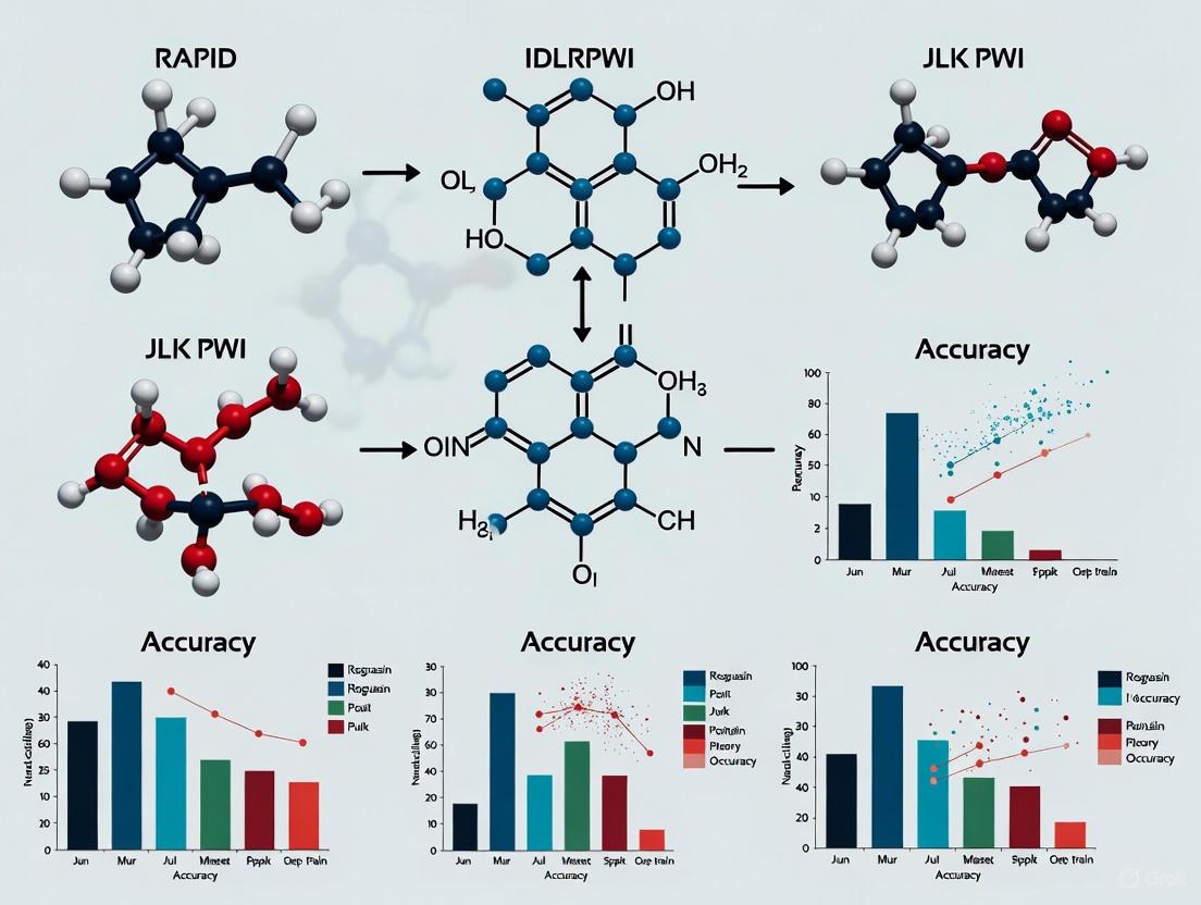
Figure 2: Automated PWI Analysis Workflow. This diagram illustrates the shared processing pipeline and methodological differences between RAPID and JLK PWI platforms for ischemic core and penumbra estimation.
Comparative Performance Data
The comparative validation study generated substantial quantitative data on the agreement between RAPID and JLK PWI:
Table 2: Volumetric Agreement Between RAPID and JLK PWI [7]
| Parameter | Concordance Correlation Coefficient | Strength of Agreement | Statistical Significance |
|---|---|---|---|
| Ischemic Core Volume | 0.87 | Excellent | p < 0.001 |
| Hypoperfused Volume | 0.88 | Excellent | p < 0.001 |
| Mismatch Volume | Not reported | Substantial to Excellent | Not reported |
Beyond volumetric agreement, the study assessed clinical decision concordance based on established trial criteria:
Table 3: Clinical Decision Concordance for EVT Eligibility [7]
| Trial Criteria | Cohen's Kappa (κ) | Strength of Agreement | Clinical Context |
|---|---|---|---|
| DAWN Criteria | 0.80-0.90 | Very High | Large vessel occlusion, 6-24 hour window |
| DEFUSE-3 Criteria | 0.76 | Substantial | Large vessel occlusion, 6-16 hour window |
These results demonstrate that JLK PWI showed excellent technical and clinical concordance with RAPID, supporting its viability as a reliable alternative for MRI-based perfusion analysis in acute stroke care [7]. The high agreement in endovascular therapy eligibility classifications is particularly significant, as this represents the most critical clinical application of this technology.
Table 4: Key Research Reagents and Solutions for Penumbra Imaging Studies
| Resource | Function/Application | Examples/Specifications |
|---|---|---|
| Automated Perfusion Software | Quantification of ischemic core and penumbra volumes | RAPID, JLK PWI, other commercial platforms |
| High-Field MRI Scanners | High-resolution perfusion and diffusion imaging | 3.0T scanners preferred for superior spatial resolution |
| CT Perfusion Protocols | Rapid assessment of cerebral hemodynamics | 256-slice scanners with standardized contrast protocols |
| Deconvolution Algorithms | Calculation of quantitative perfusion parameters | Block-circulant singular value decomposition (SVD) |
| Deep Learning Segmentation | Automated infarct core identification | JLK PWI algorithm trained on manually segmented datasets |
| Threshold Parameters | Standardized definition of core and penumbra | Tmax >6s for hypoperfusion; variable CBF/CBV for core |
| Clinical Trial Criteria | Patient selection for reperfusion therapies | DAWN, DEFUSE-3 criteria for extended window EVT |
Implications for Research and Therapeutic Development
The validation of automated perfusion analysis platforms like RAPID and JLK PWI has profound implications for both clinical practice and research. For drug development professionals, these technologies offer standardized, quantitative biomarkers for patient selection and outcome assessment in clinical trials [5]. The ability to precisely identify penumbral tissue enables more targeted testing of neuroprotective agents that aim to preserve this vulnerable region during ischemia [2].
The excellent agreement between established and emerging platforms suggests that perfusion imaging is reaching a stage of technological maturity where different software solutions can be used interchangeably in research settings, potentially accelerating multi-center trials through harmonized imaging protocols [7] [6]. Furthermore, the integration of artificial intelligence and deep learning approaches, as demonstrated in JLK PWI's core segmentation algorithm, points toward increasingly automated and precise tissue characterization that may further standardize imaging biomarkers across research sites [7].
For researchers investigating the fundamental pathophysiology of cerebral ischemia, these tools provide non-invasive methods to study the temporal and spatial evolution of ischemic injury in human subjects, bridging the gap between animal models and clinical reality [4]. The continued refinement of these technologies will likely focus on expanding applications to more challenging scenarios, such as medium vessel occlusions and posterior circulation strokes, where precise tissue characterization is even more critical [7].
The precise differentiation between ischemic core and penumbra remains the cornerstone of modern acute stroke therapy. Automated perfusion analysis software, including both established platforms like RAPID and emerging solutions like JLK PWI, provide the technological foundation for reliably identifying these critical tissue compartments in clinical and research settings. The excellent concordance between these platforms in both volumetric measurements and clinical decision-making supports their interchangeable use in appropriate contexts.
For researchers and drug development professionals, these technologies offer validated, quantitative biomarkers that can standardize patient selection and outcome assessment across clinical trials. As these platforms continue to evolve with artificial intelligence and machine learning enhancements, they promise to further refine our ability to target therapeutic interventions to those patients most likely to benefit, ultimately advancing the goal of preserving neurological function after ischemic stroke.
The Evolution from Time-Based to Tissue-Based Stroke Windows
The management of acute ischemic stroke has undergone a paradigm shift from rigid time-based windows to individualized tissue-based assessments. This transition has been facilitated by advanced neuroimaging technologies that visualize salvageable brain tissue, enabling treatment decisions based on physiological rather than chronological criteria. This review examines the evolution toward tissue-based windows, with a focused comparison of two automated perfusion analysis platforms: the established RAPID software and the emerging JLK PWI solution. We present experimental data from direct comparative studies evaluating their performance in ischemic core estimation, penumbral quantification, and endovascular therapy selection, providing researchers and clinicians with evidence-based insights for implementation in both clinical and research settings.
The concept of the ischemic penumbra—hypoperfused but potentially salvageable brain tissue surrounding the irreversibly damaged core—has fundamentally transformed acute stroke management. While time from symptom onset remains a crucial factor, the deterministic approach has been supplanted by physiological imaging that identifies patients who may benefit from reperfusion therapies beyond conventional time windows.
This transition was catalyzed by landmark clinical trials (DAWN and DEFUSE 3) that utilized automated perfusion imaging to select patients for endovascular thrombectomy (EVT) up to 24 hours after symptom onset [7] [8]. These trials established that tissue viability, rather than time alone, should determine treatment eligibility. The successful implementation of this paradigm depends on accurate, rapid, and reliable imaging biomarkers to differentiate the ischemic core from the penumbra.
Automated perfusion analysis software has become indispensable in this assessment, with RAPID achieving widespread clinical adoption. However, the evolving landscape has prompted the development of alternative platforms, including JLK PWI. This review provides a comprehensive comparison of these two systems within the broader context of tissue-based stroke windows, examining their technical approaches, performance characteristics, and clinical concordance based on recent comparative validation studies.
Technical Approaches to Perfusion Analysis
The RAPID Platform
RAPID (iSchemaView, USA) is an FDA-approved automated software package that has become the reference standard in perfusion imaging, with validation through multiple major clinical trials [8]. The platform utilizes established thresholding methods for lesion identification:
- Ischemic Core: Defined by apparent diffusion coefficient (ADC) < 620 × 10⁻⁶ mm²/s on MRI or relative cerebral blood flow (rCBF) < 30% on CT perfusion (CTP) [7] [9]
- Hypoperfused Volume: Defined by Tmax > 6 seconds, representing tissue at risk [7]
The software automatically processes perfusion data, generating volumetric outputs for core, penumbra, and mismatch calculations within 5-7 minutes, enabling rapid treatment decisions in emergency settings [8].
The JLK PWI Platform
JLK PWI (JLK Inc., Republic of Korea) is a newly developed alternative that incorporates deep learning algorithms for infarct segmentation [7]. While utilizing similar perfusion thresholds for hypoperfused volume (Tmax > 6 seconds), it employs a distinct approach to core estimation:
- Ischemic Core: Utilizes a deep learning-based infarct segmentation algorithm applied to b1000 DWI images, developed and validated on large manually segmented datasets [7]
- Perfusion Analysis: Employs a multi-step pipeline including motion correction, brain extraction, arterial input function selection, and block-circulant singular value deconvolution for calculating perfusion parameters (CBF, CBV, MTT, Tmax) [7]
Table 1: Technical Comparison of RAPID and JLK PWI Platforms
| Feature | RAPID | JLK PWI |
|---|---|---|
| Core Estimation Method | Threshold-based (ADC < 620) | Deep learning-based segmentation |
| Penumbra Estimation | Tmax > 6 seconds | Tmax > 6 seconds |
| Processing Time | 5-7 minutes | Similar automated processing |
| Primary Input | DWI/PWI or CTP | DWI/PWI or CTP |
| Regulatory Status | FDA-approved | Under validation |
Experimental Workflow for Comparative Validation
The following diagram illustrates the experimental workflow used in recent comparative studies between RAPID and JLK PWI:
Comparative Performance Analysis
Volumetric Agreement Between Platforms
A recent multicenter, retrospective study directly compared RAPID and JLK PWI in 299 patients with acute ischemic stroke who underwent PWI within 24 hours of symptom onset [7] [10]. The study population had a mean age of 70.9 years, 55.9% were male, and the median NIHSS score was 11, representing a typical stroke cohort.
Table 2: Volumetric Agreement Between RAPID and JLK PWI (n=299)
| Parameter | Concordance Correlation Coefficient (CCC) | 95% Confidence Interval | Agreement Classification |
|---|---|---|---|
| Ischemic Core Volume | 0.87 | 0.77-0.94 | Excellent |
| Hypoperfused Volume | 0.88 | 0.81-0.95 | Excellent |
| Mismatch Volume | 0.85 | 0.76-0.92 | Excellent |
The excellent agreement across all volumetric parameters demonstrates that both platforms provide comparable assessments of critical perfusion metrics [7]. Bland-Altman analysis further confirmed minimal mean differences between measurements, with narrow limits of agreement for ischemic core (-2.1 to 3.8 mL), hypoperfused volume (-15.2 to 18.7 mL), and mismatch volume (-17.1 to 19.9 mL) [7].
Clinical Decision Concordance
The ultimate test of perfusion software performance lies in its impact on clinical decision-making, particularly regarding endovascular therapy eligibility. Researchers evaluated this using the validated criteria from the DAWN and DEFUSE-3 trials:
Table 3: EVT Eligibility Concordance Based on Trial Criteria
| Trial Criteria | Patient Subgroup | Cohen's Kappa (κ) | Agreement Classification |
|---|---|---|---|
| DAWN | Anterior circulation LVO (n=123) | 0.80-0.90 | Very High |
| DEFUSE-3 | All patients (n=299) | 0.76 | Substantial |
The very high concordance in DAWN criteria application and substantial agreement for DEFUSE-3 criteria indicates that both platforms would have selected similar patients for thrombectomy in late time windows [7] [10]. This suggests that JLK PWI could serve as a reliable alternative to RAPID without significantly altering treatment decisions.
Advanced Algorithm Approaches
Beyond conventional thresholding methods, deep learning approaches have emerged as promising alternatives for ischemic core estimation. One study explored attention-gated convolutional neural networks trained on multicenter trial data, comparing three approaches [11]:
- Separate Training: Independent models for minimal and major reperfusion cases
- Pretraining with Fine-Tuning: Single model pretrained on diverse cases then fine-tuned
- Conventional Thresholding: Standard clinical method using ADC and Tmax thresholds
The pretraining approach achieved superior performance with median Dice score coefficients of 0.60 for tissue at risk and 0.57 for core, significantly outperforming both separate training (p=0.01 and p=0.04, respectively) and conventional thresholding (p=0.008 and p<0.001, respectively) [11]. This demonstrates the potential for advanced algorithms to enhance accuracy in tissue outcome prediction.
Table 4: Key Research Reagents and Resources for Perfusion Imaging Studies
| Resource | Function/Application | Examples/Specifications |
|---|---|---|
| Perfusion MRI | Spatial resolution, tissue specificity | 1.5T or 3.0T scanners (GE, Philips, Siemens); DSC-PWI with GE-EPI sequence |
| CT Perfusion | Rapid acquisition, accessibility | 256-slice CT scanner; 80 kVp, 150 mAs; iodinated contrast agent |
| Deconvolution Algorithms | Perfusion parameter calculation | Block-circulant SVD (delay-insensitive); Standard SVD |
| Arterial Input Function | Reference for perfusion calculation | Automatically selected from proximal cerebral arteries |
| Validation Reference | Ground truth for algorithm validation | Follow-up DWI (24h-7 days); manually segmented by expert neuroradiologists |
| Statistical Framework | Agreement assessment | Concordance correlation coefficient; Bland-Altman plots; Cohen's kappa |
Methodological Considerations in Perfusion Imaging
Technical Variations and Standardization Challenges
Despite excellent agreement between platforms, several technical factors contribute to inter-software variability:
- Deconvolution Methods: Delay-sensitive versus delay-insensitive algorithms affect Tmax calculations [12]
- Arterial Input Function Selection: Automated versus manual selection introduces variability [13]
- Scanner Differences: Field strength (1.5T vs. 3.0T) and manufacturer (GE, Philips, Siemens) affect perfusion values [7]
- Threshold Applicability: Optimal thresholds may vary across populations and etiologies [6]
The following diagram illustrates the key methodological considerations in perfusion analysis:
Validation Standards and Reference Selection
Determining the optimal reference standard for perfusion software validation remains challenging. Options include:
- Follow-up DWI: Obtained 24 hours to 7 days post-treatment, considered the most practical reference [11] [13]
- Final Infarct on T2-FLAIR: At 3-7 days or 30 days, used in clinical trials [11]
- Expert Manual Segmentation: Time-consuming but provides detailed ground truth [11]
- PET Imaging: Historical gold standard but impractical in acute settings [12]
Most contemporary studies utilize early follow-up DWI (within 24-72 hours) as the reference standard, balancing practical considerations with reasonable accuracy for final infarct representation [11] [13].
The evolution from time-based to tissue-based stroke windows represents one of the most significant advancements in cerebrovascular medicine. Automated perfusion analysis platforms like RAPID and JLK PWI have been instrumental in this transition by providing rapid, objective assessment of salvageable tissue.
Based on current evidence, JLK PWI demonstrates excellent technical and clinical concordance with the established RAPID platform, supporting its viability as an alternative for MRI-based perfusion analysis in acute stroke care [7] [10]. The high agreement in both volumetric measurements and EVT eligibility decisions suggests these platforms can be used interchangeably in clinical and research settings.
Future developments will likely focus on deep learning algorithms that move beyond rigid thresholds to incorporate spatial, temporal, and clinical data for more personalized tissue outcome predictions [11]. Additionally, standardization initiatives addressing technical variations across platforms will be crucial for comparing results across studies and institutions. As tissue-based selection continues to evolve, validation of new platforms against clinical outcomes rather than just reference software will be essential to advance the field and improve patient care.
Automated perfusion analysis has revolutionized the triage of patients with acute ischemic stroke, particularly in extending the treatment window for endovascular therapy (EVT) [7] [14]. RAPID (RAPID AI, CA, USA) has emerged as the established commercial platform in this field, serving as the reference standard in numerous clinical trials and routine care [7] [14]. This guide provides an objective comparison between RAPID and a newly developed alternative, JLK PWI (JLK Inc., Republic of Korea), focusing on their technical performance in ischemic core estimation within magnetic resonance perfusion-weighted imaging (PWI) [7] [10] [15]. The evaluation is contextualized within the broader thesis of advancing ischemic stroke imaging, where accurate infarct core quantification directly impacts patient selection for reperfusion therapies and clinical trial outcomes [7] [14].
Experimental Design & Methodologies
Study Population and Design
The comparative validation between RAPID and JLK PWI employed a retrospective multicenter study design conducted across two tertiary hospitals in Korea [7] [14]. The study enrolled 299 patients with acute ischemic stroke who underwent PWI within 24 hours of symptom onset [7] [15] [14]. Key inclusion criteria involved patients presenting with acute ischemic stroke who had undergone MR perfusion imaging, while exclusion criteria eliminated patients with abnormal arterial input function (n=6), severe motion artifacts (n=2), or inadequate image quality (n=11) [7] [14]. The final cohort had a mean age of 70.9 years, was 55.9% male, and presented with a median NIHSS score of 11 (IQR 5-17), representing a typical acute ischemic stroke population [7] [10] [15].
Imaging Protocols and Technical Parameters
Imaging was performed using standardized protocols across multiple scanner platforms [7] [14]:
- Scanner Distribution: 62.3% on 3.0T and 37.7% on 1.5T scanners
- Vendor Distribution: 34.1% GE, 60.2% Philips, and 5.7% Siemens systems
- Pulse Sequence: Dynamic susceptibility contrast-enhanced perfusion using gradient-echo echo-planar imaging (GE-EPI)
- Key Parameters:
- Repetition time (TR): 1,500-2,000 ms (66.7%)
- Echo time (TE): 40-50 ms (91.8%)
- Field of view (FOV): 230 × 230 mm² (94.3%)
- Slice thickness: 5 mm with no interslice gap
To minimize inter-scanner variability, all datasets underwent standardized preprocessing and normalization prior to PWI mapping in a central image laboratory [7] [14].
Automated PWI Analysis Workflow
The following diagram illustrates the comprehensive processing pipeline for automated perfusion analysis, representing the core methodology shared by both RAPID and JLK PWI platforms:
Automated PWI Processing Workflow
For infarct core estimation, the two platforms employed different approaches rooted in their proprietary algorithms [7] [14]:
- RAPID: Utilized the default threshold of ADC < 620 × 10⁻⁶ mm²/s for infarct core definition
- JLK PWI: Employed a deep learning-based infarct segmentation algorithm applied to b1000 DWI images, developed and validated using large manually segmented datasets
Both platforms defined hypoperfused regions using Tmax >6 seconds threshold and performed automated coregistration between diffusion and perfusion maps for mismatch computation [7] [14]. All segmentations underwent visual inspection to ensure technical adequacy before inclusion in the analysis.
Statistical Analysis Framework
The statistical evaluation employed multiple complementary approaches to assess agreement [7] [14]:
- Volumetric Agreement: Assessed using concordance correlation coefficients (CCC), Pearson correlation coefficients, and Bland-Altman plots for ischemic core volume, hypoperfused volume, and mismatch volume
- Clinical Decision Concordance: Evaluated using Cohen's kappa coefficient based on DAWN and DEFUSE-3 trial criteria for endovascular therapy eligibility
- Agreement Classification:
- Poor (0.0-0.2), Fair (0.21-0.40), Moderate (0.41-0.60)
- Substantial (0.61-0.80), Excellent (0.81-1.0)
Comparative Performance Data
Volumetric Agreement Analysis
The quantitative comparison between RAPID and JLK PWI demonstrated excellent agreement across key perfusion parameters essential for clinical decision-making in acute stroke [7] [10] [15].
Table 1: Volumetric Agreement Between RAPID and JLK PWI Software
| Perfusion Parameter | Concordance Correlation Coefficient (CCC) | Strength of Agreement | P-value |
|---|---|---|---|
| Ischemic Core Volume | 0.87 | Excellent | <0.001 |
| Hypoperfused Volume | 0.88 | Excellent | <0.001 |
The consistency in volumetric measurements extends across both ischemic core and hypoperfused tissue volumes, indicating that JLK PWI provides comparable quantitative assessments to the established RAPID platform [7] [15] [14]. This level of technical concordance supports the potential interoperability of these platforms in both clinical and research settings.
Clinical Decision Concordance
Beyond technical parameters, the study evaluated how software differences translated into clinical decision-making, particularly regarding endovascular therapy eligibility using established trial criteria [7] [14].
Table 2: EVT Eligibility Agreement Based on Clinical Trial Criteria
| Trial Criteria | Cohen's Kappa (κ) | Strength of Agreement | Subgroup Range |
|---|---|---|---|
| DAWN | 0.80-0.90 | Very High | κ=0.80-0.90 |
| DEFUSE-3 | 0.76 | Substantial | - |
The DAWN classification, which stratifies eligible infarct volume based on age and NIHSS into three prespecified categories, showed particularly strong concordance across subgroups (κ=0.80-0.90) [7] [15]. DEFUSE-3 criteria, which utilize a mismatch ratio ≥1.8, infarct core volume <70 mL, and absolute penumbra volume ≥15 mL, demonstrated substantial agreement (κ=0.76) [7] [14].
Research Reagent Solutions
The following table details essential materials and methodological components utilized in the comparative validation study, providing researchers with key resources for experimental replication in perfusion imaging research [7] [14].
Table 3: Essential Research Materials and Methodological Components
| Component Category | Specific Implementation | Research Function |
|---|---|---|
| Primary Software Platforms | RAPID (iSchemaView Inc.) | Established reference standard for automated perfusion analysis |
| JLK PWI (JLK Inc.) | Novel evaluation platform with deep learning segmentation | |
| Imaging Modalities | 3.0T & 1.5T MRI Scanners | Image acquisition across field strengths |
| Dynamic Susceptibility Contrast PWI | Perfusion-weighted image acquisition | |
| Diffusion-Weighted Imaging (DWI) | Infarct core delineation | |
| Analysis Methodologies | Block-Circulant SVD Deconvolution | Perfusion parameter calculation |
| Tmax >6s Threshold | Hypoperfused tissue definition | |
| Deep Learning Segmentation (JLK) | Automated infarct core delineation | |
| Validation Frameworks | DAWN Trial Criteria | EVT eligibility assessment |
| DEFUSE-3 Trial Criteria | Alternative EVT eligibility framework | |
| Concordance Correlation Coefficient | Volumetric agreement quantification |
Technical Considerations and Clinical Implications
The comparative analysis reveals several important technical considerations for researchers and clinicians working with automated perfusion platforms. The excellent volumetric agreement (CCC=0.87-0.88) between JLK PWI and RAPID indicates that these platforms can provide functionally interchangeable quantitative assessments of ischemic core and hypoperfused tissue volumes [7] [15] [14]. This technical concordance is further reinforced by the substantial to very high clinical agreement (κ=0.76-0.90) in EVT eligibility classification based on DAWN and DEFUSE-3 criteria [7] [10] [15].
From a research perspective, the different methodological approaches to infarct core estimation between the platforms—RAPID using ADC thresholding versus JLK PWI employing deep learning segmentation—represent alternative technological pathways to similar clinical endpoints [7] [14]. This methodological divergence highlights the potential for multiple valid computational approaches in perfusion analysis while maintaining consistent output for clinical decision-making.
The implications for clinical trial design are significant, particularly as stroke therapy advances toward more refined patient selection. Recent clinical trials targeting medium vessel occlusion (MeVO) have underscored the need for more precise imaging biomarkers [7] [14]. The high concordance between RAPID and JLK PWI supports the potential for standardized MRI-based perfusion assessment across multiple research platforms, facilitating more consistent patient stratification in future clinical trials [7].
In acute ischemic stroke care, the accurate estimation of the ischemic core and penumbra is paramount for treatment decisions, particularly for endovascular thrombectomy (EVT) in extended time windows. For years, the RAPID software has been the reference standard, validated by landmark clinical trials. However, the landscape of automated perfusion analysis is evolving with the emergence of new platforms. Among these, JLK PWI has recently gained U.S. Food and Drug Administration (FDA) approval, positioning itself as a potential alternative [16]. This comparison guide objectively evaluates the performance of JLK PWI against RAPID in the critical domain of ischemic core estimation accuracy, synthesizing evidence from recent, direct comparative studies to inform researchers and clinicians.
RAPID is a globally established, FDA-approved AI-based software for processing both CT and MR perfusion imaging. It utilizes a delay-insensitive deconvolution algorithm to calculate perfusion maps. For MR perfusion-weighted imaging (PWI), its ischemic core is typically defined by a default threshold of ADC < 620 × 10⁻⁶ mm²/s on diffusion-weighted imaging (DWI) [7]. Its algorithms have been extensively validated in pivotal trials like DEFUSE and DAWN.
JLK PWI is a newly developed software that also received FDA 510(k) clearance [16]. It is a fully automated platform for analyzing MR perfusion data. Its technical pipeline includes motion correction, brain extraction, and automated selection of the arterial input function. It employs a block-circulant single value deconvolution to generate quantitative maps of cerebral blood flow (CBF), cerebral blood volume (CBV), mean transit time (MTT), and Tmax [7]. A key differentiator is its use of a deep learning-based algorithm for segmenting the infarct core on b1000 DWI images, developed using large, manually segmented datasets [7]. The hypoperfused volume is defined using a Tmax > 6 s threshold, consistent with RAPID [7].
Direct, head-to-head comparisons between JLK PWI and RAPID are available from several recent studies. The table below summarizes the core findings from the most comprehensive validation study.
Table 1: Summary of Key Comparative Study between JLK PWI and RAPID
| Study Characteristic | Description | |
|---|---|---|
| Study Design | Retrospective, multicenter study [7] | |
| Patient Population | 299 patients with acute ischemic stroke from two Korean tertiary hospitals [7] | |
| Key Metric | Ischemic Core Volume Agreement | Hypoperfused Volume Agreement |
| Concordance Correlation Coefficient (CCC) | 0.87 (Excellent) [7] | 0.88 (Excellent) [7] |
| Clinical Decision Agreement (DAWN Criteria) | κ = 0.80 - 0.90 (Very High Concordance) [7] | - |
| Clinical Decision Agreement (DEFUSE-3 Criteria) | κ = 0.76 (Substantial Agreement) [7] | - |
Detailed Experimental Protocols and Methodologies
To critically appraise the data, understanding the methodology of the primary validation study is essential.
Study Population and Data Collection
The study pooled data from 299 patients who underwent PWI within 24 hours of symptom onset [7]. Patients were excluded due to abnormal arterial input function, severe motion artifacts, or inadequate images [7]. Clinical data, including demographics, vascular risk factors, and NIH Stroke Scale (NIHSS) scores, were prospectively collected using a standardized protocol [7].
Imaging Acquisition and Analysis
Imaging was performed on 3.0T and 1.5T scanners from multiple vendors (GE, Philips, Siemens) using a gradient-echo echo-planar imaging sequence for dynamic susceptibility contrast-enhanced perfusion [7]. All datasets underwent standardized preprocessing and normalization to minimize inter-scanner variability in a central image laboratory [7]. Each patient's imaging data was processed independently by both the RAPID and JLK PWI software platforms for subsequent comparison.
Statistical Analysis for Comparison
Agreement between the two platforms for volumetric parameters (ischemic core, hypoperfused volume, mismatch volume) was primarily assessed using the concordance correlation coefficient (CCC), supplemented by Pearson correlation and Bland-Altman plots [7]. The strength of agreement was classified as poor (0.0-0.2), fair (0.21-0.40), moderate (0.41-0.60), substantial (0.61-0.80), or excellent (0.81-1.0) [7]. Clinical concordance for EVT eligibility was evaluated using Cohen’s kappa (κ) based on the well-established DAWN and DEFUSE-3 trial criteria [7].
The following diagram illustrates the experimental workflow.
The Scientist's Toolkit: Key Research Reagents and Materials
In the context of computational stroke research, "research reagents" can be conceptualized as the essential software, data, and methodological components required to conduct a validation study.
Table 2: Essential Components for Perfusion Software Validation Research
| Component | Function in Research | Example from JLK vs. RAPID Study |
|---|---|---|
| Automated Perfusion Software | The primary tool for image post-processing; generates quantitative maps of core, penumbra, and mismatch volumes. | RAPID (iSchemaView) and JLK PWI (JLK Inc.) [7]. |
| Curated Patient Imaging Datasets | A well-characterized cohort of raw imaging data (e.g., DICOM files) to serve as the input for software comparison. | 299 patients' PWI and DWI data from two multicenter cohorts [7]. |
| Deep Learning Segmentation Algorithms | Provides automated, reproducible segmentation of infarct regions, reducing manual labor and inter-rater variability. | JLK's deep learning-based infarct segmentation on b1000 DWI [7]. |
| Statistical Analysis Packages (e.g., R, SPSS) | Performs quantitative agreement statistics and generates plots to objectively compare software outputs. | Used for CCC, Bland-Altman analysis, and Cohen's kappa [7] [17]. |
| Clinical Trial Criteria (DAWN/DEFUSE-3) | Acts as a standardized framework to translate volumetric data into clinically actionable decisions (EVT eligibility). | Used to calculate Cohen's kappa for treatment decision concordance [7]. |
Analysis of Performance and Clinical Applicability
The high concordance demonstrated in the key study translates into significant clinical reliability. The excellent volumetric agreement (CCC > 0.85) indicates that JLK PWI provides near-interchangeable measurements of ischemic core and hypoperfused tissue volumes compared to the established RAPID platform [7]. More importantly, the very high agreement in EVT eligibility based on DAWN criteria (κ = 0.80-0.90) suggests that, in the vast majority of cases, both software platforms would lead clinicians to the same treatment decision [7]. This is a critical finding for clinical implementation.
A potential differentiating strength of the JLK platform may lie in the sensitivity of its underlying algorithms. A separate, vendor-reported study highlighted that JLK's DWI-based solution failed to detect lesions in only 1.9% of patients, compared to a reported 61.4% for a competitor's product (identified as RAPID in the context), suggesting a particular efficacy in identifying small lesions [16]. This capability could be increasingly valuable as stroke treatment expands to include smaller, medium vessel occlusions (MeVOs). However, it is important to note that this specific claim requires further independent validation.
The emergence of JLK PWI represents a significant development in the field of automated perfusion analysis. Based on the current evidence, JLK PWI demonstrates excellent technical and substantial clinical concordance with the reference standard RAPID software for MRI-based perfusion analysis in acute ischemic stroke [7]. This supports its validity as a reliable and potential alternative for both clinical and research applications. Its recent FDA approval [16] and purported strengths in detecting smaller infarcts could foster greater competition and innovation in the market. For researchers and clinicians, these findings affirm that JLK PWI is a rigorously validated tool that can be confidently used for ischemic core estimation and EVT triage, aligning closely with the outcomes generated by the established RAPID platform.
Advantages of MR Perfusion-Weighted Imaging (PWI) over CT Perfusion
This guide provides an objective comparison between Magnetic Resonance Perfusion-Weighted Imaging (PWI) and Computed Tomography Perfusion (CTP) in the evaluation of acute ischemic stroke. Framed within the context of ongoing research comparing RAPID and JLK automated analysis platforms, we synthesize technical advantages, clinical performance data, and experimental methodologies. Quantitative analyses from recent multicenter studies demonstrate that MR PWI offers superior spatial resolution, enhanced tissue characterization, and reduced artifact susceptibility compared to CTP, supporting its role as a premium imaging modality for precise ischemic core and penumbra quantification in both clinical and research settings.
Perfusion imaging has revolutionized acute ischemic stroke management by enabling the transition from a rigid "time window" to a more personalized "tissue window" for treatment [18]. The fundamental principle involves differentiating the irreversibly damaged ischemic core from the potentially salvageable ischemic penumbra [19]. The ischemic core represents tissue with profound blood flow reduction that has incurred cellular death, while the penumbra is hypoperfused tissue that remains viable but at risk of infarction without timely reperfusion [19]. Accurate identification of this penumbra is the primary target for revascularization therapies, including endovascular thrombectomy (EVT) and thrombolysis [18] [19].
Both CT perfusion and MR perfusion-weighted imaging are utilized to quantify these tissue compartments, yet they differ significantly in their underlying technology, performance characteristics, and clinical applications. This guide details the specific advantages of MR PWI, with supporting evidence from validation studies of automated analysis platforms such as RAPID and the newer JLK software.
Technical and Physiological Advantages of MR PWI
MR PWI offers several foundational advantages over CTP that enhance its capability for precise tissue characterization in acute stroke.
Superior Spatial Resolution and Tissue Specificity
- Higher Spatial Resolution: PWI provides superior spatial resolution compared to CTP, allowing for more detailed delineation of ischemic territories, particularly in challenging anatomical regions like the posterior fossa [7].
- Enhanced Tissue Characterization: When combined with Diffusion-Weighted Imaging (DWI), PWI enables a more accurate delineation of the infarct core and penumbra. The DWI lesion is considered the MR equivalent of the infarction core, while the mismatch between the abnormal DWI signal and the perfusion deficit on PWI represents the ischemic penumbra [19]. This combined approach offers superior tissue specificity compared to CTP alone [7].
Reduced Artifact Susceptibility
- Freedom from Beam-Hardening Artifacts: Unlike CTP, PWI is free from beam-hardening artifacts, which can degrade image quality and complicate interpretation [7].
- Less Susceptibility to Contrast Timing Errors: The PWI technique is less susceptible to errors related to contrast bolus timing, contributing to more robust and reliable perfusion parameter maps [7].
Absence of Ionizing Radiation
A significant patient safety advantage of MR PWI is that it avoids exposing patients to ionizing radiation, unlike CTP [7]. This is particularly relevant for selected patient populations and research contexts where repeated imaging may be necessary.
Experimental Data and Validation Protocols
Recent comparative studies directly validate the performance of automated PWI analysis and highlight its advantages over CTP.
Multicenter Validation of JLK PWI vs. RAPID
A 2025 retrospective multicenter study directly compared a newly developed software (JLK PWI) against the established RAPID platform for MRI-based perfusion analysis [7].
Study Methodology:
- Population: 299 patients with acute ischemic stroke from two tertiary hospitals who underwent PWI within 24 hours of symptom onset.
- Image Analysis: Both RAPID and JLK PWI platforms were used to calculate ischemic core volume, hypoperfused volume (Tmax > 6 seconds), and mismatch volume.
- Core Estimation: RAPID used an ADC < 620 × 10⁻⁶ mm²/s threshold. JLK PWI utilized a deep learning-based infarct segmentation algorithm on b1000 DWI images.
- Statistical Analysis: Volumetric agreement was assessed using concordance correlation coefficients (CCC), Bland-Altman plots, and Pearson correlations. Clinical concordance for EVT eligibility was evaluated using Cohen’s kappa based on DAWN and DEFUSE-3 criteria.
Key Findings: Table 1: Volumetric Agreement Between JLK PWI and RAPID Software
| Parameter | Concordance Correlation Coefficient (CCC) | P-value |
|---|---|---|
| Ischemic Core Volume | 0.87 | < 0.001 |
| Hypoperfused Volume (Tmax > 6s) | 0.88 | < 0.001 |
Table 2: Clinical Decision Concordance for EVT Eligibility
| Trial Criteria | Cohen's Kappa (κ) | Agreement Level |
|---|---|---|
| DAWN Criteria | 0.80 - 0.90 | Very High |
| DEFUSE-3 Criteria | 0.76 | Substantial |
The study concluded that JLK PWI demonstrates high technical and clinical concordance with RAPID, supporting its use as a reliable alternative for MRI-based perfusion analysis in acute stroke care [7].
Comparative Performance of CTP Software
In contrast, CTP software platforms show greater variability in performance, particularly in specific clinical scenarios. A 2025 study assessed the accuracy of different CTP software packages in ruling out small lacunar infarcts [20].
Methodology:
- Population: 58 patients with suspected acute ischemic stroke but negative follow-up DWI-MRI.
- Software Comparison: syngo.via (Siemens) with multiple parameter settings vs. Cercare Medical Neurosuite (CMN).
- Outcome Measure: Specificity for ischemic core detection (false-positive rate).
Findings: CMN demonstrated high specificity (98.3%, zero infarct volume in 57/58 patients), whereas all syngo.via settings produced false-positive ischemic cores with median volumes ranging from 21.3 mL to 92.1 mL [20]. This highlights significant variability among CTP software and a potential limitation in reliably excluding small infarcts compared to MRI-DWI.
Visualization of Workflows and Advantages
Figure 1: Comparative Workflow and Advantage Analysis of MR PWI versus CT Perfusion
The Scientist's Toolkit: Essential Research Reagents and Materials
Table 3: Key Research Reagents and Software for Perfusion Imaging Studies
| Item | Type/Function | Research Application |
|---|---|---|
| RAPID Software | Automated perfusion analysis platform | Reference standard for ischemic core/penumbra quantification in clinical trials [18] |
| JLK PWI Software | Emerging automated PWI analysis with deep learning algorithm | Comparative validation studies against established platforms [7] |
| Gadolinium-Based Contrast Agents | MR contrast media for perfusion imaging | Essential for generating T2*-weighted perfusion maps in PWI [19] |
| Iodinated Contrast Media | CT contrast agent for perfusion imaging | Required for CTP studies to track contrast bolus through cerebral circulation [21] |
| DWI Sequences (b=1000 s/mm²) | MRI sequence for infarct core detection | Gold standard for infarct core definition in MR studies [7] [19] |
| ADC Maps | Quantitative diffusion imaging | Confirmation of DWI lesions as new ischemic areas (typically ADC < 620×10⁻⁶ mm²/s) [7] |
MR Perfusion-Weighted Imaging demonstrates distinct advantages over CT Perfusion through its superior spatial resolution, reduced artifact susceptibility, absence of ionizing radiation, and enhanced tissue characterization when combined with DWI. Validation studies show that automated PWI analysis platforms like JLK PWI achieve excellent agreement with the established RAPID standard, supporting their reliability for both clinical decision-making and research applications. While CTP remains widely used in emergency settings due to rapid acquisition and broad availability, MR PWI offers a technically advanced alternative for precise patient stratification, particularly valuable in research contexts and selected clinical scenarios where imaging precision is paramount.
Inside the Algorithms: Technical Pipelines of RAPID and JLK PWI
In acute ischemic stroke care, the accurate and rapid estimation of the ischemic core (irreversibly damaged tissue) and the hypoperfused region (tissue at risk) is critical for therapeutic decision-making, particularly for endovascular thrombectomy (EVT) in extended time windows. The RAPID software has become an established tool for this purpose, with its methodology centered on specific imaging thresholds. Its approach is frequently used as a benchmark against which emerging alternatives are validated. This guide provides an objective comparison of RAPID's methodology and performance against JLK PWI, a newly developed software, focusing on their technical approaches and concordance in a research setting. The content is framed within the context of a broader thesis on RAPID vs. JLK PWI ischemic core estimation accuracy, synthesizing findings from a recent multicenter study [7] [14].
Core Methodologies: A Technical Breakdown
RAPID's Established Framework
RAPID employs a fully automated, operator-independent pipeline for processing perfusion and diffusion data. Its methodology is built on the following core principles [22]:
- Ischemic Core Estimation: For MRI-based perfusion, RAPID typically defines the ischemic core using a fixed threshold on the Apparent Diffusion Coefficient (ADC) map, with ADC < 620 × 10⁻⁶ mm²/s as the standard criterion [7].
- Hypoperfusion Estimation: The software identifies hypoperfused tissue (penumbra) using the Tmax > 6 seconds threshold on perfusion maps. Tmax represents the time delay for blood arrival in the tissue relative to the arterial input function [7] [22].
- Automated Processing Workflow: The system automatically performs motion correction, arterial input function (AIF) selection, and deconvolution of tissue and arterial signals to calculate quantitative maps, including Cerebral Blood Flow (CBF), Cerebral Blood Volume (CBV), Mean Transit Time (MTT), and Tmax [22].
JLK PWI's Comparative Approach
JLK PWI is a newer software that mirrors RAPID's automated workflow but incorporates different technological elements in its core estimation [7] [14]:
- Ischemic Core Estimation: Instead of a fixed ADC threshold, JLK PWI utilizes a deep learning-based infarct segmentation algorithm applied to the b1000 Diffusion-Weighted Imaging (DWI) images. This algorithm was developed and validated on large, manually segmented datasets [7].
- Hypoperfusion Estimation: JLK PWI aligns with RAPID by also using the Tmax > 6 seconds threshold to define the hypoperfused tissue volume [7].
- Shared Workflow Elements: Similar to RAPID, its pipeline includes automated preprocessing, motion correction, brain extraction, and block-circulant single value deconvolution to generate perfusion maps [7].
Table 1: Core Methodology Comparison between RAPID and JLK PWI
| Feature | RAPID | JLK PWI |
|---|---|---|
| Ischemic Core Definition (MRI) | Fixed threshold: ADC < 620 × 10⁻⁶ mm²/s [7] | Deep learning-based segmentation on b1000 DWI [7] |
| Hypoperfusion Definition | Tmax > 6 seconds [7] | Tmax > 6 seconds [7] |
| Core Technology | Threshold-based, delay-insensitive deconvolution [22] | Deep learning AI, block-circulant deconvolution [7] |
| Automation Level | Fully automated | Fully automated |
Experimental Validation and Performance Data
Study Protocol and Patient Cohort
The comparative data presented here are primarily derived from a retrospective multicenter study that serves as a key piece of research for the RAPID vs. JLK PWI thesis [7] [14]:
- Study Population: 299 patients with acute ischemic stroke from two tertiary hospitals in Korea.
- Inclusion Criteria: Patients who underwent PWI within 24 hours of symptom onset.
- Key Baseline Characteristics:
- Mean Age: 70.9 years
- Male: 55.9%
- Median NIHSS Score: 11 (IQR 5–17)
- Median Time from Symptom Onset to PWI: 6.0 hours [7]
- Imaging Protocol: Perfusion MRI scans were performed on 3.0T (62.3%) or 1.5T (37.7%) scanners from multiple vendors (GE, Philips, Siemens), using dynamic susceptibility contrast-enhanced perfusion imaging [7].
- Analysis Method: All imaging data were processed by both RAPID and JLK PWI software. Agreement was assessed using Concordance Correlation Coefficients (CCC) and Bland-Altman plots for volumetric parameters. Clinical decision concordance for EVT eligibility was evaluated using Cohen’s kappa based on DAWN and DEFUSE-3 trial criteria [7] [14].
Quantitative Volumetric Agreement
The study found excellent agreement between the two software platforms for key volumetric parameters, as summarized in the table below [7].
Table 2: Volumetric Agreement between RAPID and JLK PWI
| Volumetric Parameter | Concordance Correlation Coefficient (CCC) | Strength of Agreement |
|---|---|---|
| Ischemic Core Volume | 0.87 | Excellent |
| Hypoperfused Volume (Tmax > 6s) | 0.88 | Excellent |
Clinical Decision Concordance
Beyond technical metrics, the study critically evaluated whether the software led to the same clinical treatment decisions. The agreement in EVT eligibility was high [7]:
- DAWN Criteria Concordance: Very high agreement across all patient subgroups (Cohen’s kappa, κ = 0.80–0.90).
- DEFUSE-3 Criteria Concordance: Substantial agreement (Cohen’s kappa, κ = 0.76).
Essential Research Reagents and Materials
The following table details key software and data solutions used in this field of research, as utilized in the featured study.
Table 3: Research Reagent Solutions for Perfusion Imaging Analysis
| Solution Name | Function in Research |
|---|---|
| RAPID (iSchemaView) | Established, FDA-cleared automated software for processing MR PWI and CT Perfusion data; serves as a common benchmark in comparative studies [7] [22]. |
| JLK PWI (JLK Inc.) | A newly developed, fully automated software for MRI-based perfusion analysis; often evaluated as an alternative to RAPID in validation studies [7] [14]. |
| DICOM Datasets | Standardized medical imaging files containing raw PWI and DWI data; the primary input for both RAPID and JLK PWI analysis [7]. |
| DAWN & DEFUSE-3 Criteria | Clinical trial-derived protocols used to define patient eligibility for endovascular therapy based on imaging and clinical features; a key endpoint for evaluating clinical concordance [7]. |
Workflow and Relationship Visualization
The diagram below illustrates the parallel processing workflows of RAPID and JLK PWI, highlighting their methodological convergence at the hypoperfusion stage and divergence at the core estimation stage.
The comparative analysis reveals that while RAPID and JLK PWI employ different technological approaches for ischemic core estimation—threshold-based versus deep learning-based—they demonstrate remarkable technical and clinical concordance. The excellent agreement in volumetric outputs (CCC > 0.85) and substantial to very high agreement in EVT eligibility (κ = 0.76-0.90) validate JLK PWI as a reliable alternative for MRI-based perfusion analysis in acute stroke care [7]. This high level of agreement persists despite their different core estimation methodologies, suggesting that both threshold-based and advanced AI methods can achieve clinically equivalent results in this context.
For researchers and drug development professionals, these findings indicate that the choice of software may be influenced by factors beyond pure performance, such as integration with existing hospital systems, cost, and specific research questions related to AI model interpretability versus traditional thresholding. The consistency in using Tmax > 6s for hypoperfusion measurement provides a standardized metric across platforms, facilitating more uniform patient selection in clinical trials and longitudinal research.
In acute ischemic stroke care, accurate estimation of the ischemic core (irreversibly injured tissue) and the penumbra (at-risk but salvageable tissue) is crucial for treatment decisions, particularly for endovascular thrombectomy (EVT). While computed tomography perfusion (CTP) is widely used in emergency settings, magnetic resonance perfusion-weighted imaging (PWI) offers superior spatial resolution and tissue specificity, especially when combined with diffusion-weighted imaging (DWI) [7] [14]. The RAPID platform has been an established solution in this domain. However, JLK PWI has emerged as a new competitor, leveraging a deep learning-based segmentation approach. This guide provides an objective comparison of their performance, supported by recent experimental data and detailed methodologies, framed within broader research on ischemic core estimation accuracy.
The fundamental difference between JLK PWI and RAPID lies in their technological approaches to infarct core estimation and perfusion mapping.
JLK PWI utilizes a deep learning-based infarct segmentation algorithm applied to b1000 DWI images. This algorithm was developed and validated using large, manually segmented datasets [7] [14]. Its workflow includes automated preprocessing, motion correction, brain extraction (skull stripping and vessel masking), and MR signal conversion. The software automatically selects the arterial input function and venous output function, followed by block-circulant single value deconvolution to calculate quantitative perfusion maps, including cerebral blood flow (CBF), cerebral blood volume (CBV), mean transit time (MTT), and Tmax [7]. The hypoperfused region is delineated using a threshold of Tmax >6 seconds [14].
In contrast, RAPID employs the default threshold of ADC < 620 × 10−6 mm²/s for infarct core estimation from DWI [7] [14]. This threshold-based method represents a more conventional approach to ischemic core delineation.
Table: Core Technical Approaches of JLK PWI vs. RAPID
| Feature | JLK PWI | RAPID |
|---|---|---|
| Infarct Core Estimation | Deep learning-based algorithm on b1000 DWI | Default threshold of ADC < 620 × 10⁻⁶ mm²/s |
| Hypoperfusion Delineation | Tmax >6 seconds threshold | Tmax >6 seconds threshold |
| Key Differentiator | AI-driven segmentation | Established threshold-based method |
Performance Comparison: Volumetric and Clinical Agreement
A recent retrospective multicenter study directly compared JLK PWI and RAPID, involving 299 patients with acute ischemic stroke who underwent PWI within 24 hours of symptom onset [7] [14] [10]. The study evaluated both technical agreement in volume measurements and clinical concordance in EVT eligibility.
Volumetric Agreement
The study assessed agreement for key volumetric parameters using concordance correlation coefficients (CCC), with results summarized in the table below [7] [14].
Table: Volumetric Agreement Between JLK PWI and RAPID
| Parameter | Concordance Correlation Coefficient (CCC) | Agreement Classification |
|---|---|---|
| Ischemic Core Volume | 0.87 | Excellent |
| Hypoperfused Volume | 0.88 | Excellent |
Clinical Decision Concordance
Perhaps more importantly, the study evaluated how the software agreements translated into clinical decision-making for endovascular therapy. Agreement was assessed using Cohen’s kappa (κ) based on the criteria from two major clinical trials: DAWN and DEFUSE-3 [7] [14].
Table: Agreement in EVT Eligibility Classification
| Clinical Trial Criteria | Cohen’s Kappa (κ) | Agreement Classification |
|---|---|---|
| DAWN Criteria | 0.80 - 0.90 | Very High |
| DEFUSE-3 Criteria | 0.76 | Substantial |
Detailed Experimental Protocols
To ensure reproducibility and provide critical context for the comparative data, this section outlines the key methodological details from the validation study.
Study Population and Design
The comparative analysis was a retrospective multicenter study that included patients from two tertiary hospitals in Korea [7] [14]. After initial screening and exclusion of patients due to factors like abnormal arterial input function or severe motion artifacts, 299 patients were included in the final analysis. The cohort had a mean age of 70.9 years, was 55.9% male, and had a median NIHSS score of 11, indicating moderate stroke severity. The median time from the last known well to PWI acquisition was 6.0 hours [7].
Image Acquisition and Analysis Protocol
Imaging was performed across multiple platforms to reflect clinical reality [7] [14]:
- Scanner Distribution: 62.3% of scans were on 3.0 T scanners and 37.7% on 1.5 T scanners.
- Vendor Mix: Scanners from GE (34.1%), Philips (60.2%), and Siemens (5.7%) were used.
- Sequence: Dynamic susceptibility contrast-enhanced PWI was performed using a gradient-echo echo-planar imaging (GE-EPI) sequence. To minimize inter-scanner variability, all datasets underwent standardized preprocessing and normalization prior to PWI mapping. All image analyses were performed in a central image laboratory [7].
Statistical Analysis Protocol
The analysis plan was designed to comprehensively evaluate agreement [7] [14]:
- Volumetric Agreement: Assessed using concordance correlation coefficients (CCC), Pearson correlation coefficients, and Bland–Altman plots.
- Clinical Agreement: EVT eligibility classification agreement was evaluated using Cohen’s kappa coefficient, applied separately for DAWN and DEFUSE-3 trial criteria. The magnitude of agreement for CCC and kappa was classified as per the established guidelines of Landis and Koch (1977).
Workflow and Logical Diagrams
The following diagram illustrates the automated processing pipeline of JLK PWI, which underpins its analytical capabilities.
The Scientist's Toolkit: Essential Research Reagents and Materials
For researchers aiming to conduct similar validation studies or develop algorithms in this field, the following table details key components and their functions as reflected in the cited literature.
Table: Essential Research Reagents and Materials for Perfusion Analysis Validation
| Item | Function / Relevance in Research |
|---|---|
| 3.0 T & 1.5 T MRI Scanners | Platform for acquiring high-resolution DWI and PWI data; essential for testing cross-platform robustness. |
| Multi-Vendor Scanners (GE, Philips, Siemens) | Critical for assessing the generalizability and interoperability of the analysis software across different hardware. |
| Gradient-Echo Echo-Planar Imaging (GE-EPI) Sequence | Standard MRI sequence for dynamic susceptibility contrast-enhanced PWI. |
| b1000 DWI Images | Specific input for the deep learning-based infarct segmentation algorithm in JLK PWI. |
| Manually Segmented Infarct Datasets | Gold-standard ground truth data required for training and validating deep learning models. |
| DAWN & DEFUSE-3 Trial Criteria | Benchmark clinical rulesets for standardizing the assessment of endovascular therapy eligibility. |
| Arterial Input Function (AIF) / Venous Output Function (VOF) | Crucial for deconvolution models in perfusion parameter calculation; often automated in software. |
| Block-Circulant Single Value Deconvolution | Mathematical method used to calculate hemodynamic parameters (CBF, CBV, MTT, Tmax) from raw perfusion data. |
The comparative validation indicates that JLK PWI demonstrates excellent technical agreement and substantial to very high clinical concordance with the established RAPID platform [7] [14]. Its deep learning-based approach for infarct core segmentation presents a viable and reliable alternative for MRI-based perfusion analysis in acute stroke care. This performance, combined with its recent U.S. FDA 510(k) clearance, positions JLK PWI as a competitive tool in the clinical and research landscape [23] [24] [16]. For researchers and drug development professionals, these findings highlight the ongoing evolution and validation of AI-driven methodologies in medical imaging, which are increasingly critical for patient stratification and outcomes research in acute ischemic stroke.
The accurate estimation of the ischemic core is a critical determinant in treatment decisions and prognostic predictions for acute ischemic stroke. Within this context, the comparative performance of automated perfusion analysis software represents a central theme in modern neuroimaging research. This guide provides a systematic, data-driven comparison of two prominent platforms—RAPID and JLK PWI—focusing on their technical workflows and ischemic core estimation accuracy. Framed within a broader thesis on validation research, this analysis leverages a recent multicenter study to objectively evaluate their operational protocols, quantitative outputs, and final clinical conclusions [14] [7].
Experimental Protocol and Study Design
The foundational data for this comparison are derived from a retrospective multicenter study designed to validate a new software platform against an established standard [14].
Study Population and Data Acquisition
The study incorporated 299 patients with acute ischemic stroke who underwent perfusion-weighted imaging (PWI) within 24 hours of symptom onset [14] [7]. The cohort's key characteristics are summarized in Table 1.
Table 1: Baseline Characteristics of the Study Population
| Characteristic | Summary Value |
|---|---|
| Mean Age (years) | 70.9 |
| Male Sex | 55.9% |
| Median NIHSS Score | 11 (IQR 5-17) |
| Median Time from LKW to PWI (hours) | 6.0 |
| Magnetic Field Strength | 3.0 T (62.3%), 1.5 T (37.7%) |
| Scanner Vendors | GE (34.1%), Philips (60.2%), Siemens (5.7%) |
All PWI scans were performed using dynamic susceptibility contrast-enhanced imaging with a gradient-echo echo-planar imaging (GE-EPI) sequence. To ensure consistency and minimize inter-scanner variability, all datasets underwent standardized preprocessing and normalization in a central image laboratory before quantitative perfusion analysis [14] [7].
Key Research Reagent Solutions
The following table details the essential software and methodological "reagents" crucial for replicating this comparative workflow.
Table 2: Research Reagent Solutions for Perfusion Analysis Validation
| Research Reagent | Function/Description |
|---|---|
| JLK PWI (JLK Inc.) | Automated PWI analysis software; the test platform in the validation study. |
| RAPID (RAPID AI) | Established commercial PWI analysis software; the reference platform. |
| GE-EPI Sequence | The specific MRI sequence used for acquiring all perfusion-weighted images. |
| Block-Circulant SVD | The deconvolution algorithm used by JLK PWI to calculate perfusion maps [14]. |
| Deep Learning Infarct Segmentation | JLK's algorithm for infarct core estimation on b1000 DWI images [14]. |
| DAWN/DEFUSE-3 Criteria | Standardized clinical criteria used to assess concordance in EVT eligibility. |
Comparative Workflow Analysis: From Preprocessing to Final Output
The workflows for RAPID and JLK PWI, while sharing a common goal, exhibit distinct differences in their approach to image processing and analysis. The following diagram illustrates the parallel pathways from image input to final output.
Diagram Title: RAPID vs. JLK PWI Workflow Comparison
Workflow Stage 1: Image Preprocessing
Both platforms initiate their pipelines with automated preprocessing to prepare images for analysis.
- RAPID: The specific preprocessing steps for RAPID's MRI pipeline are not exhaustively detailed in the provided results but are inferred to include essential steps like motion correction and arterial input function (AIF) selection, consistent with standard perfusion analysis [14].
- JLK PWI: Its workflow explicitly includes motion correction for acquisition artifacts, brain extraction via skull stripping, vessel masking, and conversion of the MR signal. A distinctive step is the automated selection of both the arterial input function (AIF) and venous output function (VOF) [14].
Workflow Stage 2: Ischemic Core Estimation
This stage highlights a fundamental methodological divergence between the two platforms.
- RAPID: Estimates the infarct core using a fixed apparent diffusion coefficient (ADC) threshold of < 620 × 10⁻⁶ mm²/s, a well-established and widely used method in acute stroke imaging [14].
- JLK PWI: Employs a deep learning-based infarct segmentation algorithm applied directly to the b1000 DWI images. This method was developed and validated on large, manually segmented datasets, potentially offering a more nuanced approach to core identification [14] [7].
Workflow Stage 3: Perfusion Map Generation and Final Output
Both software packages then calculate quantitative perfusion maps—including cerebral blood flow (CBF), cerebral blood volume (CBV), mean transit time (MTT), and time to maximum (Tmax)—using delay-insensitive deconvolution algorithms, with JLK PWI specified to use block-circulant singular value deconvolution [14].
The final output for both platforms is a set of quantitative volumes: the ischemic core volume (from DWI), the hypoperfused tissue volume (typically defined as Tmax > 6 seconds), and the calculated mismatch ratio between them [14]. These outputs are directly used for clinical decision-making.
Quantitative Performance and Clinical Concordance
The validation study employed robust statistical methods to evaluate agreement between RAPID and JLK PWI, assessing both technical performance and clinical utility.
Volumetric Agreement
The agreement for key volumetric parameters was quantified using concordance correlation coefficients (CCC), with results demonstrating excellent concordance [14] [7].
Table 3: Volumetric Agreement Between RAPID and JLK PWI
| Volumetric Parameter | Concordance Correlation Coefficient (CCC) | Strength of Agreement |
|---|---|---|
| Ischemic Core Volume | 0.87 (p < 0.001) | Excellent |
| Hypoperfused Volume (Tmax > 6s) | 0.88 (p < 0.001) | Excellent |
Clinical Decision-Making Concordance
The most critical test for any alternative platform is its agreement with the established standard on clinical decisions. The study evaluated this using Cohen's kappa (κ) to measure concordance in endovascular therapy (EVT) eligibility based on the criteria of two major clinical trials [14] [7].
Table 4: Clinical Decision Concordance for EVT Eligibility
| Clinical Trial Criteria | Cohen's Kappa (κ) | Strength of Agreement |
|---|---|---|
| DAWN Criteria | 0.80 - 0.90 | Very High |
| DEFUSE-3 Criteria | 0.76 | Substantial |
The experimental data from this validation study indicate that JLK PWI demonstrates high technical and clinical concordance with the established RAPID platform [14] [7]. The excellent volumetric agreement (CCC > 0.85) and substantial to very high concordance in EVT eligibility (κ = 0.76 to 0.90) provide strong evidence that JLK PWI is a reliable alternative for MRI-based perfusion analysis in acute stroke care.
This conclusion is strengthened by the rigorous study design, which was a multicenter retrospective analysis with a substantial sample size (n=299), conducted in a central laboratory to minimize variability. The divergence in core estimation methodologies—a threshold-based approach for RAPID versus a deep learning-based approach for JLK PWI—makes the high level of agreement particularly noteworthy. It suggests that while the internal workflows of the two platforms differ, their final outputs are highly consistent, supporting the clinical viability of the JLK PWI software. This independent validation is a crucial step in the broader thesis of benchmarking new analytical tools against proven standards in stroke imaging.
This guide provides an objective comparison of the RAPID and JLK PWI software platforms, focusing on their performance in implementing the DAWN and DEFUSE-3 trial criteria for endovascular therapy (EVT) selection in acute ischemic stroke. The data presented is framed within broader research on ischemic core estimation accuracy.
The DAWN and DEFUSE-3 clinical trials revolutionized stroke care by establishing that patients with large vessel occlusive stroke could benefit from endovascular thrombectomy (EVT) beyond the traditional 6-hour time window, up to 16-24 hours after symptom onset [25] [26]. Both trials utilized advanced perfusion imaging with automated software to identify patients with salvageable brain tissue despite extended time from symptom onset.
The DAWN trial criteria use a clinical-imaging mismatch approach, stratifying patients based on age, National Institutes of Health Stroke Scale (NIHSS) score, and infarct core volume [25] [27]. Specifically, it defines eligibility as: (1) age <80 years with NIHSS ≥10 and infarct volume <31 mL; (2) age <80 years with NIHSS ≥20 and infarct volume 31-51 mL; or (3) age ≥80 years with NIHSS ≥10 and infarct volume <21 mL [25].
The DEFUSE-3 trial criteria employ a core-penumbra mismatch model, requiring NIHSS ≥6, infarct core volume <70 mL, penumbra volume ≥15 mL, and a mismatch ratio (penumbra/core) ≥1.8 [25] [7]. Both protocols depend on accurate, automated imaging analysis to qualify patients for treatment, with RAPID software being used in the original trials [26].
Experimental Protocols for Software Comparison
Study Population and Design
The comparative validation data presented herein is derived from a retrospective multicenter study that included 299 patients with acute ischemic stroke who underwent perfusion-weighted imaging (PWI) within 24 hours of symptom onset [7] [10]. The mean age of participants was 70.9 years, 55.9% were male, and the median NIHSS score was 11 (IQR 5-17). The median time from last known well to PWI was 6.0 hours [7].
Patients were recruited from two tertiary hospitals in Korea, with initial screening of 318 patients. After exclusion of 19 patients due to abnormal arterial input function (n=6), severe motion artifacts (n=2), or inadequate images (n=11), 299 patients were included in the final analysis [7]. The study protocol was approved by the institutional review board of Seoul National University Bundang Hospital, and written informed consent was obtained from all patients or their legal representatives [7] [10].
Imaging Acquisition and Analysis
All perfusion MRI scans were performed on either 3.0T (62.3%) or 1.5T (37.7%) scanners from multiple vendors [7]. Dynamic susceptibility contrast-enhanced perfusion imaging was performed using a gradient-echo echo-planar imaging sequence with the following parameters: repetition time (TR) = 1,000-2,500 ms; echo time (TE) = 30-70 ms; field of view (FOV) = 210×210 mm² or 230×230 mm²; and slice thickness of 5 mm with no interslice gap [7].
Both RAPID and JLK PWI platforms performed automated preprocessing and perfusion parameter calculations through multi-step pipelines. The JLK PWI workflow included motion correction, brain extraction via skull stripping and vessel masking, and conversion of MR signal [7]. The software automatically selected arterial input function and venous output function, followed by block-circulant single value deconvolution to calculate quantitative perfusion maps, including cerebral blood flow (CBF), cerebral blood volume (CBV), mean transit time (MTT), and Tmax parameters [7].
For infarct core estimation, RAPID employed the default threshold of ADC < 620×10⁻⁶ mm²/s, while JLK PWI utilized a deep learning-based infarct segmentation algorithm applied to the b1000 diffusion-weighted imaging (DWI) images [7]. The hypoperfused region was delineated using the threshold of Tmax >6 seconds for both platforms [7].
Statistical Analysis
Agreement between the two platforms in perfusion parameter measurements (ischemic core volume, hypoperfused volume, and mismatch volume) was assessed using concordance correlation coefficients (CCC), Pearson correlation coefficients, and Bland-Altman plots [7]. The magnitude of agreement was classified as: poor (0.0-0.2), fair (0.21-0.40), moderate (0.41-0.60), substantial (0.61-0.80), and excellent (0.81-1.0) [7].
For EVT eligibility, classification agreement between RAPID and JLK software was evaluated using Cohen's kappa coefficient, applied separately for each subgroup defined by the DAWN and DEFUSE-3 trial criteria [7] [10]. The DAWN classification stratified eligible infarct volume based on age and NIHSS into three prespecified categories, while the DEFUSE-3 classification used a mismatch ratio ≥1.8, infarct core volume <70 mL, and absolute penumbra volume ≥15 mL [7].
Comparative Performance Data
Volumetric Agreement in Ischemic Core Estimation
Table 1: Volumetric Agreement Between RAPID and JLK PWI Platforms
| Parameter | Concordance Correlation Coefficient (CCC) | Pearson Correlation Coefficient | Agreement Classification |
|---|---|---|---|
| Ischemic Core Volume | 0.87 | 0.89 | Excellent |
| Hypoperfused Volume | 0.88 | 0.90 | Excellent |
| Mismatch Volume | 0.85 | 0.87 | Excellent |
JLK PWI showed excellent agreement with RAPID for all key volumetric parameters used in DAWN and DEFUSE-3 criteria implementation [7]. The concordance correlation coefficient was 0.87 (p<0.001) for ischemic core volume and 0.88 (p<0.001) for hypoperfused volume, both classified as excellent agreement [7]. The high agreement in mismatch volume (CCC=0.85) is particularly relevant for DEFUSE-3 criteria application, which requires a mismatch ratio ≥1.8 [7].
Clinical Decision Concordance for EVT Eligibility
Table 2: EVT Eligibility Concordance Based on DAWN and DEFUSE-3 Criteria
| Trial Criteria | Cohen's Kappa (κ) | Agreement Classification | Key Determining Parameters |
|---|---|---|---|
| DAWN | 0.80-0.90 | Substantial to Excellent | Age, NIHSS, infarct core volume |
| DEFUSE-3 | 0.71-0.76 | Substantial | Infarct core <70mL, mismatch ratio ≥1.8, penumbra ≥15mL |
EVT eligibility classifications based on DAWN criteria showed very high concordance across subgroups (κ=0.80-0.90), indicating that both platforms would select similar patients for treatment using DAWN criteria [7] [10]. Substantial agreement was observed using DEFUSE-3 criteria (κ=0.71-0.76) [7] [10]. This demonstrates that JLK PWI can implement both major trial protocols with high consistency compared to the established RAPID platform.
The Scientist's Toolkit: Research Reagent Solutions
Table 3: Essential Materials and Software for DAWN/DEFUSE-3 Implementation
| Resource | Type | Function in Implementation |
|---|---|---|
| RAPID Software | Automated perfusion analysis platform | Reference standard for processing CTP/PWI data; used in original trials |
| JLK PWI | Automated perfusion analysis platform | Alternative platform for MRI-based perfusion analysis; utilizes deep learning algorithms |
| CT Perfusion (CTP) | Imaging modality | Widely available method for perfusion imaging; used in many stroke centers |
| MR Perfusion (PWI) | Imaging modality | Higher spatial resolution alternative to CTP; avoids ionizing radiation |
| Tmax >6s Threshold | Processing parameter | Defines hypoperfused tissue volume in both platforms |
| ADC <620×10⁻⁶ mm²/s | Processing parameter | RAPID's threshold for infarct core definition on DWI |
| Deep Learning Segmentation | Algorithm | JLK PWI's method for infarct core definition on DWI |
Technical Advantages and Implementation Considerations
The comparative studies reveal several important technical aspects relevant to researchers and clinicians implementing DAWN and DEFUSE-3 protocols. MR perfusion-weighted imaging (PWI), used in both platforms, offers several advantages over CT perfusion, including higher spatial resolution, freedom from beam-hardening artifacts, less susceptibility to contrast timing errors, and no ionizing radiation exposure [7]. These features improve image quality, particularly in challenging regions such as the posterior fossa or in patients with small vessel disease [7].
The JLK PWI platform employs a deep learning-based infarct segmentation algorithm applied to b1000 DWI images, which was developed and validated using large manually segmented datasets [7]. This approach differs from RAPID's use of ADC thresholding (<620×10⁻⁶ mm²/s) for infarct core definition [7]. Despite these methodological differences, the high concordance between platforms suggests both can reliably implement the core volume measurements critical to DAWN and DEFUSE-3 criteria.
For researchers designing clinical trials or implementing these protocols in practice, the substantial to excellent agreement in EVT eligibility classification (κ=0.71-0.90) indicates that JLK PWI can serve as a reliable alternative to RAPID for applying both DAWN and DEFUSE-3 criteria [7] [10]. This is particularly relevant for centers utilizing MRI as their primary advanced imaging modality for acute stroke assessment.
This guide provides an objective comparison of the volumetric outputs for ischemic core, penumbra, and mismatch ratio generated by the established RAPID software and the newer JLK PWI platform, based on current clinical research.
Experimental Protocols & Methodologies
A direct comparative validation study employed a retrospective, multicenter design to evaluate the performance of JLK PWI against RAPID [7] [14] [15]. The key methodological elements are summarized below.
Study Population: The research involved 299 patients with acute ischemic stroke who underwent MRI with perfusion-weighted imaging (PWI) and diffusion-weighted imaging (DWI) within 24 hours of symptom onset [7]. The mean age of participants was 70.9 years, and the median NIHSS score was 11, indicating moderate stroke severity [14].
Image Analysis Workflow: Both software packages, RAPID and JLK PWI, are designed for fully automated processing of PWI and DWI data [28]. The core analysis pipeline, which is largely similar between the two platforms, is illustrated in the following diagram.
Key Parameter Definitions: Both platforms utilize specific, validated thresholds to define critical tissue compartments [7] [18]:
- Ischemic Core: RAPID employs an apparent diffusion coefficient (ADC) threshold of < 620 × 10⁻⁶ mm²/s. JLK PWI uses a deep learning-based segmentation algorithm applied to DWI (b=1000) images [7].
- Hypoperfused Tissue (Penumbra): Both platforms defined the hypoperfused volume using a Tmax threshold of >6 seconds [7].
- Mismatch Ratio: This was calculated as the ratio of the hypoperfused volume (Tmax >6s) to the ischemic core volume [7] [29].
Statistical Analysis: The primary analysis assessed agreement for continuous volumetric measures (ischemic core, hypoperfused volume, mismatch volume) using concordance correlation coefficients (CCC), Pearson correlations, and Bland-Altman plots. For clinical decision-making, agreement on endovascular therapy (EVT) eligibility was evaluated using Cohen’s kappa (κ) statistic, applied based on the criteria from the DAWN and DEFUSE-3 clinical trials [7] [14].
Comparative Volumetric Output Data
The study provided a direct, head-to-head comparison of the volumetric outputs generated by the two software platforms. The following table summarizes the key agreement metrics for the primary volumetric parameters.
Table 1: Volumetric Agreement between RAPID and JLK PWI
| Volumetric Parameter | Concordance Correlation Coefficient (CCC) | Strength of Agreement | P-value |
|---|---|---|---|
| Ischemic Core Volume | 0.87 | Excellent | < 0.001 |
| Hypoperfused Volume | 0.88 | Excellent | < 0.001 |
Source: Adapted from Kim et al. (2025). Frontiers in Neuroscience [7] [14].
The high concordance observed in volumetric outputs naturally translates into clinical decision-making. The study also evaluated how the use of either software would impact patient selection for endovascular thrombectomy (EVT) based on the criteria of two major clinical trials.
Table 2: Clinical Decision Concordance for EVT Eligibility
| Clinical Trial Criteria | Cohen's Kappa (κ) | Strength of Agreement |
|---|---|---|
| DAWN Criteria | 0.80 - 0.90 | Very High |
| DEFUSE-3 Criteria | 0.76 | Substantial |
Source: Adapted from Kim et al. (2025). Frontiers in Neuroscience [7] [15].
A separate study focusing on CT perfusion (CTP) found similarly excellent agreement between the JLK and RAPID platforms, with a CCC of 0.958 for ischemic core volume (using rCBF <30%) and 0.835 for hypoperfused volume (Tmax >6s) [6]. This indicates consistent performance of the JLK software across different imaging modalities.
The Scientist's Toolkit: Research Reagent Solutions
The following table details key software and data components essential for conducting or interpreting comparative research in automated perfusion analysis.
Table 3: Essential Research Materials and Software Solutions
| Item | Function in Research Context |
|---|---|
| RAPID Software | The established, FDA-cleared commercial platform used as a reference standard for comparing the performance of new perfusion analysis tools [18]. |
| JLK PWI Software | The novel software under investigation; it utilizes a deep learning-based algorithm for DWI infarct segmentation and a standardized pipeline for PWI parameter calculation [7]. |
| DICOM Datasets | Multicenter, retrospective collections of PWI and DWI images from patients with acute ischemic stroke, which serve as the primary input data for validation studies [7] [6]. |
| DAWN/DEFUSE-3 Criteria | Standardized clinical trial protocols used to translate volumetric data (core, penumbra, mismatch ratio) into binary decisions for endovascular therapy eligibility, enabling the assessment of clinical concordance [7] [18]. |
| Statistical Packages (e.g., SPSS, R) | Software used for calculating concordance metrics (CCC, Cohen's Kappa) and generating Bland-Altman plots to quantify the level of agreement between different software platforms [7] [29]. |
Visualizing Clinical Decision Pathways
The volumetric outputs generated by software like RAPID and JLK PWI are integrated into specific decision pathways to guide treatment. The logic for determining EVT eligibility based on the DEFUSE-3 criteria is mapped out below.
Current evidence demonstrates that the JLK PWI software produces volumetric outputs for ischemic core and hypoperfused tissue that have excellent technical agreement with those generated by the established RAPID platform [7]. This high technical concordance directly translates into substantial to near-perfect agreement in clinical decision-making for endovascular therapy, as defined by DAWN and DEFUSE-3 criteria [7] [15]. These findings support JLK PWI as a reliable and accurate alternative for MRI-based perfusion analysis in both research and clinical practice for acute ischemic stroke.
Challenges and Limitations in Automated Perfusion Analysis
In the field of acute ischemic stroke imaging, the accurate estimation of the ischemic core and penumbra is critical for guiding treatment decisions, particularly for endovascular therapy (EVT). Automated perfusion-weighted imaging (PWI) analysis software, such as RAPID and JLK PWI, has become indispensable in clinical and research settings for this purpose. However, the accuracy of these platforms is susceptible to several technical pitfalls that can significantly impact their volumetric assessments and subsequent clinical decisions. This guide provides a systematic comparison of how RAPID and JLK PWI address common challenges including motion artifacts, arterial input function (AIF) selection, and overall image quality, framing this discussion within the broader context of ischemic core estimation accuracy research. The analysis draws upon recent multicenter validation studies to offer evidence-based insights for researchers and drug development professionals working to optimize stroke imaging protocols and validate novel therapeutic approaches.
Technical Approaches to Common Pitfalls
Motion Artifacts: Mitigation Strategies
Patient motion during MR perfusion acquisition can introduce significant errors in final cerebral blood flow (CBF) maps by creating misalignment between control and label images. These artifacts manifest as artificial hyper- or hypointense perfusion signals that can distort the true ischemic core and penumbra volumes [30].
RAPID's Approach: The established RAPID platform incorporates automated registration and motion correction as part of its processing pipeline. This methodology aligns with principles demonstrated in large-scale clinical implementations where realignment of individual control and label imaging volumes is performed before quantitative analysis [30].
JLK PWI's Approach: JLK PWI implements a multi-step preprocessing pipeline that specifically includes motion correction to address acquisition artifacts [7] [14]. This automated approach to motion mitigation demonstrates the platform's capacity to handle a common source of technical variability in perfusion imaging.
Table: Motion Artifact Mitigation Strategies in PWI Platforms
| Platform | Motion Correction Method | Technical Basis | Validation Approach |
|---|---|---|---|
| RAPID | Automated registration and realignment | Control/label image realignment using statistical parametric mapping | Clinical implementation across multiple sites |
| JLK PWI | Multi-step preprocessing with motion correction | Automated pipeline addressing acquisition artifacts | Visual inspection of technical adequacy post-segmentation |
Arterial Input Function Selection: Algorithmic Differences
The selection of an appropriate arterial input function (AIF) is crucial for accurate deconvolution of perfusion parameters, as it represents the contrast agent concentration input to the brain tissue. Inaccurate AIF selection can lead to substantial errors in cerebral blood flow (CBF) and time-to-maximum (Tmax) calculations [7].
Comparative Analysis: Both RAPID and JLK PWI employ fully automated algorithms for AIF selection, eliminating inter-observer variability that can occur with manual selection. According to validation studies, JLK PWI automatically selects the arterial input function and venous output function as part of its processing pipeline, followed by block-circulant single value deconvolution for calculating quantitative perfusion maps [7] [14]. This automated approach to AIF selection represents a critical methodological similarity between the platforms that contributes to their high concordance.
The exclusion criteria used in the comparative validation study further highlight the importance of proper AIF selection, with patients excluded due to abnormal arterial input function [7], indicating that both platforms require adequate AIF for reliable operation.
Image Quality and Reconstruction
Image quality in perfusion MRI is influenced by multiple factors including spatial resolution, signal-to-noise ratio, and reconstruction algorithms. These factors directly impact the accuracy of ischemic tissue delineation.
Spatial Resolution Advantages: Magnetic resonance perfusion-weighted imaging offers several technical advantages over computed tomography perfusion, including higher spatial resolution and freedom from beam-hardening artifacts [7] [14]. These characteristics are particularly beneficial for imaging challenging regions such as the posterior fossa or in patients with small vessel disease.
Reconstruction Methods: While the search results don't detail specific reconstruction algorithms for the PWI platforms, recent advances in deep learning reconstruction for medical imaging suggest potential future directions. Studies of CT reconstruction have demonstrated that deep learning algorithms can reduce noise magnitude significantly (-27% ± 3%) while improving spatial resolution and detectability compared to traditional iterative reconstruction methods [31]. Although these findings come from CT imaging, they highlight the potential for advanced reconstruction techniques in perfusion imaging more broadly.
Diagram Title: PWI Processing Pipeline and Critical Pitfall Points
Quantitative Comparison of Platform Performance
Volumetric Agreement in Multicenter Validation
A direct comparative validation study between JLK PWI and RAPID evaluated 299 patients with acute ischemic stroke from multiple centers, providing robust evidence of their technical concordance [7] [14].
Table: Volumetric Agreement Between JLK PWI and RAPID
| Parameter | Concordance Correlation Coefficient (CCC) | Statistical Significance | Agreement Classification |
|---|---|---|---|
| Ischemic Core Volume | 0.87 | p < 0.001 | Excellent |
| Hypoperfused Volume (Tmax > 6s) | 0.88 | p < 0.001 | Excellent |
| Mismatch Volume | Reported in study | p < 0.001 | Substantial to Excellent |
The excellent agreement observed across key volumetric parameters demonstrates that both platforms produce highly comparable estimates of critically hypoperfused tissue, despite potential differences in their underlying algorithms for motion correction and AIF selection.
Clinical Decision Concordance
Beyond technical agreement, the crucial test for any perfusion platform lies in its consistency in guiding clinical treatment decisions. The study evaluated this using established trial criteria for endovascular therapy eligibility [7] [15].
Table: EVT Eligibility Concordance Based on Trial Criteria
| Eligibility Criteria | Cohen's Kappa (κ) | Agreement Level | Clinical Impact |
|---|---|---|---|
| DAWN Criteria | 0.80-0.90 (across subgroups) | Very High | Consistent patient selection for thrombectomy |
| DEFUSE-3 Criteria | 0.76 | Substantial | Reliable mismatch identification |
The high concordance in EVT eligibility decisions underscores the clinical reliability of both platforms and suggests that technical differences in their approach to image quality optimization and artifact mitigation do not translate into significantly different treatment recommendations.
Experimental Protocols for Methodological Validation
Multicenter Study Design
The primary validation study employed a retrospective multicenter design including 299 patients from two tertiary hospitals in Korea [7] [14]. Patients underwent PWI within 24 hours of symptom onset, with a median time from last known well to PWI of 6.0 hours. The study population had a mean age of 70.9 years, with 55.9% male participants and a median NIHSS score of 11, representing a typical acute stroke cohort.
Exclusion Criteria: The study applied specific quality control exclusions, removing patients with:
- Abnormal arterial input function (n=6)
- Severe motion artifacts (n=2)
- Inadequate images (n=11)
These exclusions highlight the very real impact of the technical pitfalls discussed in this guide and demonstrate how researchers should account for them in study design.
Image Acquisition and Analysis Protocol
MRI Acquisition Parameters: All perfusion MRI scans were performed on either 3.0T (62.3%) or 1.5T (37.7%) scanners from multiple vendors [7]. Dynamic susceptibility contrast-enhanced perfusion imaging used a gradient-echo echo-planar imaging sequence with the following parameters:
- Repetition Time (TR): 1,000–1,500 ms (6.3%), 1,500–2,000 ms (66.7%), or 2,000–2,500 ms (27.0%)
- Echo Time (TE): 30–40 ms (1.0%), 40–50 ms (91.8%), or 60–70 ms (7.2%)
- Field of View: 210 × 210 mm² (5.7%) or 230 × 230 mm² (94.3%)
- Slice Thickness: 5 mm with no interslice gap
Analysis Methodology: To minimize inter-scanner variability, all datasets underwent standardized preprocessing and normalization prior to PWI mapping. All image analyses were performed in a central image laboratory, ensuring consistent application of quality control measures [7] [14].
The Scientist's Toolkit: Essential Research Reagents and Materials
Table: Key Experimental Components for PWI Validation Studies
| Component | Specification | Research Function | Considerations |
|---|---|---|---|
| PWI Acquisition Sequence | Gradient-echo echo-planar imaging (GE-EPI) | Dynamic susceptibility contrast-enhanced perfusion weighting | Optimize for TR/TE parameters based on scanner capabilities |
| Ischemic Core Segmentation | JLK: Deep learning-based algorithm on b1000 DWI; RAPID: ADC < 620 × 10⁻⁶ mm²/s | Quantification of irreversibly infarcted tissue | Different methodological approaches require validation against follow-up imaging |
| Hypoperfusion Threshold | Tmax > 6 seconds | Identification of critically hypoperfused tissue | Standardized threshold enables cross-platform comparisons |
| Deconvolution Algorithm | Block-circulant singular value decomposition | Calculation of quantitative perfusion parameters | Delay-insensitive method reduces errors in low-flow conditions |
| Validation Reference | Early follow-up DWI (within 3 hours of CTP) | Ground truth for ischemic core validation | Minimizes confounding from infarct growth between scans |
| Statistical Framework | Concordance correlation coefficients (CCC) & Bland-Altman plots | Quantification of volumetric agreement | Provides comprehensive assessment beyond simple correlation |
The comparative validation of RAPID and JLK PWI reveals excellent technical agreement in ischemic core and hypoperfused volume estimation, with very high concordance in clinical decision-making for endovascular therapy. While both platforms employ automated solutions to mitigate common pitfalls like motion artifacts and AIF selection errors, researchers should remain vigilant about image quality factors that could impact volumetric assessments in individual cases. The methodological approaches and experimental protocols outlined in this guide provide a framework for rigorous validation of perfusion analysis software, ensuring that technical differences between platforms do not unduly influence research outcomes or clinical decisions in stroke imaging.
Addressing Inter-Scanner and Inter-Vendor Variability in MRI Data
In multi-center clinical trials and research studies, quantitative Magnetic Resonance Imaging (qMRI) is a powerful tool for non-invasive tissue characterization and outcome measurement. However, its reliability is fundamentally challenged by inter-scanner and inter-vendor variability. This variability arises from differences in hardware (e.g., magnetic field homogeneity, gradient performance, radiofrequency coils) and software (e.g., sequence implementation, reconstruction algorithms) across MRI systems from different vendors like GE, Siemens, and Philips. Such discrepancies can introduce systematic biases in quantitative measurements, potentially obscuring true biological effects, compromising study power, and hindering the generalizability of findings. This guide objectively compares the performance of two automated software platforms, RAPID and JLK PWI, in estimating the ischemic core in acute stroke, with a specific focus on their robustness to this variability within the context of multi-vendor, multi-center research.
Experimental Protocols for Multi-Scanner Validation
Robust validation of quantitative imaging biomarkers requires carefully designed experiments that assess accuracy, precision, and reproducibility across different scanning environments.
Phantom Studies for Technical Validation
Phantom studies are the cornerstone for evaluating technical performance, as they eliminate biological variability and provide ground truth references.
- ISMRM/NIST System Phantom: A widely adopted tool for multi-scanner validation contains arrays of vials with varying concentrations of NiCl₂ and MnCl₂, providing a range of known T1 and T2 relaxation times [32]. The phantom also includes MR-accessible thermometers to monitor and account for temperature fluctuations, a critical factor influencing relaxation times [32].
- Standardized Acquisition Protocols: Studies employ standardized quantitative sequences, such as 2D fast spin-echo inversion recovery (IR) for T1 mapping and multi-echo spin-echo (MESE) for T2 mapping, executed across multiple scanners [32]. The NIST-led studies provide a model for protocol harmonization.
- Stimulated Echo Compensation: For T2 measurements, the use of tools like the StimFit toolbox to compensate for stimulated echoes has been shown to significantly improve accuracy, particularly in the renal T2 range, underscoring the importance of appropriate post-processing corrections [32].
- Transient-State Relaxometry (TSR): Emerging rapid quantitative techniques, like MR Fingerprinting, have been evaluated in traveling-head studies. These involve scanning the same phantom or volunteers on multiple 3T systems from different manufacturers. Such studies have demonstrated excellent cross-vendor reproducibility in phantoms but reveal vendor-related biases in vivo (e.g., ~100 ms for T1, ~2 ms for T2) attributed to factors like magnetization transfer effects and B1+ inhomogeneities [33].
Clinical Software Comparison Studies
Clinical software validation requires large, multi-center patient cohorts to assess performance under real-world conditions.
- Retrospective Multicenter Study Design: A comparative study of RAPID and JLK PWI retrospectively included 299 patients with acute ischemic stroke from two tertiary hospitals [7] [10]. All patients underwent PWI within 24 hours of symptom onset.
- Imaging Acquisition: Scans were performed on a mix of 1.5T and 3T scanners from GE, Philips, and Siemens, using dynamic susceptibility contrast-enhanced perfusion imaging with a gradient-echo echo-planar imaging (GE-EPI) sequence [7].
- Software Analysis Workflow:
- RAPID: A widely established platform, it uses a fixed ADC threshold (< 620 × 10⁻⁶ mm²/s) for infarct core estimation from DWI [7].
- JLK PWI: A newly developed software that employs a deep learning-based algorithm for infarct core segmentation on DWI (b1000 images) and calculates perfusion maps (e.g., Tmax) using a pipeline that includes motion correction, brain extraction, and block-circulant singular value deconvolution [7]. The hypoperfused volume is delineated at Tmax > 6 seconds [7].
- Outcome Measures: The primary comparisons were the volumetric agreement for ischemic core, hypoperfused volume, and mismatch volume, as well as the concordance in endovascular therapy (EVT) eligibility based on DAWN and DEFUSE-3 trial criteria [7] [10].
Quantitative Performance Data
The following tables summarize the key experimental data from the cited studies, highlighting the performance of different platforms and the scale of multi-scanner variability.
Table 1: Inter-Scanner Variability of T1 and T2 Measurements from the ISMRM/NIST Phantom Study (13 Scanners) [32]
| Metric | Performance Outcome | Notes |
|---|---|---|
| Accuracy (vs. Reference) | Excellent correlation with reference T1 & T2 values | Pearson's correlation and accuracy error used |
| Short-term Reproducibility | Limits of Agreement < 10% | Assessed via Bland-Altman plots and precision error |
| Inter-Scanner Agreement | Median Coefficient of Variation (CV) < 7% | Good overall agreement across 13 scanners |
| Agreement in Renal Range | Inter-scanner CV < 5% for both T1 & T2 | High consistency in a clinically relevant range |
Table 2: Comparative Performance of RAPID vs. JLK PWI in Acute Stroke (n=299 patients) [7] [10]
| Performance Metric | Ischemic Core Volume | Hypoperfused Volume | EVT Eligibility (DAWN Criteria) | EVT Eligibility (DEFUSE-3 Criteria) |
|---|---|---|---|---|
| Volumetric Agreement | CCC = 0.87 | CCC = 0.88 | - | - |
| Clinical Decision Concordance | - | - | κ = 0.80 – 0.90 | κ = 0.76 |
Table 3: Inter-Vendor Performance of Deep Learning for DWI Lesion Segmentation [34]
| Model Training & Testing Scenario | Median Dice Score | Comparison to Radiologist |
|---|---|---|
| Model A (Siemens) on Internal Test | 0.858 | Non-inferior |
| Model B (GE) on Internal Test | 0.857 | Non-inferior |
| Model A on External Test (GE data) | Lower Performance | Lower than radiologist |
| Model B on External Test (Siemens data) | Lower Performance | Lower than radiologist |
| Fine-Tuned Model A on External Test | 0.832 | Non-inferior |
| Fine-Tuned Model B on External Test | 0.846 | Non-inferior |
Table 4: Intra- and Inter-Scanner Variability of Automated Brain Volumetry [35]
| Variability Type | Average Coefficient of Variation (CV) | Mean Dice Similarity Coefficient (DSC) |
|---|---|---|
| Intra-Scanner | < 2% | > 0.88 |
| Inter-Scanner (Same Vendor) | Closer to intra-scanner performance | Not specified |
| Inter-Scanner (Different Vendors) | < 5% | > 0.88 |
Visualizing the Multi-Scanner Validation Workflow
The following diagram illustrates a standardized workflow for conducting a multi-vendor, multi-center validation study for quantitative MRI, as implemented in the cited research.
The Scientist's Toolkit: Key Research Reagents & Materials
This section details the essential tools and materials referenced in the experimental protocols for conducting robust multi-scanner MRI studies.
Table 5: Essential Research Reagents and Solutions for Multi-Scanner MRI Studies
| Item Name | Function / Description | Example Use Case |
|---|---|---|
| ISMRM/NIST MRI System Phantom | A standardized phantom with vials of known T1/T2 relaxation times and MR-accessible thermometers to provide ground truth data and control for temperature. | Assessing accuracy and inter-scanner variability of quantitative sequences (T1/T2 mapping) [32]. |
| StimFit Toolbox | A software tool for post-processing T2 maps that corrects for stimulated echo effects, improving measurement accuracy. | Enhancing T2 estimation accuracy in multi-echo spin-echo data, particularly for specific T2 ranges [32]. |
| icobrain dm | An FDA-cleared, CE-labeled medical device software for automated volumetric analysis of brain structures from MRI scans. | Evaluating intra- and inter-scanner variability of brain volumetry in neurodegenerative disease studies [35]. |
| Arterial Input Function (AIF) | A crucial component in perfusion modeling; a signal-time curve from a major artery used to deconvolve tissue signals and calculate hemodynamic parameters. | Automatically selected by perfusion software (e.g., JLK PWI, RAPID) to generate CBF, CBV, MTT, and Tmax maps [7]. |
| Deep Learning Segmentation Model | A convolutional neural network (e.g., U-Net) trained to segment pathological lesions or anatomical structures. Used for fully automated, reproducible analysis. | Segmenting acute ischemic lesions on DWI; performance and generalizability across scanner vendors must be validated [34]. |
Implications for Research and Development
The empirical data demonstrates that while inter-scanner and inter-vendor variability presents a significant challenge, it can be managed and quantified through rigorous methodology. The high concordance between RAPID and JLK PWI suggests that, for perfusion analysis in stroke, well-validated software can provide consistent results [7] [10]. However, the performance drop of vendor-specific deep learning models on external data highlights that algorithmic generalizability cannot be assumed and must be actively tested and improved via techniques like transfer learning [34]. For structural volumetry and relaxometry, harmonized protocols and post-processing corrections can yield excellent reproducibility, though vendor-related biases in advanced quantitative techniques (e.g., TSR) necessitate careful consideration in multi-center study design [32] [33] [35].
For researchers and drug developers, these findings underscore the necessity of:
- Phantom Validation: Establishing a baseline for scanner performance and sequence accuracy before initiating a multi-center trial.
- Protocol Harmonization: Striving for consistent acquisition parameters across sites and vendors to minimize variability at the source.
- Centralized Analysis: Using standardized, validated software platforms for image processing to reduce a major source of variability.
- Statistical Correction: Employing statistical models, such as Gaussian process regression, to regress out scanner-specific effects from the data when confounding is unavoidable [36].
By systematically addressing these factors, the scientific community can enhance the reliability and reproducibility of MRI biomarkers, accelerating their translation into clinical trials and patient care.
Limitations of Thresholding Methods and AI Model Generalizability
Image thresholding is a fundamental technique in digital image processing, serving as the simplest method for segmenting images by converting grayscale images into binary form. This process classifies each pixel as either black or white based on its intensity value relative to a predetermined threshold [37] [38]. In medical imaging, particularly in acute ischemic stroke care, thresholding plays a crucial role in isolating specific anatomical structures and pathological regions within complex scan data, enabling quantitative analysis of conditions like infarct core and hypoperfused tissue volumes [37] [39].
The application of thresholding extends across various imaging modalities, including computed tomography perfusion (CTP) and magnetic resonance perfusion-weighted imaging (PWI), where it facilitates the delineation of ischemic regions by applying intensity-based segmentation algorithms to perfusion parameter maps [7] [6]. However, the fundamental limitations of thresholding methods become particularly pronounced in medical imaging contexts, where variations in scanning protocols, patient populations, and imaging hardware can significantly impact the reliability and generalizability of the resulting segmentations [40] [41]. These challenges form a critical foundation for understanding the comparative performance of automated perfusion analysis software platforms like RAPID and JLK PWI in acute stroke imaging.
Fundamental Limitations of Thresholding Methods
Technical Constraints of Thresholding Algorithms
Thresholding methods, while computationally efficient, face several inherent technical limitations that impact their performance in medical image analysis. Global thresholding techniques, which apply a single threshold value across an entire image, perform adequately only when images exhibit uniform lighting conditions and clear foreground-background separation [37] [38]. However, they struggle significantly with images featuring uneven illumination, low contrast, or complex backgrounds, commonly encountered in medical imaging environments [39]. The sensitivity to noise represents another critical limitation, as minor variations in brightness caused by imaging artifacts, shadows, or reflections can substantially degrade thresholding performance, particularly in detailed or textured medical images [39].
The static and rule-based nature of traditional thresholding algorithms further constrains their utility in clinical applications. Unlike adaptive artificial intelligence models, thresholding approaches lack the capacity to learn from data or improve over time, functioning only within the narrow conditions for which they were specifically designed [39]. This limitation becomes particularly problematic when analyzing complex medical images containing multiple overlapping structures or heterogeneous tissue characteristics, where simple intensity-based separation often proves insufficient for accurate segmentation [37].
Challenges in Medical Imaging Applications
In acute stroke imaging, thresholding techniques face specific challenges related to biological variability and technical imaging factors. The epidemiology of stroke, including its frequency, contributing factors, and death rates, differs markedly across ethnic groups [6]. For example, compared to North Americans and Europeans, the incidence of intracranial arterial disease is higher among Asians [6]. This variability raises important questions about whether ischemic core thresholds derived from studies on Caucasian populations may lead to inaccurate estimations in Asian patients, potentially due to differences in pre-stroke development of leptomeningeal collaterals after chronic intracranial stenosis [6].
Additionally, disparities in ischemic core volume estimation may be influenced by various comorbidities, such as hypertension and diabetes, as well as delays in both seeking and receiving timely stroke treatment [6]. Scanner variability across institutions introduces another layer of complexity, as differences in imaging protocols, reconstruction algorithms, and contrast administration can significantly impact perfusion parameter calculations and subsequent thresholding applications [7] [6].
AI Model Generalizability in Healthcare
Conceptual Framework of Model Generalizability
Generalizability represents a critical property of artificial intelligence algorithms in healthcare, referring to their ability to maintain performance when applied to new data from different locations or populations than those used for training [42]. An algorithm demonstrates good generalizability when it performs equally well across multiple hospitals, not just in the original institution where it was trained [42]. This capability is essential for clinical implementation, as models that cannot generalize beyond their training environment have limited utility in real-world healthcare settings with diverse patient populations and imaging protocols [40] [41].
The generalizability spectrum encompasses several distinct levels, each with different implications for clinical deployment. Internal generalizability refers to performance consistency within the context where the model was trained, such as different patient subsets within the same hospital [42]. Temporal generalizability describes model performance on new data collected from the same institution over time [42]. External generalizability, the most rigorous standard, indicates consistent performance across completely different healthcare settings, such as between hospitals in different countries or healthcare systems [41] [42]. For acute stroke imaging software like RAPID and JLK PWI, achieving external generalizability is particularly challenging due to variations in patient populations, imaging protocols, and clinical practices across institutions [41].
Barriers to Generalizability in Medical AI
Multiple significant barriers impede the achievement of robust generalizability in medical artificial intelligence applications. Overfitting represents perhaps the most common challenge, occurring when an algorithm becomes so closely fit to its training data that it learns noise and irrelevant patterns that do not transfer to new datasets [40] [42]. This phenomenon is exemplified by an algorithm trained to distinguish between wolves and huskies that failed when deployed because it had learned to associate wolves with snowy backgrounds present in the training images rather than the actual animal characteristics [42]. Similar issues occur in healthcare when algorithms learn to classify medical conditions based on scanner-specific artifacts rather than true pathological features [40].
Dataset shift presents another substantial barrier, occurring when the demographic composition or clinical characteristics of a patient population change over time or differ across locations [42]. This includes population variability, where patients at one hospital may not represent those in another location due to differences in age, ethnicity, comorbidities, or genetic factors [41]. Healthcare disparities further compound these challenges, as variations in access to healthcare services, quality of care, and healthcare infrastructure can significantly impact model performance [41]. Additionally, clinical practice variations, including differences in local treatment guidelines, healthcare systems, and cultural factors, introduce variability that models must accommodate to maintain generalizability [41].
Underspecification represents a less recognized but equally important barrier to generalizability in medical AI. This concept defines the inability of a model development pipeline to ensure that the resulting algorithm has encoded the true underlying logic of the system rather than superficial patterns in the training data [40]. Unlike overfitting, which represents a training phase issue, underspecification manifests when a single AI pipeline produces multiple models with comparable performance on identically distributed test sets but varying levels of generalizability to new environments [40]. This phenomenon is particularly problematic in medical imaging, where models may learn to leverage confounding factors or scanner-specific artifacts that correlate with outcomes in the training data but lack clinical relevance [40].
Comparative Analysis of RAPID vs JLK PWI Software
Experimental Protocols and Methodologies
The comparative evaluation of RAPID and JLK PWI software for ischemic core estimation followed rigorous experimental protocols in multiple research studies. A retrospective multicenter study included 299 patients with acute ischemic stroke who underwent PWI within 24 hours of symptom onset at two tertiary hospitals in Korea [7]. The mean age of participants was 70.9 years, 55.9% were male, and the median National Institutes of Health Stroke Scale (NIHSS) score was 11 (IQR 5-17) [7]. The median time from the last known well to PWI was 6.0 hours, representing a typical acute stroke imaging scenario [7].
All perfusion MRI scans were performed on either 3.0T (62.3%) or 1.5T (37.7%) scanners from multiple vendors (34.1% GE, 60.2% Philips, 5.7% Siemens), all equipped with an 8-channel head coil [7]. Dynamic susceptibility contrast-enhanced perfusion imaging was performed using a gradient-echo echo-planar imaging sequence with standardized parameters: repetition time (TR) = 1,000-2,500 ms; echo time (TE) = 30-70 ms; field of view (FOV) = 210-230 mm²; and slice thickness of 5 mm with no interslice gap [7]. This protocol ensured consistent image acquisition across multiple scanning platforms.
For infarct core estimation, each software platform employed distinct methodological approaches. RAPID utilized the default threshold of ADC < 620 × 10⁻⁶ mm²/s, consistent with its established clinical implementation [7]. JLK PWI employed a deep learning-based infarct segmentation algorithm applied to the b1000 DWI images, which was developed and validated in previous studies using large manually segmented datasets [7]. The JLK PWI workflow incorporated automated preprocessing steps including motion correction, brain extraction via skull stripping and vessel masking, and conversion of MR signal [7]. The software automatically selected the arterial input function and venous output function, followed by block-circulant singular value deconvolution and calculation of quantitative perfusion maps, including cerebral blood flow (CBF), cerebral blood volume (CBV), mean transit time (MTT), and time to maximum (Tmax) [7].
Quantitative Performance Comparison
Volumetric agreement between RAPID and JLK PWI software platforms was systematically evaluated using multiple statistical measures across several studies. The results demonstrated remarkable technical concordance between the two platforms for both ischemic core and hypoperfused volume estimation.
Table 1: Volumetric Agreement Between RAPID and JLK PWI for Ischemic Core Estimation
| Software Comparison | Measurement Parameter | Concordance Correlation Coefficient (CCC) | Probability Value | Study Reference |
|---|---|---|---|---|
| JLK PWI vs RAPID | Ischemic Core Volume | 0.87 | p < 0.001 | Multicenter PWI Study [7] |
| JLK PWI vs RAPID | Hypoperfused Volume (Tmax > 6s) | 0.88 | p < 0.001 | Multicenter PWI Study [7] |
| JLK-CTP vs RAPID | Ischemic Core (rCBF < 30%) | 0.958 (95% CI: 0.949-0.966) | Not specified | CTP Study [6] |
| JLK-CTP vs RAPID | Hypoperfused Volume (Tmax > 6s) | 0.835 (95% CI: 0.806-0.863) | Not specified | CTP Study [6] |
The excellent agreement observed in both PWI and CTP studies indicates that JLK PWI demonstrates high technical concordance with the established RAPID platform for quantitative perfusion parameter estimation [7] [6]. The concordance correlation coefficients for ischemic core volume (0.87 for PWI; 0.958 for CTP) and hypoperfused volume (0.88 for PWI; 0.835 for CTP) all fall within the "excellent" agreement range according to established statistical criteria [7].
Clinical Decision Concordance
Beyond technical volumetric agreement, the clinical decision concordance between RAPID and JLK PWI represents a critical metric for evaluating functional equivalence in acute stroke care settings. Treatment eligibility classification based on established clinical trial criteria demonstrated substantial to excellent agreement between the two platforms.
Table 2: Clinical Decision Agreement for Endovascular Therapy Eligibility
| Clinical Trial Criteria | Eligibility Classification Agreement (Cohen's κ) | Agreement Level | Study Reference |
|---|---|---|---|
| DAWN Criteria | κ = 0.80-0.90 | Very High Concordance | Multicenter PWI Study [7] |
| DEFUSE-3 Criteria | κ = 0.76 | Substantial Agreement | Multicenter PWI Study [7] |
| DAWN Criteria Subgroups | κ = 0.85-0.91 | Very High Concordance | Preprint PWI Study [10] |
| DEFUSE-3 Criteria | κ = 0.71 | Substantial Agreement | Preprint PWI Study [10] |
The very high concordance for DAWN criteria classifications (κ = 0.80-0.90) across multiple subgroups indicates that both software platforms would recommend endovascular thrombectomy for similar patient populations based on these established clinical criteria [7] [10]. The substantial agreement for DEFUSE-3 criteria (κ = 0.76 and κ = 0.71) further supports the clinical interoperability of these platforms, though with slightly lower concordance potentially due to the more complex multivariate nature of DEFUSE-3 eligibility requirements [7].
The Research Toolkit for Perfusion Imaging Analysis
Essential Research Reagents and Solutions
Conducting rigorous comparative evaluations of automated perfusion analysis software requires specific technical resources and methodological components. The following table details key "research reagent solutions" essential for performing valid and generalizable comparisons between platforms like RAPID and JLK PWI.
Table 3: Essential Research Reagents for Perfusion Software Comparison Studies
| Research Reagent | Function & Purpose | Implementation Example |
|---|---|---|
| Multicenter Patient Cohorts | Provides demographic and clinical diversity to assess generalizability across populations | 299 patients from two tertiary hospitals in Korea with acute ischemic stroke [7] |
| Standardized Imaging Protocols | Ensures consistent image acquisition across multiple scanners and sites | Dynamic susceptibility contrast-enhanced PWI with specified TR/TE parameters and slice thickness [7] |
| Statistical Agreement Metrics | Quantifies technical and clinical concordance between software platforms | Concordance correlation coefficients (CCC), Bland-Altman plots, Cohen's kappa [7] [6] |
| Clinical Trial Criteria Frameworks | Enables evaluation of clinical decision concordance beyond technical metrics | DAWN and DEFUSE-3 endovascular therapy eligibility criteria [7] |
| Reference Standard Validation | Provides ground truth for algorithm performance assessment | Early follow-up DWI infarct volumes compared to baseline ischemic core predictions [6] |
| Preprocessing Pipelines | Standardizes image preparation before software analysis | Motion correction, brain extraction, arterial input function selection [7] |
These research reagents collectively enable comprehensive validation studies that assess both the technical performance and clinical utility of automated perfusion analysis software. The multicenter design incorporating diverse patient populations is particularly crucial for evaluating generalizability across different healthcare settings and patient demographics [41]. Similarly, the use of established clinical trial criteria as evaluation frameworks ensures that software comparisons reflect real-world clinical decision-making requirements rather than purely technical metrics [7].
The comparative analysis of RAPID and JLK PWI automated perfusion software reveals both remarkable technical concordance and important considerations regarding the limitations of thresholding methods and AI model generalizability in acute stroke imaging. Both platforms demonstrated excellent agreement in ischemic core volume estimation (CCC = 0.87 for PWI; 0.958 for CTP) and hypoperfused tissue quantification (CCC = 0.88 for PWI; 0.835 for CTP), supporting their potential interoperability in clinical practice [7] [6]. Furthermore, the very high concordance in endovascular therapy eligibility classification based on DAWN criteria (κ = 0.80-0.90) indicates that both systems would recommend similar treatment decisions for the majority of patients [7].
The fundamental limitations of traditional thresholding methods highlight the necessity for advanced analytical approaches in medical image analysis. While basic thresholding techniques struggle with variations in imaging protocols, biological diversity, and complex pathological presentations [37] [38] [39], sophisticated software platforms incorporating delay-insensitive algorithms [6] and deep learning segmentation [7] demonstrate improved robustness across diverse clinical scenarios. Nevertheless, challenges in AI model generalizability persist, particularly when deploying models across healthcare systems with different patient populations, imaging protocols, and clinical practices [40] [41].
Future developments in automated perfusion analysis should prioritize enhanced generalizability through multi-center training datasets [41], explicit addressing of underspecification issues [40], and continuous performance monitoring in real-world clinical implementations. By directly confronting the limitations of thresholding methods and systematically evaluating generalizability across diverse clinical environments, researchers and clinicians can advance the field toward more reliable, equitable, and clinically effective automated perfusion analysis tools for acute stroke care.
Strategies for Optimizing Software Performance in Multicenter Studies
In the field of acute ischemic stroke care, the precise and rapid estimation of the ischemic core and penumbra through perfusion imaging is a critical determinant of patient eligibility for endovascular therapy (EVT). This process relies heavily on the performance of automated perfusion analysis software. In multicenter research, which is essential for validating software across diverse populations and scanner platforms, optimizing this software's performance is paramount to ensuring reliable, consistent, and clinically actionable results.
The cornerstone of modern stroke treatment in extended time windows is the identification of a salvageable "mismatch" between the irreversibly infarcted core and the hypoperfused tissue at risk. Software platforms like RAPID have become the established reference in this domain, underpinning major clinical trials. However, the emergence of new software, such as JLK PWI, necessitates rigorous comparative validation to establish its clinical viability. This guide objectively compares the performance of JLK PWI against RAPID, framing the analysis within the broader thesis of ischemic core estimation accuracy. The data and methodologies presented are drawn from recent, peer-reviewed multicenter studies, providing a robust evidence base for researchers and clinicians.
Comparative Software Performance Data
Quantitative data from recent studies provide a direct comparison of the volumetric agreement and clinical decision-making concordance between JLK PWI and the established RAPID platform.
Table 1: Volumetric Agreement between JLK PWI and RAPID in a Multicenter Study (n=299) [7] [10] [15]
| Perfusion Parameter | Concordance Correlation Coefficient (CCC) | Strength of Agreement | P-value |
|---|---|---|---|
| Ischemic Core Volume | 0.87 | Excellent | < 0.001 |
| Hypoperfused Volume (Tmax > 6s) | 0.88 | Excellent | < 0.001 |
Table 2: Clinical Decision-Making Agreement for Endovascular Therapy [7] [10] [15]
| Clinical Trial Criteria | Cohen's Kappa (κ) | Strength of Agreement |
|---|---|---|
| DAWN Criteria | 0.80 - 0.90 | Very High |
| DEFUSE-3 Criteria | 0.76 | Substantial |
A separate multicenter study of 362 patients comparing two CT perfusion (CTP) software packages, Viz.ai and RAPID, found that while there were statistically significant differences in their core and penumbra volume estimates, these did not generally translate into a significant difference in DEFUSE-3 thrombectomy eligibility. This highlights that volumetric differences must be contextualized within their clinical impact [43].
Experimental Protocols for Validation
The high level of agreement between JLK PWI and RAPID was demonstrated through a meticulously designed retrospective multicenter study. The following workflow details the key experimental protocols employed.
Study Population and Image Acquisition
The study included 299 patients with acute ischemic stroke (AIS) who underwent PWI within 24 hours of symptom onset. The cohort had a mean age of 70.9 years, was 55.9% male, and had a median NIHSS score of 11, representing a typical stroke population [7] [15]. To ensure generalizability, imaging data were retrospectively collected from two tertiary hospitals in Korea using multiple MRI scanner vendors (34.1% GE, 60.2% Philips, 5.7% Siemens) and field strengths (62.3% 3.0T, 37.7% 1.5T) [7]. This multicenter, multi-vendor design is crucial for testing software robustness against real-world variability.
Automated PWI Analysis Workflow
Both software platforms employ sophisticated, fully automated pipelines to calculate perfusion maps, though their underlying methodologies for core estimation differ.
- RAPID Platform: This established software uses a default threshold of Apparent Diffusion Coefficient (ADC) < 620 × 10⁻⁶ mm²/s on DWI to define the ischemic core [7] [15].
- JLK PWI Platform: This newer software utilizes a deep learning-based infarct segmentation algorithm applied to the b1000 DWI images, which was developed and validated on large, manually segmented datasets [7] [10]. For both platforms, the hypoperfused tissue was defined using a Tmax > 6 seconds threshold.
Statistical Evaluation
Agreement was assessed on two levels:
- Technical/Volumetric: Concordance correlation coefficients (CCC), Pearson correlations, and Bland-Altman plots were used to evaluate agreement for ischemic core, hypoperfused, and mismatch volumes. CCC values were interpreted as follows: >0.80 "Excellent," 0.61-0.80 "Substantial," 0.41-0.60 "Moderate," 0.21-0.40 "Fair," and 0.0-0.20 "Poor" [7] [15].
- Clinical: Cohen's kappa coefficient was used to evaluate the agreement in EVT eligibility based on the well-defined DAWN and DEFUSE-3 trial criteria [7] [10].
The Scientist's Toolkit
The following table details key reagents, software, and analytical tools essential for conducting a rigorous multicenter comparison of perfusion analysis software.
Table 3: Essential Research Reagents and Solutions for Multicenter Perfusion Software Validation
| Item Name | Function / Description | Example / Specification |
|---|---|---|
| Multi-vendor MRI Scanners | Acquires raw perfusion-weighted (PWI) and diffusion-weighted (DWI) imaging data. | 1.5T and 3.0T scanners from major vendors (GE, Philips, Siemens) [7]. |
| Standardized Imaging Protocol | Ensures consistency and comparability of image data across multiple clinical sites. | Dynamic susceptibility contrast-enhanced PWI with specified TR/TE ranges and slice thickness [7]. |
| Reference Standard Software | Serves as the benchmark for comparative performance analysis. | RAPID (iSchemaView Inc.) [7] [43]. |
| Validated Clinical Criteria | Provides objective, clinically-relevant endpoints for evaluating software impact. | DAWN and DEFUSE-3 trial criteria for endovascular therapy eligibility [7] [15]. |
| Statistical Analysis Software | Performs quantitative agreement and correlation analysis. | Tools for calculating Concordance Correlation Coefficient (CCC), Bland-Altman plots, and Cohen's Kappa [7] [10]. |
| Deep Learning Segmentation Model | Automates the segmentation of infarct core from DWI, offering a potential alternative to threshold-based methods. | JLK PWI's algorithm trained on manually segmented datasets [7]. |
The excellent technical agreement and substantial clinical decision-making concordance between JLK PWI and RAPID, as demonstrated in a large, multicenter, multi-vendor patient cohort, strongly supports the reliability of JLK PWI as a viable alternative for MRI-based perfusion analysis in acute stroke care [7] [10] [15]. For researchers conducting multicenter studies, this validation underscores the importance of a comprehensive optimization strategy that includes robust testing across diverse scanner platforms, the use of standardized clinical endpoints, and the application of rigorous statistical measures of agreement beyond simple correlation. The finding that significant volumetric differences between software platforms may not always translate into clinically meaningful discrepancies in patient selection further emphasizes that performance optimization must be evaluated with the ultimate clinical use case in mind [43].
In the evolving landscape of acute ischemic stroke management, automated perfusion analysis software has become indispensable for treatment decisions, particularly for endovascular thrombectomy (EVT) eligibility. While computed tomography perfusion (CTP) is widely used in emergency settings, magnetic resonance perfusion-weighted imaging (PWI) offers superior spatial resolution and tissue specificity, especially when combined with diffusion-weighted imaging (DWI) [7]. Despite these advantages, the performance of automated PWI analysis platforms can vary, particularly in specific clinical scenarios where technical limitations are most apparent.
This article focuses on two particularly challenging domains for automated perfusion analysis: small ischemic lesions and strokes located in the posterior circulation. Within the broader context of research comparing RAPID and JLK PWI ischemic core estimation accuracy, we examine how these platforms perform in these discrepancy-prone scenarios, providing researchers and clinicians with experimental data and methodological insights to inform their work.
Technical Foundations of PWI Analysis
Physiological Basis of Perfusion Imaging
In acute ischemic stroke, cerebral perfusion pressure (CPP) reductions trigger a cascade of hemodynamic changes. The cerebral vasculature initially compensates through vasodilation, maintaining cerebral blood flow (CBF) while increasing cerebral blood volume (CBV) and mean transit time (MTT) [44]. As ischemia progresses, CBF falls, potentially leading to irreversible injury. The fundamental relationship between these parameters is defined by the central volume theorem: MTT = CBV/CBF [44].
Perfusion imaging aims to identify the "ischemic penumbra"—tissue that is functionally impaired but potentially salvageable—which often surrounds a core of irreversibly infarcted tissue. Accurate differentiation between these regions is critical for treatment decisions, especially in late time windows [44].
PWI Analysis Methodologies: RAPID vs. JLK PWI
Table 1: Core Methodological Differences Between RAPID and JLK PWI Platforms
| Analysis Component | RAPID Approach | JLK PWI Approach |
|---|---|---|
| Infarct Core Estimation | ADC < 620 × 10⁻⁶ mm²/s [7] | Deep learning-based segmentation on b1000 DWI [7] |
| Hypoperfusion Threshold | Tmax > 6 seconds [7] | Tmax > 6 seconds [7] |
| Algorithm Foundation | Conventional threshold-based | Artificial intelligence-driven |
| Technical Advantages | Established validation in clinical trials [7] | Enhanced detection of smaller lesions [45] |
Figure 1: Comparative Workflow of RAPID and JLK PWI Analysis Platforms. Both platforms process DWI and PWI data but employ different methodological approaches for ischemic core estimation, culminating in treatment decision support.
Experimental Comparison: Methodology and Performance Metrics
Study Design and Patient Population
A recent retrospective multicenter study directly compared RAPID and JLK PWI platforms using data from 299 patients with acute ischemic stroke who underwent PWI within 24 hours of symptom onset [7] [10]. The study population had a mean age of 70.9 years, with 55.9% male participants, and a median NIHSS score of 11 (IQR 5-17) [7]. The median time from last known well to PWI was 6.0 hours [7].
Imaging was performed on both 3.0T (62.3%) and 1.5T (37.7%) scanners from multiple vendors, reflecting real-world clinical diversity [7]. All datasets underwent standardized preprocessing and normalization before PWI mapping to minimize inter-scanner variability, with analyses conducted in a central image laboratory [7].
Volumetric Agreement Assessment
Table 2: Quantitative Comparison of Volumetric Agreement Between RAPID and JLK PWI
| Parameter | Concordance Correlation Coefficient (CCC) | Pearson Correlation | Statistical Significance |
|---|---|---|---|
| Ischemic Core Volume | 0.87 | 0.89 | p < 0.001 [7] |
| Hypoperfused Volume | 0.88 | 0.90 | p < 0.001 [7] |
| Mismatch Volume | Not reported | 0.85 | p < 0.001 [10] |
The excellent agreement in volumetric parameters (CCC > 0.85 for both ischemic core and hypoperfused volume) indicates that both platforms generally produce comparable results in most clinical scenarios [7]. However, specific challenging cases reveal more nuanced differences in performance.
Small Lesion Detection: A Critical Divergence Point
Experimental Evidence for Detection Discrepancies
While overall volumetric agreement between RAPID and JLK PWI is excellent, significant differences emerge in the detection of small ischemic lesions. A comparative study of 414 acute ischemic stroke patients at Chonnam National University Hospital revealed striking disparities in failure rates for lesion detection [45].
RAPID failed to detect lesions in 61.4% of cases, whereas JLK's solution had a substantially lower failure rate of only 1.9% [45]. This performance gap is particularly relevant for small infarcts, where JLK's deep learning-based approach demonstrated superior detection capabilities compared to RAPID's threshold-based method [45].
Clinical Implications of Small Lesion Detection
The growing emphasis on medium vessel occlusion (MeVO) strokes in recent clinical trials highlights the importance of accurate small lesion detection [7]. As the stroke community expands acute stroke procedures to address smaller infarcts in smaller vessels, JLK PWI's strength in detecting smaller lesions may offer substantial advantages over competitors like RapidAI [45].
This technical capability aligns with evolving treatment paradigms that require precise visualization of smaller ischemic cores for appropriate patient selection and treatment planning.
Posterior Circulation Strokes: Technical Challenges and Solutions
Anatomical and Technical Complexities
Posterior circulation strokes, particularly those affecting the brainstem and cerebellum, present unique challenges for perfusion imaging. These regions are susceptible to beam-hardening artifacts in CTP and partial volume effects in PWI, potentially compromising assessment accuracy [7].
PWI offers inherent advantages in posterior fossa imaging due to its freedom from bone-related beam-hardening artifacts that frequently plague CTP evaluations in this region [7]. The higher spatial resolution of PWI improves image quality in challenging anatomical areas like the posterior fossa, though variations in software performance persist [7].
Figure 2: Technical Challenges and Solutions in Posterior Circulation Stroke Assessment. Posterior circulation strokes present unique imaging difficulties that affect assessment accuracy, though PWI offers specific advantages in this domain.
Platform Performance in Posterior Circulation Assessment
While the available studies primarily focus on anterior circulation strokes, the technical advantages of PWI over CTP in posterior fossa imaging are well-established [7]. The higher spatial resolution of PWI and its freedom from beam-hardening artifacts make it particularly suitable for evaluating posterior circulation strokes [7].
Both RAPID and JLK PWI benefit from these inherent PWI advantages, though direct comparative data specifically addressing posterior circulation strokes requires further investigation. The integration of DWI with PWI further enhances tissue specificity in these challenging anatomical regions [7].
The Scientist's Toolkit: Essential Research Reagents
Table 3: Key Research Reagents and Methodological Components for Perfusion Software Validation
| Research Component | Function/Role in Validation | Implementation Examples |
|---|---|---|
| Multicenter Patient Cohorts | Provides diverse clinical and imaging scenarios for robust testing | 299 patients from two tertiary hospitals [7] |
| Reference Standard Definitions | Establishes ground truth for algorithm validation | DAWN/DEFUSE-3 criteria for EVT eligibility [7] |
| Statistical Agreement Metrics | Quantifies reliability and consistency between platforms | Concordance correlation coefficients, Bland-Altman plots [7] |
| Spatial Agreement Analysis | Evaluates voxel-level precision in lesion identification | Dice similarity coefficient, positive predictive value [46] |
| Clinical Endpoint Correlations | Connects imaging findings to patient outcomes | Follow-up infarct volume on DWI, functional outcomes [46] |
The comparative analysis between RAPID and JLK PWI platforms reveals both excellent overall agreement and specific, clinically relevant divergences. While these platforms demonstrate strong concordance in general volumetric parameters (CCC > 0.87) and EVT eligibility assessments (κ = 0.80-0.90 for DAWN criteria) [7], their performance meaningfully differs in critical scenarios.
Small lesion detection represents a significant divergence point, with JLK PWI demonstrating substantially lower failure rates (1.9% versus 61.4% for RAPID) in a 414-patient sample [45]. For posterior circulation strokes, both platforms benefit from PWI's inherent technical advantages over CTP, though direct comparative data remains limited.
These findings underscore the importance of context in selecting perfusion analysis software, particularly as stroke management evolves to include smaller vessel occlusions and more complex anatomical presentations. Researchers and clinicians should consider these scenario-specific performance characteristics when designing studies or implementing these tools in clinical practice.
Head-to-Head Validation: Assessing Technical and Clinical Concordance
Multicenter Study Designs for Comparative Software Validation
The validation of clinical software through multicenter study designs is a critical methodology for establishing robust performance and clinical credibility. This approach is particularly vital in acute medical fields such as ischemic stroke, where software output directly influences time-sensitive treatment decisions. This guide examines the methodological framework for comparative software validation, focusing on a specific case study that evaluates the performance of JLK PWI against the established RAPID platform in estimating ischemic core volume in acute stroke patients [7] [10]. The design, execution, and analysis of such studies provide a template for rigorous software comparison that balances scientific thoroughness with clinical practicality.
Experimental Protocol & Methodological Framework
Core Study Design Elements
- Study Design: The foundational protocol employed a retrospective, multicenter cohort design, enabling researchers to analyze existing data collected from multiple institutions [7]. This approach enhances the generalizability of findings across different clinical environments and patient populations.
- Patient Cohort: The study enrolled 299 patients with acute ischemic stroke who underwent perfusion-weighted imaging (PWI) within 24 hours of symptom onset [7] [15]. The mean age was 70.9 years, with 55.9% male participants, and a median NIH Stroke Scale (NIHSS) score of 11 (IQR 5-17), representing a clinically relevant patient population [7].
- Data Source Heterogeneity: To ensure robustness, data was aggregated from multiple centers with different imaging equipment. Scanners included 3.0 T (62.3%) and 1.5 T (37.7%) systems from multiple vendors (GE: 34.1%, Philips: 60.2%, Siemens: 5.7%) [7]. This technical diversity strengthens the validation by testing software performance across real-world variations in imaging technology.
Imaging Protocol and Analysis
All participants underwent dynamic susceptibility contrast-enhanced perfusion imaging using a gradient-echo echo-planar imaging sequence with standardized parameters [7]. The image analysis workflow encompassed several critical stages, visualized in the following experimental workflow:
Software-Specific Methodologies:
- RAPID: Employed the default threshold of ADC < 620 × 10⁻⁶ mm²/s for infarct core estimation [7].
- JLK PWI: Utilized a deep learning-based infarct segmentation algorithm applied to b1000 DWI images, with hypoperfused regions delineated using Tmax >6 seconds threshold [7]. The software performed automated preprocessing including motion correction, brain extraction, and arterial input function selection, followed by block-circulant single value deconvolution to calculate perfusion maps [7].
Statistical Validation Framework
The analytical approach incorporated multiple statistical methods to evaluate different aspects of software performance:
- Volumetric Agreement: Assessed using concordance correlation coefficients (CCC), Bland-Altman plots, and Pearson correlation coefficients for ischemic core volume, hypoperfused volume, and mismatch volume [7] [10].
- Clinical Decision Concordance: Evaluated using Cohen's kappa coefficient based on DAWN and DEFUSE-3 trial criteria for endovascular therapy eligibility [7] [15].
- Agreement Classification: Statistical agreement was classified as: poor (0.0-0.2), fair (0.21-0.40), moderate (0.41-0.60), substantial (0.61-0.80), and excellent (0.81-1.0) [7].
Key Experimental Results & Comparative Analysis
Quantitative Volumetric Agreement
Table 1: Volumetric Agreement Between JLK PWI and RAPID Platforms
| Parameter | Concordance Correlation Coefficient (CCC) | Strength of Agreement | P-value |
|---|---|---|---|
| Ischemic Core Volume | 0.87 | Excellent | <0.001 |
| Hypoperfused Volume | 0.88 | Excellent | <0.001 |
The quantitative analysis revealed excellent agreement between JLK PWI and RAPID across all key volumetric parameters [7] [15]. The high concordance correlation coefficients (0.87 for ischemic core, 0.88 for hypoperfused volume) indicate strong technical alignment between the two platforms in quantifying critical stroke imaging biomarkers [7].
Clinical Decision Concordance
Table 2: EVT Eligibility Agreement Based on Clinical Trial Criteria
| Clinical Criteria | Cohen's Kappa (κ) | Strength of Agreement | Subgroup Range (κ) |
|---|---|---|---|
| DAWN Criteria | 0.80-0.90 | Very High | 0.85-0.91 [10] |
| DEFUSE-3 Criteria | 0.76 | Substantial | N/A |
The clinical decision concordance analysis demonstrated that both platforms would have led to similar treatment decisions in the majority of cases [7] [15]. The very high agreement using DAWN criteria (κ=0.80-0.90) across subgroups is particularly noteworthy, as it suggests consistent performance regardless of specific patient characteristics [7].
The Scientist's Toolkit: Essential Research Reagents & Materials
Table 3: Key Research Materials and Methodological Components
| Component | Specification/Function | Research Application |
|---|---|---|
| Patient Cohort | 299 patients with acute ischemic stroke [7] | Provides biological data for validation against clinical ground truth |
| Multicenter Imaging Data | Data from 2 tertiary hospitals with different scanner types [7] | Tests software robustness across technical variations |
| RAPID Software | Established commercial platform (RAPID AI, CA, USA) [7] | Serves as reference standard for comparative validation |
| JLK PWI Software | Novel perfusion analysis platform (JLK Inc., Republic of Korea) [7] | Target software under evaluation |
| Statistical Analysis Package | CCC, Bland-Altman, Cohen's kappa [7] | Provides quantitative framework for agreement assessment |
| Clinical Trial Criteria | DAWN & DEFUSE-3 eligibility criteria [7] | Enables translation of technical results to clinical relevance |
Discussion & Research Implications
The multicenter validation framework demonstrated in this case study offers a robust methodology for comparative software evaluation that balances scientific rigor with practical implementation. The retrospective design utilizing existing patient data facilitates efficient recruitment of adequate sample sizes, while the multicenter approach incorporating diverse imaging equipment strengthens the external validity of the findings [7].
The excellent technical agreement (CCC=0.87-0.88) combined with substantial to very high clinical decision concordance (κ=0.76-0.90) suggests that JLK PWI performs comparably to the established RAPID platform for MRI-based perfusion analysis in acute stroke [7] [10] [15]. This level of validation is essential for clinical adoption of new software tools, particularly in time-sensitive applications like stroke care where software output directly influences treatment decisions.
This study design also highlights the importance of evaluating both technical performance (volumetric agreement) and clinical impact (treatment decision concordance), as strong technical correlation does not necessarily guarantee equivalent clinical utility. The methodology provides a template for validating other clinical software tools where comparative performance against established standards is a prerequisite for implementation.
The accurate quantification of ischemic core and penumbra volumes is a cornerstone of modern acute ischemic stroke care, directly influencing treatment decisions and patient outcomes. Automated perfusion analysis software has become indispensable for providing rapid, objective assessments in emergency settings. This comparative guide evaluates the volumetric agreement between two prominent perfusion-weighted imaging (PWI) analysis platforms—the established RAPID software and the newly developed JLK PWI. We focus specifically on their performance in estimating ischemic core volume and hypoperfused tissue volume using concordance correlation coefficients (CCC) and Bland-Altman analysis, providing researchers and clinicians with objective experimental data to inform their selection of perfusion analysis tools.
Technical Comparison of RAPID and JLK PWI Platforms
Core Algorithmic Differences
| Feature | RAPID | JLK PWI |
|---|---|---|
| Infarct Core Estimation | ADC < 620 × 10⁻⁶ mm²/s threshold [14] | Deep learning-based segmentation on b1000 DWI [14] [7] |
| Hypoperfusion Threshold | Tmax > 6 s [14] [7] | Tmax > 6 s [14] [7] |
| Perfusion Map Calculation | Proprietary deconvolution algorithm | Block-circulant singular value deconvolution [14] [7] |
| Technical Approach | Established commercial platform | AI-driven approach with automated preprocessing pipeline [14] |
| Image Registration | Automated co-registration of diffusion and perfusion lesions | Automated co-registration of JLK-DWI to perfusion maps [14] |
Technical Workflow
The following diagram illustrates the comparative technical workflows of RAPID and JLK PWI platforms:
Experimental Protocol and Study Design
Study Population and Imaging Parameters
The validation study employed a retrospective multicenter design involving 299 patients with acute ischemic stroke who underwent PWI within 24 hours of symptom onset [14]. Patients were recruited from two tertiary hospitals in Korea, with the study protocol approved by the institutional review board of Seoul National University Bundang Hospital (IRB# B-1710-429-102) [14] [7]. Written informed consent was obtained from all patients or their legal representatives.
Key Inclusion/Exclusion Criteria:
- Initial Screening: 318 patients met initial inclusion criteria [14]
- Exclusions: 19 patients excluded due to abnormal arterial input function (n=6), severe motion artifacts (n=2), or inadequate images (n=11) [14]
- Final Cohort: 299 patients included in the final analysis [14] [7]
Baseline Characteristics:
- Mean age: 70.9 years [14] [7]
- Gender: 55.9% male [14] [7]
- Median NIHSS score: 11 (IQR 5-17) [14] [7]
- Median time from last known well to PWI: 6.0 hours [14] [7]
Imaging Protocol: All perfusion MRI scans were performed on either 3.0T (62.3%) or 1.5T (37.7%) scanners from multiple vendors (GE: 34.1%, Philips: 60.2%, Siemens: 5.7%) [14]. Dynamic susceptibility contrast-enhanced perfusion imaging was performed using a gradient-echo echo-planar imaging (GE-EPI) sequence with the following parameters:
- Repetition time (TR): 1,000-1,500 ms (6.3%), 1,500-2,000 ms (66.7%), or 2,000-2,500 ms (27.0%) [14]
- Echo time (TE): 30-40 ms (1.0%), 40-50 ms (91.8%), or 60-70 ms (7.2%) [14]
- Field of view (FOV): 210 × 210 mm² (5.7%), or 230 × 230 mm² (94.3%) [14]
- Slice thickness: 5 mm with no interslice gap [14]
To minimize inter-scanner variability, all datasets underwent standardized preprocessing and normalization prior to PWI mapping, with all image analyses conducted in a central image laboratory [14].
Statistical Analysis Framework
The study employed a comprehensive statistical approach to evaluate volumetric agreement:
Primary Agreement Metrics:
- Concordance Correlation Coefficients (CCC): Assessed volumetric agreement for ischemic core, hypoperfused volume, and mismatch volume [14] [7]
- Bland-Altman Plots: Evaluated limits of agreement and systematic biases between platforms [14] [7]
- Pearson Correlation Coefficients: Provided additional measures of linear association [14]
Clinical Decision Concordance:
- Cohen's Kappa (κ): Assessed agreement in endovascular therapy (EVT) eligibility based on DAWN and DEFUSE-3 trial criteria [14] [7]
- Interpretation Guidelines: Agreement magnitudes were classified as: poor (0.0-0.2), fair (0.21-0.40), moderate (0.41-0.60), substantial (0.61-0.80), and excellent (0.81-1.0) [7]
Results and Quantitative Comparison
Volumetric Agreement Analysis
| Parameter | Concordance Correlation Coefficient (CCC) | 95% Confidence Interval | Agreement Classification |
|---|---|---|---|
| Ischemic Core Volume | 0.87 [14] [7] | p < 0.001 [14] [7] | Excellent [7] |
| Hypoperfused Volume | 0.88 [14] [7] | p < 0.001 [14] [7] | Excellent [7] |
| Mismatch Volume | Reported in study | Reported in study | Substantial to Excellent |
The excellent agreement observed for both ischemic core (CCC=0.87) and hypoperfused volume (CCC=0.88) indicates that JLK PWI provides highly comparable volumetric assessments to the established RAPID platform [14] [7]. These correlation values suggest strong technical concordance between the two platforms, supporting the potential for JLK PWI to serve as a viable alternative for clinical and research applications.
Clinical Decision Concordance
| Trial Criteria | Cohen's Kappa (κ) | Agreement Classification |
|---|---|---|
| DAWN Criteria | 0.80-0.90 [14] [7] | Substantial to Excellent [7] |
| DEFUSE-3 Criteria | 0.76 [14] [7] | Substantial [7] |
The very high concordance in EVT eligibility classifications based on DAWN criteria (κ=0.80-0.90) demonstrates strong clinical agreement between the two platforms [14] [7]. The substantial agreement observed using DEFUSE-3 criteria (κ=0.76) further supports the clinical utility of JLK PWI for treatment decision-making in acute ischemic stroke [14] [7].
Research Reagent Solutions and Essential Materials
| Research Tool | Function/Application | Specifications |
|---|---|---|
| JLK PWI Software | Automated PWI analysis platform | Deep learning-based infarct segmentation; Tmax >6s for hypoperfusion [14] |
| RAPID Software | Reference standard PWI analysis | ADC < 620×10⁻⁶ mm²/s for ischemic core; Tmax >6s for hypoperfusion [14] |
| 3.0T/1.5T MRI Scanners | Perfusion-weighted image acquisition | Multi-vendor support (GE, Philips, Siemens); 8-channel head coil [14] |
| GE-EPI Sequence | Dynamic susceptibility contrast PWI | TR: 1,000-2,500 ms; TE: 30-70 ms; Slice thickness: 5mm [14] |
| Statistical Analysis Package | Volumetric agreement assessment | Concordance correlation coefficients, Bland-Altman plots, Cohen's kappa [14] [7] |
Discussion and Research Implications
The comparative validation of RAPID and JLK PWI demonstrates excellent technical agreement in quantifying ischemic core and hypoperfused volumes, alongside substantial to excellent concordance in clinical treatment decisions. These findings position JLK PWI as a reliable alternative for MRI-based perfusion analysis in acute stroke care [14] [7].
For the research community, these results highlight several important considerations. The high volumetric agreement (CCC=0.87-0.88) suggests that JLK PWI could be effectively employed in both observational studies and clinical trials where precise quantification of ischemic tissue is paramount [14] [7]. The substantial agreement in EVT eligibility (κ=0.76-0.90) further supports its potential utility in patient selection for interventional studies [14] [7].
Future research directions should include:
- Prospective validation in diverse populations and healthcare settings
- Direct comparison with PET-based penumbra assessment, considered the historical reference standard for penumbra identification [47]
- Evaluation of performance in medium vessel occlusion (MeVO) strokes, which represent an emerging frontier in endovascular therapy [14]
- Investigation of the impact of different ischemic core segmentation approaches (threshold-based vs. deep learning) on clinical outcomes
The methodological framework established in this comparison—employing concordance correlation coefficients, Bland-Altman analysis, and clinical decision concordance metrics—provides a robust template for future validation studies of novel perfusion analysis platforms.
Excellent Agreement in Ischemic Core and Hypoperfused Volume
The accurate estimation of ischemic core and hypoperfused tissue volumes is a critical determinant in therapeutic decision-making for acute ischemic stroke. Perfusion-weighted imaging (PWI), particularly when combined with diffusion-weighted imaging (DWI), offers superior spatial resolution and tissue specificity for identifying salvageable penumbra versus irreversibly infarcted core [7]. While computed tomography perfusion (CTP) has been widely adopted in emergency settings, magnetic resonance perfusion-weighted imaging (PWI) provides distinct advantages, including freedom from beam-hardening artifacts and reduced susceptibility to contrast timing errors, which are particularly beneficial for imaging posterior fossa strokes and patients with small vessel disease [7].
Within this clinical context, automated perfusion analysis software has become indispensable for standardizing assessments and expediting treatment decisions. The RAPID software has emerged as an established platform used in major clinical trials. Recently, JLK PWI has been developed as a new automated analysis platform, necessitating rigorous comparative validation to establish its clinical viability [7] [10]. This analysis evaluates the performance of JLK PWI against the reference standard RAPID platform, focusing on volumetric agreement and its impact on endovascular therapy eligibility decisions.
Methodological Framework
Study Design and Participant Recruitment
This comparative analysis is based on a retrospective multicenter study that included 299 patients with acute ischemic stroke who underwent PWI within 24 hours of symptom onset [7] [10]. The study pooled data from two tertiary hospitals in Korea, with patients recruited between January 2015 and April 2024 [7]. This design ensures a diverse representation of stroke presentations and enhances the generalizability of the findings.
Key inclusion criteria encompassed clinical and imaging parameters essential for robust validation:
- Confirmed diagnosis of acute ischemic stroke
- PWI performed within 24 hours of symptom onset
- Availability of complete imaging data for processing by both software platforms
Exclusion criteria were applied to maintain analytical integrity, excluding patients with:
- Abnormal arterial input function (n=6)
- Severe motion artifacts (n=2)
- Inadequate image quality (n=11) [7]
The final cohort of 299 patients had a mean age of 70.9 years, with 55.9% male participants, and a median National Institutes of Health Stroke Scale (NIHSS) score of 11 (IQR 5-17), representing a typical stroke population [7] [10].
Imaging Acquisition Parameters
All perfusion MRI scans were performed using clinical MRI systems with standardized protocols to ensure consistency across the multicenter design:
| Parameter | Specifications |
|---|---|
| Magnetic Field Strength | 3.0 T (62.3%) and 1.5 T (37.7%) |
| Scanner Vendors | GE (34.1%), Philips (60.2%), Siemens (5.7%) |
| Pulse Sequence | Gradient-echo echo-planar imaging (GE-EPI) |
| Repetition Time (TR) | 1,000–1,500 ms (6.3%), 1,500–2,000 ms (66.7%), or 2,000–2,500 ms (27.0%) |
| Echo Time (TE) | 30–40 ms (1.0%), 40–50 ms (91.8%), or 60–70 ms (7.2%) |
| Field of View (FOV) | 210 × 210 mm² (5.7%) or 230 × 230 mm² (94.3%) |
| Slice Thickness | 5 mm with no interslice gap [7] |
To minimize inter-scanner variability, all datasets underwent standardized preprocessing and normalization before PWI mapping in a central image laboratory [7].
Automated Perfusion Analysis Platforms
RAPID Software
The RAPID platform (iSchemaView, Menlo Park, CA) represents the currently established commercial standard for automated perfusion analysis. For infarct core estimation, RAPID employs a default threshold of ADC < 620 × 10⁻⁶ mm²/s [7]. The platform utilizes fully automated processing that includes motion correction, brain extraction, and delay-insensitive deconvolution to calculate perfusion parameters including cerebral blood flow (CBF), cerebral blood volume (CBV), mean transit time (MTT), and Tmax [48].
JLK PWI Software
The JLK PWI platform (JLK Inc., Republic of Korea) implements a multi-step processing pipeline with several distinctive technical features:
- Motion correction to address acquisition artifacts
- Brain extraction via skull stripping and vessel masking
- Automatic selection of arterial input function (AIF) and venous output function
- Block-circulant singular value decomposition (cSVD) for delay-insensitive deconvolution
- Deep learning-based infarct segmentation algorithm applied to b1000 DWI images, developed and validated using large manually segmented datasets [7]
For hypoperfused tissue delineation, JLK PWI uses a threshold of Tmax > 6 seconds, consistent with RAPID and other commercial platforms [7]. All segmentations and resulting images underwent visual inspection to ensure technical adequacy before inclusion in the analysis.
Statistical Analysis Framework
The statistical evaluation employed multiple complementary approaches to assess agreement between the two platforms:
Volumetric agreement for ischemic core, hypoperfused volume, and mismatch volume was assessed using:
- Concordance correlation coefficients (CCC) with classification as poor (0.0-0.2), fair (0.21-0.40), moderate (0.41-0.60), substantial (0.61-0.80), and excellent (0.81-1.0)
- Bland-Altman plots to evaluate systematic biases and limits of agreement
- Pearson correlation coefficients to measure linear associations [7]
Clinical decision concordance for endovascular therapy (EVT) eligibility was evaluated using:
Subgroup analyses were conducted for patients with anterior circulation large vessel occlusion to assess performance in clinically relevant populations [7].
Key Experimental Findings
Volumetric Agreement Assessments
The comparative analysis demonstrated strong agreement between JLK PWI and RAPID across all key volumetric parameters essential for clinical decision-making in acute stroke.
Table 1: Volumetric Agreement Between JLK PWI and RAPID Software
| Parameter | Concordance Correlation Coefficient (CCC) | Strength of Agreement | P-value |
|---|---|---|---|
| Ischemic Core Volume | 0.87 | Excellent | <0.001 |
| Hypoperfused Volume (Tmax >6s) | 0.88 | Excellent | <0.001 |
| Mismatch Volume | Not reported | Substantial to Excellent | Not reported |
The excellent correlation for both ischemic core and hypoperfused volumes indicates that JLK PWI provides quantitatively similar tissue classifications to the established RAPID platform [7]. These findings are particularly notable given the different algorithmic approaches employed by the two platforms, with JLK PWI utilizing a deep learning-based segmentation method compared to RAPID's conventional thresholding approach [7].
Bland-Altman analysis, which assesses agreement between two quantitative measurements by plotting differences against averages, revealed minimal systematic bias between the platforms, with narrow limits of agreement for both core and hypoperfusion volumes [7]. This suggests that the two software packages can be used interchangeably in clinical practice without significant volumetric discrepancies.
Clinical Decision Concordance
Beyond technical agreement, the critical test for any new perfusion analysis platform is its consistency in identifying patients who qualify for endovascular therapy based on established clinical trial criteria.
Table 2: EVT Eligibility Concordance Based on Clinical Trial Criteria
| Clinical Criteria | Cohen's Kappa (κ) | Strength of Agreement | Clinical Context |
|---|---|---|---|
| DAWN Criteria | 0.80-0.90 (across subgroups) | Very High | Tissue-based eligibility for 6-24 hour window |
| DEFUSE-3 Criteria | 0.76 | Substantial | Mismatch-based eligibility for 6-16 hour window |
The very high concordance for DAWN criteria across subgroups (κ=0.80-0.90) demonstrates that JLK PWI consistently classifies patients according to the same clinical thresholds as RAPID [7]. The substantial agreement for DEFUSE-3 criteria (κ=0.76) further supports the clinical interoperability of the two platforms, though with slightly lower concordance potentially reflecting differences in how mismatch ratios are calculated [7] [10].
These findings are particularly significant because previous studies have shown that different perfusion software can yield meaningfully different patient selections. For instance, one comparative study found that RAPID detected fewer patients with favorable mismatch profiles than semi-automated software, potentially underselecting patients who would achieve functional independence [48].
Complementary Validation in CT Perfusion
Supporting evidence for the JLK analytical platform comes from a parallel validation study in computed tomography perfusion, which demonstrated remarkable concordance between JLK-CTP and RAPID for both ischemic core volumes (concordance correlation coefficient ρ=0.958) and hypoperfused tissue volumes at Tmax >6s (ρ=0.835) [6]. This cross-modality consistency strengthens the validity of the underlying algorithms and processing methods employed by the JLK platform.
Technical Workflow and Signaling Pathways
The following diagram illustrates the comprehensive processing pipeline implemented by JLK PWI for perfusion analysis, from image acquisition through to clinical decision support:
Figure 1: JLK PWI Automated Analysis Workflow
This workflow highlights several technically sophisticated components essential for accurate perfusion analysis. The block-circulant singular value decomposition (cSVD) method provides delay-insensitive deconvolution, which is particularly important for robust parameter estimation in patients with collateral circulation or distal occlusions [7] [6]. The deep learning-based infarct segmentation represents an advancement over conventional thresholding approaches, potentially offering improved accuracy in heterogeneous infarct patterns [7].
The automatic AIF/VOF selection eliminates manual intervention, standardizing what has traditionally been a source of inter-operator variability in perfusion analysis [7]. This automation is particularly valuable in acute stroke settings where time-critical decisions must be made rapidly and consistently.
The Scientist's Toolkit
Table 3: Essential Research Reagents and Solutions for Perfusion Imaging Validation
| Tool/Resource | Function/Purpose | Implementation in Current Study |
|---|---|---|
| JLK PWI Software | Automated MRI perfusion analysis with deep learning segmentation | Investigational platform for ischemic core and hypoperfusion volume estimation |
| RAPID Software | Reference standard automated perfusion analysis | Established platform for comparative validation |
| Clinical MRI Systems | Image acquisition with standardized stroke protocols | Multicenter deployment with 1.5T and 3.0T systems from multiple vendors |
| DSC-PWI Sequence | Dynamic susceptibility contrast perfusion imaging | Gradient-echo echo-planar imaging sequence for PWI data acquisition |
| DAWN/DEFUSE-3 Criteria | Clinical decision frameworks for endovascular therapy | Reference standards for evaluating clinical decision concordance |
| Deep Learning Segmentation Model | Automated infarct core delineation on DWI | JLK PWI's algorithm trained on manually segmented datasets |
This comprehensive comparative analysis demonstrates that JLK PWI achieves excellent technical agreement with the established RAPID platform for both ischemic core (CCC=0.87) and hypoperfused volume (CCC=0.88) estimation in acute ischemic stroke. Furthermore, the software shows very high clinical concordance for endovascular therapy eligibility classification based on DAWN (κ=0.80-0.90) and DEFUSE-3 (κ=0.76) criteria.
These findings support the use of JLK PWI as a reliable alternative for MRI-based perfusion analysis in acute stroke care, with potential implications for standardizing stroke imaging workflows across healthcare systems. The consistency between JLK PWI and RAPID, despite their different algorithmic approaches, strengthens confidence in the robustness of automated perfusion analysis for critical therapeutic decisions in acute ischemic stroke.
The advent of automated perfusion imaging analysis has fundamentally transformed the triage of patients with acute ischemic stroke, particularly by extending the treatment window for endovascular therapy (EVT) [7]. Clinical trials such as DAWN and DEFUSE-3 established that patients with proximal large vessel occlusions could benefit from thrombectomy up to 24 hours after symptom onset when selected using specific perfusion imaging criteria [7] [49]. While computed tomography perfusion (CTP) is widely used in emergency settings, magnetic resonance perfusion-weighted imaging (PWI) offers superior spatial resolution, enhanced tissue specificity, and freedom from ionizing radiation [7]. When combined with diffusion-weighted imaging (DWI), PWI enables more accurate delineation of the infarct core and penumbra, which is crucial for identifying patients who may benefit from treatment despite extended time windows [7]. This comparison guide evaluates the concordance between the established RAPID platform and the newly developed JLK PWI software in determining EVT eligibility based on DAWN and DEFUSE-3 criteria, providing critical insights for researchers and clinicians relying on MRI-based perfusion analysis.
Methodology: Comparative Study Design and Analytical Approach
Study Population and Design
This retrospective multicenter study included 299 patients with acute ischemic stroke who underwent PWI within 24 hours of symptom onset [7] [10]. Patients were recruited from two tertiary hospitals in Korea: Seoul National University Bundang Hospital (216 patients between January 2019 and April 2024) and Chonnam National University Hospital (102 patients between January 2015 and December 2015) [7]. After pooling datasets and excluding patients due to abnormal arterial input function (n=6), severe motion artifacts (n=2), or inadequate images (n=11), the final analysis included 299 patients [7]. The study protocol received approval from the institutional review board, and written informed consent was obtained from all patients or their legal representatives [7] [10].
Table 1: Baseline Characteristics of the Study Population
| Characteristic | Value |
|---|---|
| Mean Age (years) | 70.9 |
| Male Sex | 55.9% |
| Median NIHSS Score | 11 (IQR 5-17) |
| Median Time from Last Known Well to PWI (hours) | 6.0 |
| Anterior Circulation Large Vessel Occlusion | Included subgroup |
Imaging Protocols and Acquisition Parameters
All perfusion MRI scans were performed on either 3.0 T (62.3%) or 1.5 T (37.7%) scanners from multiple vendors (GE: 34.1%, Philips: 60.2%, Siemens: 5.7%) equipped with 8-channel head coils [7]. Dynamic susceptibility contrast-enhanced perfusion imaging was performed using a gradient-echo echo-planar imaging sequence with the following parameters: repetition time (TR) = 1,000-2,500 ms; echo time (TE) = 30-70 ms; field of view (FOV) = 210×210 mm² or 230×230 mm²; and slice thickness of 5 mm with no interslice gap [7]. To minimize inter-scanner variability, all datasets underwent standardized preprocessing and normalization prior to PWI mapping, with all image analyses conducted in a central image laboratory [7].
Automated PWI Analysis Platforms
RAPID Platform
The RAPID platform (RAPID AI, CA, USA) employed its default threshold of ADC < 620 × 10⁻⁶ mm²/s for infarct core estimation [7]. This established commercial software has been widely validated in clinical trials and routine practice for automated perfusion analysis in acute stroke.
JLK PWI Platform
JLK PWI (JLK Inc., Republic of Korea) utilized a deep learning-based infarct segmentation algorithm applied to the b1000 DWI images, which was developed and validated in previous studies using large manually segmented datasets [7]. The software performs automated preprocessing and perfusion parameter calculations through a multi-step pipeline illustrated in the experimental workflow below.
Evaluation of EVT Eligibility Criteria
Endovascular therapy eligibility was assessed using the standardized criteria from the DAWN and DEFUSE-3 clinical trials [7] [10]:
- DAWN Criteria: Stratified eligible infarct volume based on age and NIHSS scores into three prespecified categories [7]
- DEFUSE-3 Criteria: Required a mismatch ratio ≥1.8, infarct core volume <70 mL, and an absolute volume of penumbra ≥15 mL [7] [49]
Statistical Analysis
Agreement between the two platforms was assessed using multiple statistical approaches [7] [10]:
- Volumetric Agreement: Evaluated using concordance correlation coefficients (CCC), Pearson correlation coefficients, and Bland-Altman plots for ischemic core volume, hypoperfused volume, and mismatch volume
- Clinical Decision Concordance: Assessed using Cohen's kappa coefficient for EVT eligibility classification based on DAWN and DEFUSE-3 criteria
- Agreement Interpretation: Classified as poor (0.0-0.2), fair (0.21-0.40), moderate (0.41-0.60), substantial (0.61-0.80), or excellent (0.81-1.0) [7]
Figure 1: Experimental workflow comparing RAPID and JLK PWI platforms for EVT eligibility assessment
Results: Quantitative Comparison of Volumetric and Clinical Agreement
Volumetric Agreement Between Platforms
The study demonstrated excellent agreement between JLK PWI and RAPID for all key volumetric parameters essential for stroke assessment [7] [10]. The concordance correlation coefficients (CCC) were 0.87 (p<0.001) for ischemic core volume and 0.88 (p<0.001) for hypoperfused volume, indicating excellent agreement according to established classification criteria [7] [15]. These strong correlations were consistent across the multicenter patient population and supported by additional statistical measures including Pearson correlations and Bland-Altman plots [7].
Table 2: Volumetric Agreement Between RAPID and JLK PWI Platforms
| Parameter | Concordance Correlation Coefficient (CCC) | Statistical Significance | Agreement Classification |
|---|---|---|---|
| Ischemic Core Volume | 0.87 | p < 0.001 | Excellent |
| Hypoperfused Volume | 0.88 | p < 0.001 | Excellent |
| Mismatch Volume | Reported | p < 0.001 | Excellent |
Clinical Decision Concordance for EVT Eligibility
The most critical finding from a clinical perspective was the high level of agreement in EVT eligibility classification between the two platforms [7] [10]. When applying DAWN criteria, the agreement was very high across all subgroups, with Cohen's kappa coefficients ranging from κ=0.80-0.90 [7] [15]. For DEFUSE-3 criteria, substantial agreement was observed with κ=0.76 in the primary analysis [7], while the preprint reported κ=0.71 [10], both representing substantial agreement according to standard interpretations.
Table 3: EVT Eligibility Concordance Based on Clinical Trial Criteria
| Eligibility Criteria | Cohen's Kappa (κ) | Agreement Classification | Key Criteria Parameters |
|---|---|---|---|
| DAWN Criteria | 0.80-0.90 | Very High | Age/NIHSS stratified infarct volume |
| DEFUSE-3 Criteria | 0.76 (0.71 in preprint) | Substantial | Mismatch ratio ≥1.8, core <70 mL, penumbra ≥15 mL |
Subgroup Analysis and Additional Findings
Subgroup analyses were conducted for patients with anterior circulation large vessel occlusion, confirming consistent performance across clinically relevant populations [7]. The deep learning-based approach used by JLK PWI for infarct core estimation demonstrated potential advantages over conventional thresholding methods, aligning with previous research showing that deep learning models with fine-tuning lead to better performance for predicting tissue at risk and ischemic core compared to conventional thresholding methods [11].
Figure 2: Clinical decision pathway for EVT eligibility using DAWN/DEFUSE-3 criteria
The Scientist's Toolkit: Essential Research Reagents and Solutions
Table 4: Key Research Reagents and Analytical Solutions for Perfusion Imaging Studies
| Tool/Reagent | Specification/Function | Application in Study |
|---|---|---|
| 3.0T & 1.5T MRI Scanners | Multi-vendor compatibility (GE, Philips, Siemens) | PWI image acquisition with standardized protocols |
| 8-Channel Head Coils | Signal reception optimization | Consistent image quality across sites |
| Gradient-Echo Echo-Planar Imaging Sequence | Dynamic susceptibility contrast-enhanced PWI | Hemodynamic parameter mapping |
| Gadolinium-Based Contrast Agents | DSC-PWI tracer | Cerebral blood flow characterization |
| RAPID Software (iSchemaView) | Reference standard automated analysis | Benchmark for comparison studies |
| JLK PWI Software (JLK Inc.) | Deep learning-based perfusion analysis | Experimental platform with novel algorithms |
| DICOM Format | Standardized medical imaging format | Data exchange and analysis compatibility |
| Montreal Neurological Institute Template | Standardized brain space | Spatial normalization for analysis |
Discussion: Implications for Clinical Practice and Research
The excellent technical and clinical concordance between JLK PWI and RAPID supports JLK PWI as a reliable alternative for MRI-based perfusion analysis in acute stroke care [7] [15]. The high agreement in EVT eligibility classification based on both DAWN (κ=0.80-0.90) and DEFUSE-3 criteria (κ=0.76) indicates that clinical decision-making would be consistent between the two platforms, which is crucial for implementing standardized treatment protocols across healthcare institutions [7].
The volumetric agreement between the platforms (CCC=0.87 for ischemic core, CCC=0.88 for hypoperfused volume) demonstrates that JLK PWI achieves comparable performance to the established RAPID system in quantifying critical stroke imaging biomarkers [7] [10]. This is particularly noteworthy given that JLK PWI employs a deep learning-based approach for infarct segmentation rather than relying solely on conventional thresholding methods [7]. Previous research has shown that deep learning models with fine-tuning can lead to better performance for predicting tissue at risk and ischemic core compared to conventional thresholding methods [11], suggesting potential avenues for future improvement in automated perfusion analysis.
From a research perspective, the consistent performance across multiple centers and scanner platforms supports the robustness of both systems for multicenter clinical trials [7]. As stroke research increasingly focuses on more refined patient populations, such as those with medium vessel occlusions [7], the combined spatial precision and tissue specificity of PWI-DWI provided by both platforms may enhance patient stratification and inform more personalized treatment strategies [7].
Within the broader thesis of RAPID versus JLK PWI ischemic core estimation accuracy research, this validation study demonstrates that both platforms show excellent agreement in both volumetric measurements and clinical decision-making for endovascular therapy eligibility. The high concordance rates across all evaluated parameters position JLK PWI as a viable alternative to RAPID for MRI-based perfusion analysis in acute stroke settings. For researchers and clinicians, this evidence supports the interchangeability of these platforms for determining EVT eligibility based on DAWN and DEFUSE-3 criteria, potentially facilitating more widespread implementation of perfusion-based patient selection in acute stroke care. Future research directions should focus on further validating these findings in prospective settings and exploring the potential advantages of deep learning-based approaches for specific stroke subtypes and challenging clinical scenarios.
Automated perfusion imaging analysis is vital for acute ischemic stroke treatment decisions. This guide objectively compares the performance of a newly developed software, JLK PWI, against the established RAPID platform. We focus on inter-rater reliability, quantified by kappa statistics, in classifying patient eligibility for endovascular therapy. Data synthesized from recent multicenter studies demonstrates that JLK PWI achieves substantial to almost perfect agreement with RAPID, supporting its viability as a reliable alternative for MRI-based perfusion analysis in both clinical and research settings.
In acute ischemic stroke, accurately identifying the ischemic core and penumbra is essential for extending the treatment window for endovascular therapy. The advent of automated perfusion imaging software like RAPID has standardized this process. However, the emergence of new platforms necessitates rigorous comparison to ensure diagnostic consistency and reliability.
Kappa statistics serve as a fundamental metric for this purpose, measuring inter-rater reliability beyond what would be expected by chance alone. This guide provides a structured framework for interpreting kappa values within the specific context of comparing JLK PWI and RAPID, detailing the experimental protocols that yield these statistics and their implications for clinical decision-making.
Experimental Protocols for Software Comparison
The comparative data presented herein is primarily derived from a 2025 retrospective multicenter study designed to evaluate the performance of JLK PWI against RAPID [7].
Study Population and Imaging
- Cohort: The study included 299 patients with acute ischemic stroke from two tertiary hospitals in Korea. Patients underwent perfusion-weighted imaging within 24 hours of symptom onset [7].
- Imaging Parameters: Dynamic susceptibility contrast-enhanced PWI was performed on 1.5T or 3.0T scanners. Standardized preprocessing, including motion correction and brain extraction, was applied to all datasets in a central image laboratory to minimize inter-scanner variability [7].
- Software Platforms:
- RAPID (RAPID AI, CA, USA): The established commercial software used a threshold of ADC < 620 × 10⁻⁶ mm²/s for infarct core estimation on DWI and Tmax > 6 seconds to define hypoperfused volume on PWI [7].
- JLK PWI (JLK Inc., Republic of Korea): The newly developed software utilized a deep learning-based algorithm for infarct segmentation on DWI and a similar deconvolution method for PWI analysis, also applying a Tmax > 6 seconds threshold for hypoperfusion [7].
Methodologies for Agreement Assessment
The study employed a multi-faceted statistical approach to evaluate agreement:
- Volumetric Agreement: The concordance correlation coefficient (CCC), Pearson correlation, and Bland-Altman plots were used to compare the continuous outputs of ischemic core volume, hypoperfused volume, and mismatch volume between the two platforms [7].
- Clinical Decision Agreement: The core of the kappa analysis involved evaluating agreement on endovascular therapy eligibility. This was assessed using Cohen’s kappa coefficient based on the criteria from the pivotal DAWN and DEFUSE-3 clinical trials [7].
Quantitative Comparison of RAPID and JLK PWI Performance
The following tables summarize the key quantitative findings from the comparative studies, focusing on volumetric agreement and clinical decision concordance.
- Table 1: Volumetric Agreement between RAPID and JLK Platforms This table displays the agreement for key perfusion parameters as reported in the PWI study [7] and a corresponding CTP analysis [6].
| Perfusion Parameter | Software Comparison | Concordance Correlation Coefficient (CCC) | Strength of Agreement |
|---|---|---|---|
| Ischemic Core Volume | JLK PWI vs. RAPID [7] | 0.87 | Excellent |
| Hypoperfused Volume (Tmax > 6s) | JLK PWI vs. RAPID [7] | 0.88 | Excellent |
| Ischemic Core Volume (rCBF < 30%) | JLK-CTP vs. RAPID [6] | 0.96 | Excellent |
- Table 2: Kappa Statistics for Clinical Decision Concordance This table summarizes the agreement on endovascular therapy eligibility, which is the primary clinical output. Data is sourced from the 2025 PWI study [7].
| Clinical Trial Criteria | Software Comparison | Cohen's Kappa (κ) | Strength of Agreement |
|---|---|---|---|
| DAWN Criteria | JLK PWI vs. RAPID | 0.80 - 0.90 | Substantial to Almost Perfect |
| DEFUSE-3 Criteria | JLK PWI vs. RAPID | 0.76 | Substantial |
The Scientist's Toolkit: Key Reagents and Materials
The following tools and definitions are essential for conducting and interpreting validation studies for automated perfusion software.
- Table 3: Essential Research Reagent Solutions
Item Function in PerAnalysis Perfusion-Weighted Imaging (PWI) MRI technique that tracks a contrast bolus to generate maps of cerebral blood flow and volume, identifying brain tissue at risk [7]. Diffusion-Weighted Imaging (DWI) MRI sequence that detects water molecule diffusion restriction, used to identify the irreversibly infarcted core [7]. Arterial Input Function (AIF) The concentration-time curve of contrast agent in a major brain artery, crucial for deconvolution algorithms to calculate quantitative perfusion maps [7]. Deconvolution Algorithm A mathematical process (e.g., block-circulant singular value decomposition) used to calculate hemodynamic parameters like Tmax and CBF from the raw PWI data [7] [6]. Cohen’s Kappa Statistic A chance-corrected measure of agreement between two raters for categorical data, calculated as (observed agreement - expected agreement) / (1 - expected agreement) [50].
Interpreting Kappa Statistics: A Framework for Clinical Research
Proper interpretation of kappa values is critical. While general guidelines exist, their application in healthcare research often demands a more rigorous threshold.
- Table 4: Standard and Recommended Kappa Interpretation Guidelines
Value of κ Strength of Agreement (Landis & Koch, 1977) [51] Strength of Agreement (Fleiss et al., 2003) [51] Recommendation for Healthcare Research < 0.00 Poor Poor Unacceptable 0.00 - 0.20 Slight Poor Unacceptable 0.21 - 0.40 Fair Poor Unacceptable 0.41 - 0.60 Moderate Fair to Good Minimum Threshold 0.61 - 0.80 Substantial Fair to Good Adequate for many applications 0.81 - 1.00 Almost Perfect Excellent Ideal; high confidence in data
The standard interpretation by Landis and Koch has been critiqued as too lenient for health research. A kappa of 0.61, labeled "substantial," still implies that nearly 40% of the data may be attributable to chance or error, which could be critical in a clinical setting [52] [51]. Therefore, many methodologies recommend treating kappa values below 0.60 as indicative of inadequate agreement and values above 0.75 as representing excellent agreement in healthcare studies [52] [51]. The substantial to almost perfect kappa values (0.76-0.90) observed in the JLK PWI vs. RAPID comparison fall within this more stringent "excellent" range, providing high confidence in the reliability of the classifications.
Based on the synthesized experimental data, JLK PWI demonstrates excellent volumetric agreement and substantial to near-perfect clinical decision concordance with the established RAPID platform. The kappa statistics, interpreted within a rigorous framework for healthcare research, indicate that the two software packages achieve a high level of reliability in classifying patients for endovascular therapy based on DAWN and DEFUSE-3 criteria. This supports JLK PWI as a clinically viable and reliable alternative for automated perfusion analysis in acute ischemic stroke.
Conclusion
The comparative validation between RAPID and JLK PWI demonstrates a high degree of technical and clinical concordance, establishing JLK PWI as a reliable alternative for MRI-based perfusion analysis in acute stroke. The excellent agreement in ischemic core volumes and substantial agreement in endovascular therapy eligibility decisions underscore the maturity of automated software platforms. For future research, the focus should shift towards refining imaging biomarkers for emerging challenges like medium vessel occlusions, validating these platforms in more diverse patient populations, and further integrating artificial intelligence to improve accuracy for smaller lesions. This progress is pivotal for standardizing stroke imaging workflows in clinical practice and enriching patient stratification in future clinical trials, ultimately advancing personalized medicine in cerebrovascular disease.
References
- 1. Ischemic penumbra [https://www.stroke-manual.com/ischemic-penumbra/]
- 2. Targeting ischemic penumbra: part I [https://pmc.ncbi.nlm.nih.gov/articles/PMC2896002/]
- 3. Penumbra, the basis of neuroimaging in acute stroke ... [https://www.sciencedirect.com/science/article/abs/pii/S0022510X09008703]
- 4. Imaging the physiological evolution of the ischemic penumbra ... [https://pmc.ncbi.nlm.nih.gov/articles/PMC6125975/]
- 5. Data Science of Stroke Imaging and Enlightenment of the ... [https://pmc.ncbi.nlm.nih.gov/articles/PMC4350425/]
- 6. Comparison of two automated CT perfusion software ... [https://www.frontiersin.org/journals/neuroscience/articles/10.3389/fnins.2024.1398889/full]
- 7. Comparative validation of automated perfusion analysis ... [https://pmc.ncbi.nlm.nih.gov/articles/PMC12620487/]
- 8. The Application of Software “Rapid Processing of Perfusion ... [https://www.mdpi.com/2076-3425/12/11/1451]
- 9. Comparison of Two Software Packages for Perfusion Imaging [https://www.mdpi.com/2073-4409/11/16/2547]
- 10. Comparison of Automated MRI Perfusion Analysis Software [https://www.medrxiv.org/content/10.1101/2025.08.07.25333241v1]
- 11. Tissue at Risk and Ischemic Core Estimation Using Deep ... [https://pmc.ncbi.nlm.nih.gov/articles/PMC8191664/]
- 12. Expanding the therapeutic window in acute ischemic stroke ... [https://www.oaepublish.com/articles/2574-1209.2020.99]
- 13. Accuracy of CT perfusion ischemic core volume and location ... [https://journals.plos.org/plosone/article?id=10.1371/journal.pone.0272276]
- 14. Comparative validation of automated perfusion analysis ... [https://www.frontiersin.org/journals/neuroscience/articles/10.3389/fnins.2025.1660870/full]
- 15. Comparative validation of automated perfusion analysis ... [https://pubmed.ncbi.nlm.nih.gov/41255820/]
- 16. JLK, 'AI Perfusion Imaging Stroke Solution' Approved by ... [https://en.topdaily.kr/articles/3285]
- 17. Validation of a novel CT perfusion software compared to ... [https://www.nature.com/articles/s41598-025-96340-9]
- 18. The Application of Software “Rapid Processing of Perfusion ... [https://pmc.ncbi.nlm.nih.gov/articles/PMC9688896/]
- 19. MR and CT Perfusion in Acute Ischemic Stroke [https://evtoday.com/articles/2009-sept/0909_08-php]
- 20. Assessing the accuracy of automated CT perfusion ... [https://pmc.ncbi.nlm.nih.gov/articles/PMC12615249/]
- 21. The Role of CT Perfusion in the Evaluation ... [https://www.mdpi.com/2075-1729/15/11/1693]
- 22. Real-time Diffusion-Perfusion Mismatch Analysis in Acute Stroke [https://pmc.ncbi.nlm.nih.gov/articles/PMC2975404/]
- 23. JLK secures FDA 510(k) clearance for stroke solution - KBR [https://www.koreabiomed.com/news/articleView.html?idxno=25417]
- 24. Group secured 510K Clearance with JLK - JLK PWI [https://productadvisor.com/archives/1831]
- 25. Comparison between collateral status and DEFUSE 3 or ... [https://pmc.ncbi.nlm.nih.gov/articles/PMC9615982/]
- 26. DEFUSE 3 Shows Effectiveness of Stroke Thrombectomy ... [https://evtoday.com/news/DEFUSE-3-shows-effectiveness-of-stroke-thrombectomy-to-16-hours-utility-of-perfusion-imaging]
- 27. Thrombectomy in DAWN- and DEFUSE-3-Ineligible Patients [https://pmc.ncbi.nlm.nih.gov/articles/PMC8204769/]
- 28. Applications of artificial intelligence for DWI and PWI data ... [https://www.sciencedirect.com/science/article/abs/pii/S0899707121003880]
- 29. Optimal definition for PWI/DWI mismatch in acute ischemic ... [https://pmc.ncbi.nlm.nih.gov/articles/PMC3985735/]
- 30. A Fast, Effective Filtering Method for Improving Clinical ... [https://pmc.ncbi.nlm.nih.gov/articles/PMC3397381/]
- 31. Comparison of image quality of two versions of deep-learning ... [https://pmc.ncbi.nlm.nih.gov/articles/PMC9826773/]
- 32. T1 and T2 standardization in the ISMRM/NIST MR phantom [https://pmc.ncbi.nlm.nih.gov/articles/PMC12255571/]
- 33. Repeatability and reproducibility of joint T1-T2 transient ... [https://www.sciencedirect.com/science/article/pii/S1053811925004744]
- 34. Inter-vendor performance of deep learning in segmenting ... [https://www.nature.com/articles/s41598-021-91467-x]
- 35. Inter- and Intra-Scanner Variability of Automated Brain ... [https://www.frontiersin.org/journals/aging-neuroscience/articles/10.3389/fnagi.2021.746982/full]
- 36. Correction of Inter-Scanner and Within-Subject Variance in ... [https://pubmed.ncbi.nlm.nih.gov/24791746/]
- 37. Image Thresholding in Image Processing [https://encord.com/blog/image-thresholding-image-processing/]
- 38. Thresholding (image processing) [https://en.wikipedia.org/wiki/Thresholding_(image_processing)]
- 39. Thresholding in Image Processing Explained [https://www.ultralytics.com/blog/thresholding-in-image-processing]
- 40. Toward Generalizability in the Deployment of Artificial ... [https://pmc.ncbi.nlm.nih.gov/articles/PMC8637230/]
- 41. Generalizability assessment of AI models across hospitals ... [https://www.nature.com/articles/s41467-024-52618-6]
- 42. What is Generalizability? | A-Z of AI for Healthcare [https://www.owkin.com/a-z-of-ai-for-healthcare/generalizability]
- 43. Multicenter comparison using two AI stroke CT perfusion ... [https://pubmed.ncbi.nlm.nih.gov/38703875/]
- 44. MR Perfusion Imaging in Acute Ischemic Stroke - PMC [https://pmc.ncbi.nlm.nih.gov/articles/PMC3135980/]
- 45. JLK seeks FDA clearance for MR brain perfusion imaging ... [https://www.koreabiomed.com/news/articleView.html?idxno=24830]
- 46. Agreement between estimated computed tomography ... [https://insightsimaging.springeropen.com/articles/10.1186/s13244-022-01334-0]
- 47. Validation of MRI Determination of the Penumbra by PET ... [https://jnm.snmjournals.org/content/58/2/187]
- 48. MRI software for diffusion-perfusion mismatch analysis may ... [https://pmc.ncbi.nlm.nih.gov/articles/PMC8794935/]
- 49. DEFUSE 3 - A New DAWN in Extending the Stroke Window? [https://journalfeed.org/article-a-day/2018/defuse-3-a-new-dawn-in-extending-the-stroke-window/]
- 50. Kappa Statistics - an overview [https://www.sciencedirect.com/topics/nursing-and-health-professions/kappa-statistics]
- 51. Kappa Coefficient Interpretation: Best Reference - Datanovia [https://www.datanovia.com/en/blog/kappa-coefficient-interpretation/]
- 52. Interrater reliability: the kappa statistic - PMC [https://pmc.ncbi.nlm.nih.gov/articles/PMC3900052/]