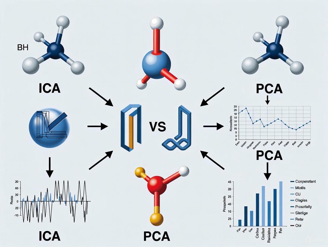
ICA vs PCA for EEG Artifact Removal: A Comprehensive Guide for Biomedical Research
Electroencephalogram (EEG) data is notoriously susceptible to contamination from physiological and non-physiological artifacts, posing a significant challenge in neuroscience research and drug development.
ICA vs PCA for EEG Artifact Removal: A Comprehensive Guide for Biomedical Research
Abstract
Electroencephalogram (EEG) data is notoriously susceptible to contamination from physiological and non-physiological artifacts, posing a significant challenge in neuroscience research and drug development. This article provides a systematic comparison of two predominant blind source separation (BSS) techniques for artifact removal: Independent Component Analysis (ICA) and Principal Component Analysis (PCA). We explore their foundational principles, methodological applications, and comparative performance in isolating and removing artifacts such as ocular, muscle, and cardiac interference. Aimed at researchers and scientists, this review synthesizes current evidence, addresses practical troubleshooting, and discusses the emerging role of hybrid and deep learning methods to guide the selection and optimization of preprocessing pipelines for clean and reliable EEG data analysis.
EEG Artifacts and Blind Source Separation: Unveiling the Core Principles of ICA and PCA
The Critical Challenge of Artifacts in EEG Analysis for Clinical and Research Applications
Electroencephalography (EEG) is a crucial tool in clinical neurology and neuroscience research, providing millisecond-scale temporal resolution for studying brain dynamics. However, a significant challenge persists: EEG signals are exceptionally vulnerable to contamination by artifacts originating from both physiological and non-physiological sources. These unwanted signals can mimic pathological activity, obscure genuine neural phenomena, and ultimately compromise diagnostic validity and research conclusions [1].
Artifacts are broadly categorized as physiological or extrinsic. Physiological artifacts arise from bodily processes, including ocular artifacts from eye blinks and movements, muscle artifacts from head and facial muscle activity, and cardiac artifacts from heart electrical activity or pulse [1]. Extrinsic artifacts stem from environmental interference, instrument noise, or electrode issues [1]. The problem is particularly acute in emerging applications using wearable EEG systems and in mobile brain imaging paradigms, where motion artifacts introduce additional complexity [2] [3].
This article objectively compares the performance of two principal computational approaches for artifact management: Principal Component Analysis (PCA) and Independent Component Analysis (ICA), with additional context on modern hybrid techniques. We present supporting experimental data to guide researchers and clinicians in selecting appropriate methodologies for their specific applications.
Technical Foundations of PCA and ICA
Principal Component Analysis (PCA)
PCA is a multivariate analysis technique based on linear transformation. It decomposes a set of potentially correlated variables into a set of linearly uncorrelated variables called Principal Components (PCs). These components are estimated as projections onto the eigenvectors of the data's covariance or correlation matrix [4].
The primary objective of PCA is dimensionality reduction and noise reduction by retaining the fewest components that account for the most variance in the original data with minimal information loss [4]. In EEG applications, PCA's performance is highly dependent on the pre-normalization method applied to the data [4].
Independent Component Analysis (ICA)
ICA extends beyond PCA by separating multivariate signals into statistically independent components. Unlike PCA, which finds uncorrelated components, ICA finds components that are independent, a stronger statistical condition measured using higher-order statistics [4] [5].
ICA operates on the assumption that the recorded EEG signals represent linear mixtures of independent source signals from the brain and artifactual sources. The algorithm searches for a linear transformation that minimizes statistical dependency and mutual information between components [4]. In practice, ICA decomposition often begins with a whitening step using PCA or Singular Value Decomposition (SVD) to improve signal-to-noise ratio before identifying independent sources [4].
Table 1: Fundamental Differences Between PCA and ICA
| Feature | Principal Component Analysis (PCA) | Independent Component Analysis (ICA) |
|---|---|---|
| Primary Objective | Variance maximization, dimensionality reduction | Statistical independence maximization, source separation |
| Statistical Basis | 2nd-order statistics (covariance) | Higher-order statistics |
| Component Nature | Orthogonal, uncorrelated | Statistically independent |
| Output Ordering | By explained variance (decreasing) | No inherent ordering |
| Assumptions | Linear correlations exist in data | Sources are statistically independent and non-Gaussian |
Figure 1: Typical ICA Workflow for EEG Artifact Removal. ICA typically incorporates PCA as a preprocessing whitening step before performing independent component decomposition.
Comparative Performance Analysis: PCA vs. ICA
Signal Separation Efficacy
Direct comparisons in controlled studies reveal fundamental performance differences. In PET imaging research, which shares similar multivariate analysis challenges with EEG, PCA demonstrated superior stability and created better results both qualitatively and quantitatively compared to ICA in simulated images. PCA effectively extracted signals from noise and showed insensitivity to noise type, magnitude, and correlation when proper pre-normalization was applied [4].
Conversely, ICA excels in specific artifact separation tasks. ICA has proven particularly effective at isolating stereotyped artifacts including ocular movements (EOG), muscle activity (EMG), cardiac signals (ECG), and channel noise [6]. These artifacts often have statistical properties distinct from cerebral activity, making them good candidates for ICA separation.
Impact of Data Dimensionality Reduction
A critical consideration in ICA implementation is whether to apply preliminary dimension reduction by PCA. Evidence indicates that PCA rank reduction adversely affects subsequent ICA decomposition quality. One study demonstrated that reducing data rank by PCA to retain 95% of original variance decreased the mean number of recovered 'dipolar' independent components from 30 to 10 per dataset and reduced median component stability from 90% to 76% [6].
Furthermore, PCA rank reduction increased uncertainty in equivalent dipole positions and spectra of neural sources, and decreased the number of subjects represented in component clusters corresponding to specific brain activities [6]. These findings suggest that for EEG data, PCA rank reduction should be avoided or carefully tested before application as an ICA preprocessing step [6].
Table 2: Quantitative Performance Comparison of PCA and ICA for EEG Artifact Removal
| Performance Metric | PCA | ICA | Experimental Context |
|---|---|---|---|
| Stability | High | Variable, sensitive to noise levels | PET simulation study [4] |
| Dipolar Components | N/A | 30/set (no PCA reduction) | 72-channel EEG study [6] |
| Dipolar Components | N/A | 10/set (with PCA 95% variance) | 72-channel EEG study [6] |
| Component Stability | N/A | 90% (no PCA reduction) | EEG reliability assessment [6] |
| Component Stability | N/A | 76% (with PCA 95% variance) | EEG reliability assessment [6] |
| Motion Artifact Handling | Limited | Improved with specialized preprocessing | Mobile EEG during running [2] |
Advanced Methodologies and Experimental Protocols
Modern Preprocessing Techniques for Mobile EEG
Recent research addresses motion artifacts in ecological settings using advanced preprocessing methods before ICA:
iCanClean: This algorithm uses canonical correlation analysis (CCA) and reference noise signals to detect and correct noise subspaces. It can utilize dual-layer EEG setups where outward-facing noise electrodes provide reference artifact recordings [2] [7]. Optimal parameters determined through parameter sweeps include a 4-second window length and R² threshold of 0.65, which improved good-quality brain components from 8.4 to 13.2 (+57%) [7].
Artifact Subspace Reconstruction (ASR): ASR employs a sliding-window PCA approach to identify and remove high-variance artifacts based on a calibration period [2]. Studies recommend a threshold parameter (k) of 20-30, with lower values producing more aggressive cleaning [2].
Experimental Protocol for Method Comparison
A rigorous comparison between artifact removal approaches for mobile EEG during running employed this methodology:
- Data Acquisition: EEG recorded during dynamic jogging and static standing versions of a Flanker task [2].
- Preprocessing: Compared no preprocessing, ASR, and iCanClean with pseudo-reference noise signals [2].
- Evaluation Metrics:
Results demonstrated that both iCanClean and ASR improved component dipolarity and reduced power at gait frequencies. However, only iCanClean consistently identified the expected P300 amplitude differences between congruent and incongruent flanker conditions during running [2].
Figure 2: Experimental Framework for Comparing Motion Artifact Removal Methods. Modern pipelines evaluate methods based on multiple criteria including component quality, spectral features, and functional neural metrics.
Table 3: Research Reagent Solutions for EEG Artifact Processing
| Tool/Resource | Type | Primary Function | Application Context |
|---|---|---|---|
| EEGLAB | Software Environment | ICA implementation and component visualization | General EEG analysis [5] |
| MNE-Python | Software Library | ICA decomposition with multiple algorithms | Python-based EEG/MEG analysis [8] |
| ICLabel | Automated Classifier | Component classification using neural networks | Identifying brain vs. artifact components [7] |
| iCanClean | Algorithm | Motion artifact removal using CCA | Mobile EEG with motion artifacts [7] |
| Artifact Subspace Reconstruction (ASR) | Algorithm | PCA-based artifact removal | High-amplitude artifact correction [2] |
| Dual-Layer EEG Caps | Hardware | Reference noise recording | Mobile EEG studies [7] |
The critical challenge of artifacts in EEG analysis requires methodological choices informed by empirical evidence. PCA offers stability and effectiveness for general noise reduction, particularly in high-noise environments, and serves as a valuable whitening step for ICA. However, ICA provides superior separation of specific physiological artifacts when sufficient high-quality data is available and dimensionality is preserved.
Emerging approaches like iCanClean and ASR demonstrate that hybrid methods combining reference signals with advanced statistical techniques can address the persistent problem of motion artifacts in mobile settings. Future research directions should include standardized benchmarking across artifact types, development of real-time processing pipelines for clinical applications, and adaptive algorithms that automatically adjust to individual differences in artifact characteristics.
For researchers and clinicians, selection between PCA, ICA, and advanced hybrid methods should be guided by specific application requirements, data quality, and the particular artifact types most prevalent in their recording environments.
Defining Blind Source Separation (BSS) in Signal Processing
Blind Source Separation (BSS) is a computational technique for separating a set of source signals from a mixture of observed signals, with little to no prior knowledge about the source signals or the mixing process. [9] This method is highly underdetermined but can yield useful solutions under a variety of conditions, making it invaluable in fields ranging from audio processing to biomedical signal analysis. [9] The classical example is the "cocktail party problem," where the goal is to isolate a single speaker's voice from a mixture of multiple conversations. [9]
In the context of electroencephalogram (EEG) artifact removal, BSS is a crucial unsupervised learning technique. [10] The core problem is modeled as:
X(t) = A · S(t) + N [10]
Where X(t) is the vector of observed mixed signals, S(t) is the vector of unknown source signals, A is the unknown mixing matrix, and N represents noise. [10] The objective is to find a demixing matrix W that approximates the inverse of A to recover the original sources: Ŝ(t) = W · X(t). [9] [10]
Theoretical Comparison: ICA vs. PCA as BSS Techniques
While both Independent Component Analysis (ICA) and Principal Component Analysis (PCA) are used for BSS, their underlying principles and objectives differ significantly, as outlined in the table below.
Table 1: Fundamental Differences Between PCA and ICA for Blind Source Separation
| Feature | Principal Component Analysis (PCA) | Independent Component Analysis (ICA) |
|---|---|---|
| Core Principle | Orthogonal transformation to decompose correlated variables into spatially orthogonal, uncorrelated components (Principal Components). [10] | Decomposes multivariate data into statistically independent components (ICs) by minimizing mutual information or maximizing non-Gaussianity. [10] |
| Primary Goal | Variance maximization; reduce data dimensionality. [11] | Statistical independence; separate latent sources. [9] [11] |
| Statistical Criterion | Identifies uncorrelated components. [10] | Identifies independent, non-Gaussian components (assumed for sources). [10] |
| Assumptions on Sources | Sources are uncorrelated. | Sources are statistically independent. [10] |
| Limitations in EEG | Limited by frequency band interference and voltage similarity between artifact and neural signal; does not provide a complete solution for artifact removal. [10] | Computationally complex; can suffer from permutation problem; may introduce signal distortion; manual component selection is often required. [10] |
Experimental Comparison in EEG Artifact Removal
Recent research directly compares the performance of ICA and other BSS-inspired methods for cleaning motion artifacts from EEG data during physical activities like running. The table below summarizes key performance metrics from a 2025 study.
Table 2: Experimental Performance of Artifact Removal Methods in Mobile EEG [2]
| Method | ICA Decomposition Quality (Component Dipolarity) | Power Reduction at Gait Frequency | Recovery of Expected P300 ERP Congruency Effect |
|---|---|---|---|
| ICA alone | Reduced quality due to motion artifact contamination. [2] | Not Applicable (Baseline) | Not Reported |
| ICA + Artifact Subspace Reconstruction (ASR) | Improved recovery of dipolar brain components. [2] | Significant reduction. [2] | Produced ERPs similar to standing task; did not capture the full P300 effect. [2] |
| ICA + iCanClean | Most effective in producing dipolar brain components. [2] | Significant reduction. [2] | Effectively captured the expected P300 congruency effect. [2] |
Detailed Experimental Protocols
The following workflow visualizes the methodology used in the aforementioned comparative study on motion artifact removal during running.
EEG Artifact Removal Workflow
1. Data Acquisition: EEG is recorded from participants during dynamic activities (e.g., an adapted Flanker task during jogging) and a static control condition (the same task during standing). [2]
2. Preprocessing & Application of BSS Methods: The recorded EEG is then processed using different methods for comparison: [2]
- ICA-only: The raw data is decomposed via ICA without specific motion artifact preprocessing.
- ICA with Artifact Subspace Reconstruction (ASR): This method uses a sliding-window Principal Component Analysis (PCA) to identify and remove high-variance, high-amplitude artifacts from continuous EEG based on a calibration period. A key parameter is the standard deviation threshold
k, with lower values (e.g., 10-30) leading to more aggressive cleaning. [2] - ICA with iCanClean: This approach leverages canonical correlation analysis (CCA) to detect and subtract noise subspaces from the scalp EEG. It uses either dedicated noise sensors or, more commonly, "pseudo-reference" noise signals created from the raw EEG (e.g., by applying a notch filter below 3 Hz). A critical parameter is the correlation criterion
R²(e.g., 0.65), which determines which noise components are subtracted. [2]
3. Performance Evaluation: The cleaned data is evaluated against three key metrics to assess the success of the underlying BSS process (ICA): [2]
- ICA Component Dipolarity: Measures the quality of the ICA decomposition. Physiological brain sources are dipolar, so a higher number of dipolar components indicates a more successful separation. [2]
- Spectral Power at Gait Frequency: A successful method will significantly reduce power at the frequency of the stepping motion and its harmonics. [2]
- Stimulus-locked Event-Related Potential (ERP) Components: The ultimate test is whether neural signatures, like the P300 wave during a cognitive task, can be recovered in the dynamic condition, matching those found in the static, artifact-free condition. [2]
The Scientist's Toolkit: Essential Research Reagents and Solutions
Table 3: Key Tools and Algorithms for BSS in EEG Research
| Tool/Algorithm | Function in BSS Research |
|---|---|
| Independent Component Analysis (ICA) | The core BSS algorithm for decomposing multichannel EEG into statistically independent sources, enabling the identification and removal of artifactual components. [9] [10] |
| Principal Component Analysis (PCA) | Used for dimensionality reduction and as a preprocessing step for whitening data before ICA. Also the core of ASR for artifact removal. [2] [10] |
| Artifact Subspace Reconstruction (ASR) | An adaptive, data-driven method for removing high-amplitude motion artifacts from continuous EEG in real-time, improving the subsequent performance of ICA. [2] |
| iCanClean Algorithm | A method that uses canonical correlation analysis (CCA) to identify and subtract motion artifact subspaces from EEG signals, particularly effective with pseudo-reference or dual-layer sensor noise signals. [2] |
| ICLabel | A classifier (often an EEGLAB plugin) that automates the labeling of ICA components as 'brain', 'muscle', 'eye', 'heart', 'line noise', or 'other', streamlining the component selection process. [2] |
| JADE Algorithm | A specific ICA algorithm (Joint Approximate Diagonalization of Eigenmatrices) used for separating mixed signals, including images and multidimensional data. [9] |
| FastICA Algorithm | A computationally efficient ICA algorithm that maximizes the non-Gaussianity of components to achieve separation. [10] |
Blind Source Separation is a powerful framework for unraveling mixed signals, with ICA being a particularly effective method for the non-trivial problem of EEG artifact removal. While PCA serves as a foundational technique for decorrelation and dimensionality reduction, ICA's pursuit of statistical independence makes it superior for isolating neural signals from complex artifacts like those generated during motion. Experimental evidence confirms that ICA's performance is significantly enhanced when combined with advanced preprocessing methods like iCanClean or ASR, which aggressively target motion-based noise. For researchers in drug development and neuroscience, this comparison underscores that a hybrid BSS approach is often essential for achieving the signal fidelity required to study brain dynamics in ecologically valid, real-world settings.
In electroencephalography (EEG) research, the analysis of neural signals is consistently challenged by the presence of various artifacts—unwanted signals originating from non-neural sources. These artifacts can stem from physiological processes like eye movements and muscle activity, or from technical issues such as electrode displacement and environmental interference. Effective artifact removal is therefore a critical preprocessing step to ensure the validity of subsequent neural signal analysis. Within this domain, Principal Component Analysis (PCA) has established itself as a fundamental, variance-based linear algebra technique for dimensionality reduction and artifact mitigation. This guide provides a comparative objective performance analysis of PCA against other prevalent methods, with a specific focus on its application in EEG artifact removal research.
PCA operates on a simple yet powerful principle: it transforms the original, potentially correlated variables (EEG channels or time points) into a new set of linearly uncorrelated variables called principal components. These components are ordered such that the first few retain most of the variation present in the original dataset. Artifact removal is achieved by projecting the data onto this new component space, identifying and discarding components that predominantly represent artifacts, and then reconstructing the signal from the remaining components. This method contrasts with Independent Component Analysis (ICA), which separates signals based on statistical independence rather than variance. The ensuing sections will evaluate PCA's efficacy using quantitative data, detail standard experimental protocols, and situate its performance within the modern EEG preprocessing toolkit.
Theoretical Foundations and Comparative Frameworks
Core Mechanism of PCA
PCA is a statistical procedure that uses an orthogonal transformation to convert a set of observations of possibly correlated variables into a set of values of linearly uncorrelated variables called principal components. This transformation is defined in such a way that:
- The first principal component has the largest possible variance.
- Each succeeding component, in turn, has the highest variance possible under the constraint that it is orthogonal to the preceding components.
The mathematical foundation of PCA lies in the eigenvalue decomposition of the data covariance matrix or singular value decomposition (SVD) of the data matrix itself. The principal components are the eigenvectors of the covariance matrix, and the eigenvalues represent the magnitude of variance captured by each component. In EEG applications, artifacts often manifest as high-variance events, causing them to be captured within the first few principal components, which can then be removed prior to signal reconstruction.
PCA vs. ICA: A Fundamental Comparison
While both PCA and ICA are blind source separation techniques, their underlying objectives and mechanisms differ significantly, as outlined in the table below.
Table 1: Fundamental Comparison of PCA and ICA
| Feature | Principal Component Analysis (PCA) | Independent Component Analysis (ICA) |
|---|---|---|
| Separation Criterion | Maximizes explained variance; components are orthogonal and uncorrelated | Maximizes statistical independence; components are non-Gaussian and independent |
| Mathematical Foundation | Eigenvalue decomposition, Singular Value Decomposition | Information-theoretic measures, entropy maximization |
| Component Ordering | Components are ordered by variance explained | No inherent ordering of components |
| Assumptions | Data lies on a linear subspace; Gaussianity is optimal | Source signals are statistically independent and non-Gaussian |
| Primary Strength | Efficient dimensionality reduction; optimal for Gaussian data | Effective separation of non-Gaussian sources (neural signals, artifacts) |
| Typical Artifact Targets | Large-amplitude transient artifacts, electrode pops | Ocular, cardiac, and muscle artifacts |
The selection between PCA and ICA often depends on the specific artifact characteristics and analysis goals. PCA is particularly effective for removing large-amplitude, transient artifacts that dominate the variance in the signal, whereas ICA is often more effective for separating persistent physiological artifacts like eye blinks and muscle activity that have distinct temporal structures but may not always be the highest-variance sources [12].
Performance Evaluation and Quantitative Comparison
Efficacy in Removing Specific Artifact Types
Different artifact removal techniques demonstrate variable performance depending on the nature of the contaminating signal. The following table summarizes quantitative performance metrics for PCA and other common methods across various artifact types.
Table 2: Performance Comparison of Artifact Removal Techniques for Different Artifact Types
| Artifact Type | Method | Key Performance Metrics | Reported Efficacy | Limitations |
|---|---|---|---|---|
| Ocular (EOG) | PCA | Reduces amplitude by ~70-80% in frontal channels | Effective for large-amplitude blinks | Risk of removing neural activity with similar variance [12] |
| ICA | Component correlation with EOG >0.9 | High accuracy in separating blink components | Requires manual component inspection [12] | |
| Muscle (EMG) | PCA | Broadband power reduction in 20-100 Hz range | Moderate efficacy | Less effective for diffuse muscle noise [3] |
| ICA | Selectively reduces EMG components | Good for focal EMG artifacts | Performance decreases with channel count reduction [3] | |
| Motion Artifacts | PCA | Power reduction at gait frequency harmonics | Limited effectiveness | Motion artifacts often non-stationary [2] |
| ASR | η (artifact reduction): 86% ±4.13, SNR improvement: 20 ±4.47 dB | High effectiveness for motion | Requires calibration data [13] | |
| Large Transients | PCA | Near-complete removal of spike artifacts | Excellent for large-amplitude transients | May oversmooth neural signals [12] |
Impact on Signal Integrity and Subsequent Analyses
Beyond mere artifact removal, a critical consideration is how these methods affect the integrity of the underlying neural signals and their impact on downstream analyses.
Table 3: Impact on Signal Integrity and Analysis Outcomes
| Method | Residual Artifact | Neural Signal Preservation | Effect on ERP Decoding | Component Dipolarity |
|---|---|---|---|---|
| PCA | Low for large artifacts | Moderate (risk of neural component removal) | Minor improvement in SNR | Not applicable |
| ICA | Low for physiological artifacts | High when properly classified | Minimal effect on decoding accuracy [14] | High for brain components [2] |
| ASR | Moderate for motion | Variable (depends on threshold) | Limited data available | Improved with optimal k (10-30) [2] |
| iCanClean | Low for motion | High with proper noise reference | Enables P300 detection during running [2] | Superior to ASR in locomotion [2] |
Notably, research has shown that for event-related potential decoding, extensive artifact correction through ICA combined with artifact rejection did not significantly improve support vector machine performance in the vast majority of cases across multiple paradigms including N170, mismatch negativity, N2pc, P3b, and N400 [14]. This suggests that while artifact removal is crucial for minimizing confounds, its impact on multivariate pattern analysis may be less pronounced than previously assumed.
Experimental Protocols and Methodologies
Standardized PCA Workflow for EEG Artifact Removal
The implementation of PCA for EEG artifact removal follows a systematic protocol to ensure reproducible results. The following diagram illustrates the standard workflow for PCA-based artifact removal in EEG processing:
PCA-Based Artifact Removal Workflow
Step-by-Step Protocol:
Data Preparation and Filtering: Begin with raw EEG data, typically referenced to a common average or mastoid reference. Apply a bandpass filter (e.g., 1-40 Hz) to remove extreme frequency components that might dominate variance calculations. A high-pass filter of 1-2 Hz is particularly important to improve stationarity [12] [15].
Covariance Matrix Calculation: The preprocessed EEG data matrix ( X ) of dimensions ( channels \times time points ) is used to compute the covariance matrix ( \Sigma = \frac{1}{N-1} XX^T ), where ( N ) represents the number of time points. This matrix captures the variance and covariance relationships between different EEG channels.
Eigenvalue Decomposition: Perform eigenvalue decomposition on the covariance matrix ( \Sigma ) to obtain eigenvectors (principal components) and eigenvalues (variance explained by each component). The components are ordered by decreasing eigenvalue magnitude.
Component Selection and Projection: Identify components representing artifacts through various criteria:
- Variance thresholding: Discard components explaining an exceedingly high percentage of total variance, characteristic of large artifacts.
- Visual inspection: Examine component topographies and time courses for patterns typical of known artifacts.
- Automated detection: Use algorithmic approaches to flag components with specific spatial or temporal characteristics matching artifact templates.
Signal Reconstruction: Project the data back to the original sensor space using only the retained components, effectively reconstructing the EEG signal without the artifact-dominated components. The reconstructed data constitutes the cleaned EEG output for subsequent analysis.
Integrated PCA-ICA Protocol for Comprehensive Cleaning
For optimal artifact removal, PCA is often combined with ICA in a sequential processing pipeline. The protocol below, adapted from a standardized EEG preprocessing framework, highlights this integrative approach [12]:
Integrated PCA-ICA Artifact Removal Protocol
This hybrid approach leverages the strengths of both methods:
- ICA is applied first to remove ocular artifacts, which it excels at identifying due to their consistent temporal structure [12].
- PCA is subsequently employed to target large-amplitude, transient artifacts (e.g., from muscle vibrations or electrode movement) that violate stationarity assumptions and may not be cleanly separated by ICA alone [12].
This sequential protocol ensures that large transient artifacts do not compromise ICA decomposition, while also capitalizing on ICA's superior ability to isolate ocular artifacts without distorting neural signals that might share similar variance profiles.
Table 4: Key Software and Analytical Resources for EEG Artifact Removal Research
| Resource | Type | Primary Function | Application Context |
|---|---|---|---|
| EEGLAB | Software toolbox | Interactive MATLAB environment for EEG processing | Standard platform for implementing PCA, ICA, and ASR [2] [12] |
| AMICA Plugin | Algorithm plugin | Adaptive Mixture ICA for enhanced source separation | Alternative to Infomax ICA; includes sample rejection options [15] |
| clean_rawdata | EEGLAB plugin | Automatic artifact removal using ASR | Handles large-amplitude artifacts using PCA-based reconstruction [2] |
| ICLabel | EEGLAB plugin | Automated IC classification | Classifies ICA components as brain, muscle, eye, etc. [2] |
| iCanClean | Standalone algorithm | Motion artifact removal using CCA | Leverages noise references for optimal motion artifact cleaning [2] |
The comparative analysis presented in this guide demonstrates that PCA remains a valuable tool in the EEG preprocessing arsenal, particularly for addressing large-amplitude, transient artifacts that dominate signal variance. Its computational efficiency and straightforward interpretation make it well-suited for initial data cleaning stages and for integration with more complex methods like ICA.
However, PCA's limitations in handling physiological artifacts with complex temporal structures and its potential for removing neural activity sharing variance characteristics with artifacts necessitate a nuanced application. For contemporary EEG research, particularly involving mobile setups and real-world applications, the integration of PCA within a hybrid processing pipeline represents the most effective strategy. Researchers should consider the specific artifact profile of their dataset, the critical neural features of interest, and available computational resources when selecting and parameterizing artifact removal approaches.
Future developments in deep learning-based artifact removal show promise for handling unknown artifacts and adapting to multi-channel contexts [16], but established linear methods like PCA and ICA will continue to provide foundational approaches for ensuring EEG data quality in both clinical and research settings.
Electroencephalography (EEG) provides unparalleled temporal resolution for studying brain dynamics but is notoriously susceptible to contamination by various artifacts. Source separation techniques are fundamental for isolating neural signals from these unwanted interferences. Among the most prominent methods are Independent Component Analysis (ICA) and Principal Component Analysis (PCA), which leverage distinct statistical principles to decompose complex multichannel recordings. ICA separates mixed signals into components that are statistically independent, a property often exhibited by biologically distinct brain and artifact sources [5]. In contrast, PCA separates signals based on orthogonality and variance, identifying components that are uncorrelated and account for the greatest variance in descending order [17] [18]. This guide provides an objective, data-driven comparison of ICA and PCA, focusing on their application in EEG artifact removal for research and clinical applications.
Theoretical Foundations: ICA vs. PCA
The mathematical divergence between ICA and PCA underlies their differing performance in source separation. PCA is a linear dimensionality reduction technique that projects data onto a new set of axes (principal components) that are orthogonal and ranked by the amount of variance they explain from the original dataset [17] [18]. This makes PCA highly effective for data compression and removing correlated noise, but it does not necessarily separate physiologically distinct sources.
ICA, conversely, is a blind source separation (BSS) technique that assumes the observed multichannel EEG data is a linear mixture of underlying source signals. Its objective is to find a demixing matrix that recovers these sources by maximizing their statistical independence—a stronger condition than mere uncorrelation [5]. ICA algorithms achieve this by optimizing higher-order statistics, such as making the distributions of the extracted components as non-Gaussian as possible. This core theoretical difference makes ICA particularly suited for separating sources like brain activity, eye blinks, and muscle noise, which are often generated by physiologically independent processes.
Experimental Protocols for Performance Comparison
Common Validation Methodologies
Researchers typically evaluate the performance of artifact removal algorithms using both simulated and real-world EEG data. Key methodological approaches include:
- Ground-Truth Simulation: Studies often create datasets by adding known artifact templates (e.g., from eye movement or muscle recordings) to clean EEG baseline data. The accuracy of an algorithm is measured by how well it recovers the original, uncontaminated signal [19] [2].
- Recovery of Evoked Responses: In event-related potential (ERP) studies, the efficacy of an artifact removal method is gauged by its ability to recover known ERP components, such as the P300, after the data has been artificially contaminated or recorded in challenging conditions (e.g., during motion) [2].
- Signal Quality Metrics: Quantitative indices like Signal-to-Noise Ratio (SNR), residual variance, and component dipolarity (a measure of how well a component's scalp map can be explained by a single equivalent brain dipole) are standard measures for comparing performance [19] [20] [15].
Workflow for Method Comparison
The following diagram illustrates a typical experimental workflow for comparing ICA and PCA in an EEG artifact removal pipeline.
Comparative Performance Data
Extensive research has quantified the performance of ICA and PCA across various artifact types and experimental conditions. The tables below summarize key findings from the literature.
Table 1: Overall Performance Comparison of ICA and PCA for Common EEG Artifacts
| Artifact Type | ICA Performance | PCA Performance | Key Supporting Evidence |
|---|---|---|---|
| Ocular (Eye Blinks) | Highly effective; reliably separates and removes blinks without distorting underlying neural signals [21] [5]. | Moderate; can remove high-variance blink artifacts but often with greater loss of neural signal [21] [20]. | ICA outperformed PCA in separating and removing ocular artifacts, with results comparing favorably [21]. |
| Muscle (EMG) | Effective, particularly with advanced algorithms like AMICA; separates high-frequency muscle noise into distinct components [5] [15]. | Limited; muscle artifacts are often distributed across multiple principal components, making clean removal difficult [17]. | ICA was more effective at isolating EMG artifacts, while PCA risked removing more neural signal [20]. |
| Gradient (EEG-fMRI) | New ICA-based methods developed specifically for 3T spiral in-out and EPI sequences outperform PCA, AAS, and TS methods, especially in theta and alpha bands [19]. | Modest performance; PCA-based methods show limited efficacy compared to advanced ICA methods for gradient artifacts [19]. | ICA-based methods recovered ERPs and resting EEG with high SNR (>4) below the beta band, outperforming PCA [19]. |
| Motion (Locomotion) | Robust when combined with preprocessing (e.g., AMICA sample rejection); improves component dipolarity and preserves ERPs during running [2] [15]. | Not commonly used as a primary method; artifact subspace reconstruction (ASR), which uses PCA, is effective but may over-clean [2]. | ICA decompositions were significantly improved with automatic sample rejection, even in high-motion environments [15]. |
Table 2: Quantitative Recovery of Neural Signals After Artifact Removal
| Metric / Condition | ICA Results | PCA Results | Experimental Context |
|---|---|---|---|
| Signal-to-Noise Ratio (SNR) in Beta Band | Modest (SNR ~1) [19]. | Modest (SNR ~1) [19]. | Gradient artifact removal during fMRI [19]. |
| Residual Variance in Brain Components | Lower residual variance, indicating cleaner separation of brain and non-brain sources [15]. | Higher residual variance, suggesting less complete separation [18]. | Component analysis after decomposition [18] [15]. |
| P300 ERP Congruency Effect | Recovered effectively during running with iCanClean (a CCA/ICA method) [2]. | Not specifically reported for PCA; ASR (PCA-based) produced similar ERP latencies but not the amplitude effect [2]. | Flanker task during static standing vs. jogging [2]. |
| Component Dipolarity | Higher number of dipolar brain components, indicating superior source separation physiologically plausible for brain activity [2] [15]. | Fewer dipolar components, as PCA separates by variance, not physiological reality [2]. | Quality assessment of decomposition during locomotion [2]. |
Implementing ICA effectively requires specific tools and considerations. The following table outlines key "research reagents" for conducting ICA in EEG studies.
Table 3: Essential Tools and Considerations for ICA Implementation
| Item | Function / Description | Examples & Notes |
|---|---|---|
| High-Density EEG | Provides sufficient spatial sampling for ICA to resolve independent sources. | 64+ channels are recommended; many studies use 32-256 channels [15] [22]. |
| ICA Algorithms | The computational engine that performs the source separation. | Infomax (runica), AMICA, FastICA, SOBI. AMICA is often among the best-performing for EEG [5] [15]. |
| Component Classification Tools | Automatically labels independent components as brain or artifact. | ICLabel is a standard EEGLAB plugin that uses a trained neural network to classify components [2] [22]. |
| Preprocessing Pipeline | Data preparation steps critical for a successful ICA decomposition. | Includes bad channel removal, high-pass filtering (e.g., 1-2 Hz), and bad segment rejection [5] [15]. |
| Computational Resources | ICA is computationally intensive, scaling with channel count and data length. | High-performance workstations or computer clusters are recommended for large datasets or using algorithms like AMICA [5]. |
Optimization and Advanced Hybrid Techniques
Preprocessing for Optimal ICA
The quality of ICA decomposition is highly dependent on data preprocessing and quality. Research shows that moderate automatic cleaning (e.g., 5-10 iterations of sample rejection within the AMICA algorithm) generally improves decomposition quality across both stationary and mobile EEG protocols [15]. Furthermore, applying a high-pass filter (e.g., 1 Hz or 2 Hz) before ICA computation increases data stationarity and improves component stability, though the resulting demixing matrix can often be successfully applied back to the less aggressively filtered data to preserve low-frequency neural content [22].
Beyond Pure ICA: Hybrid Methods
To overcome the limitations of any single method, researchers have developed powerful hybrid processing techniques:
- ICA-Wavelet Combinations: Wavelet-based thresholding is applied to the time courses of artifact-laden independent components, rather than directly to raw EEG data. This allows for more precise removal of artifact elements within a component while preserving the underlying neural signal [23].
- ICA-EMD Fusion: Empirical Mode Decomposition (EMD) adaptively decomposes a signal into oscillatory modes. These modes can be used to denoise ICA components before signal reconstruction, helping to address the "mode-mixing" problem of EMD [23].
- iCanClean: This method leverages canonical correlation analysis (CCA) to identify and subtract noise subspaces from the EEG that are highly correlated with reference noise signals, either from dedicated sensors or derived from the EEG itself. It has been shown to be highly effective for motion artifact removal during locomotion [2].
The logical relationship between core and hybrid methods is illustrated below.
The experimental data consistently demonstrates that ICA generally outperforms PCA for the removal of common physiological artifacts from EEG, particularly when the goal is to preserve the integrity of the underlying neural signal for cognitive or clinical analysis. Its strength lies in its foundation on statistical independence, which aligns well with the properties of biologically distinct sources. PCA, while excellent for dimensionality reduction and handling certain structured noises, often falls short in cleanly separating brain activity from artifacts like muscle or eye movement due to its sole reliance on variance and orthogonality.
The future of EEG artifact removal lies not in a single algorithm, but in systematic pipelines and intelligent hybrid methods. Best practices include using high-density EEG systems, ensuring rigorous data preprocessing, and leveraging advanced ICA algorithms like AMICA combined with automated component classification. For challenging recording environments, such as mobile brain-body imaging or simultaneous EEG-fMRI, hybrid approaches like iCanClean or ICA combined with wavelet denoising show significant promise. As the field moves towards more naturalistic neuroscience, the development and validation of these robust, automated artifact removal strategies will be paramount for generating reliable, high-fidelity neural data.
In electroencephalography (EEG) artifact removal research, the choice of signal separation algorithm can profoundly impact the quality of resultant neural data and the validity of subsequent neuroscientific or clinical conclusions. Two predominant theoretical frameworks employed for this purpose are Principal Component Analysis (PCA) and Independent Component Analysis (ICA). While both are linear transformation techniques, their underlying objectives and theoretical foundations are fundamentally distinct. PCA is grounded in the principle of orthogonality, seeking components that are uncorrelated and that maximize variance in the data. In contrast, ICA is rooted in the principle of statistical independence, a much stronger condition, aiming to separate mixed sources by maximizing the independence of their component distributions, often measured through higher-order statistics like kurtosis [17] [24]. This guide provides a structured, objective comparison of these frameworks, focusing on their application in EEG artifact removal, to aid researchers in selecting and implementing the most appropriate methodology for their specific investigative needs.
Theoretical Foundations: A Comparative Analysis
The core distinction between PCA and ICA lies in their statistical objectives and the type of data structure they seek to exploit. The following table delineates their fundamental theoretical differences.
Table 1: Core Theoretical Differences Between PCA and ICA
| Feature | Principal Component Analysis (PCA) | Independent Component Analysis (ICA) |
|---|---|---|
| Primary Objective | Variance maximization and dimensionality reduction [17] | Blind source separation of mixed signals [17] [25] |
| Component Relation | Uncorrelated (2nd-order statistics) [24] | Statistically independent (higher-order statistics) [24] |
| Statistical Basis | Orthogonality; diagonalizes the covariance matrix [17] | Non-Gaussianity; minimizes mutual information [17] [24] |
| Component Order | Ordered by explained variance (from largest to smallest) | No inherent order; components are considered equally [25] |
| Assumption on Sources | Assumes Gaussian or low-dimensional data | Requires non-Gaussian, independent source signals [24] |
The Principle of Orthogonality in PCA
PCA operates by identifying a new set of orthogonal axes (principal components) in the data. The first component aligns with the direction of maximum variance, with each subsequent component capturing the next highest variance under the constraint of orthogonality to all previous components [17]. This process is equivalent to an eigenvalue decomposition of the data's covariance matrix, effectively identifying and ranking the directions of correlation. In the context of EEG, this allows PCA to compress data and isolate major patterns of variance. However, a key limitation is that uncorrelatedness (orthogonality) does not imply independence. Artifacts and neural signals can have complex, higher-order statistical dependencies that PCA, focusing only on second-order correlations, may fail to separate effectively [17] [23].
The Principle of Statistical Independence in ICA
ICA is designed to solve the "blind source separation" problem. It assumes that the observed multichannel EEG signal is a linear mixture of underlying source signals originating from the brain and various artifacts. The goal of ICA is to find a linear transformation that makes the output components (the estimated sources) as statistically independent as possible [25]. Independence is a stricter condition than uncorrelatedness, as it involves the entire probability distribution and relies on higher-order statistics (e.g., kurtosis). This enables ICA to separate sources based on the morphology of their time-course distributions, making it particularly powerful for isolating stereotypical artifacts like eye blinks, muscle activity, and line noise from neural signals, even when their frequency spectra overlap [17] [26].
Figure 1: A theoretical workflow comparing the decomposition objectives of PCA and ICA when applied to a multichannel EEG signal.
Experimental Protocols in EEG Artifact Removal
The application of PCA and ICA in research follows well-established, yet distinct, experimental protocols. The following workflow generalizes the common steps for both methods in the context of EEG cleaning.
Figure 2: A generalized experimental workflow for artifact removal from EEG using PCA or ICA, highlighting key methodological differences.
Detailed Methodologies
3.1.1 PCA-Based Artifact Removal After standard EEG preprocessing (filtering, bad channel removal), PCA is applied to the multichannel data. The resulting components are orthogonal and ordered by the amount of variance they explain. Artifacts with high amplitude, such as large eye movements or channel pops, often occupy the first few components due to their high variance. The experimental protocol involves:
- Decomposition: Performing Singular Value Decomposition (SVD) on the data matrix [17].
- Identification: Inspecting the component time courses and topographies. Components with topographies dominated by frontal regions (for ocular artifacts) or exceptionally high variance are flagged as artifacts. A scree plot can aid in identifying components with disproportionate variance [17].
- Removal: Projecting the data back to the sensor space while excluding the components identified as artifacts.
3.1.2 ICA-Based Artifact Removal ICA has become a gold standard for artifact removal in EEG. Its protocol is more focused on the statistical properties of the components:
- Decomposition: Using an algorithm (e.g., FastICA, Infomax) to estimate independent components from the preprocessed EEG data [17] [24].
- Identification: This critical step uses component features to classify them as brain or non-brain:
- Automated Classification: Tools like ICLabel employ trained classifiers that use features such as time-course autocorrelation, power spectral density, and topography to label components as brain, eye, muscle, heart, line noise, or channel noise [27] [2].
- Feature-Based Detection: Components can be identified based on known characteristics: ocular artifacts have a frontal scalp map and a high-amplitude, low-frequency time course; muscle artifacts have a high-frequency, broadband spectral profile [28].
- Removal: The artifact components are subtracted from the data, and the remaining components are projected back to create the cleaned EEG signal [28].
Performance Comparison: Quantitative and Qualitative Data
The efficacy of PCA and ICA has been quantitatively compared across numerous studies. The following table synthesizes key performance metrics from experimental data.
Table 2: Experimental Performance Comparison in EEG Artifact Removal
| Metric | PCA Performance | ICA Performance | Supporting Evidence |
|---|---|---|---|
| Artifact Separation | Effective for high-variance artifacts (e.g., large eye blinks); struggles with sources that have overlapping variance with neural signals [23]. | Superior for separating biologically independent sources like ocular, muscle, and cardiac artifacts, even with spectral overlap [17] [26]. | A study on depression EEG classification found ICA outperformed PCA in separating artifacts from neural data, leading to better subsequent analysis [23]. |
| Signal Distortion | Risk of removing neural signals that are correlated with or contribute high variance to the data. | Lower signal distortion when correctly identifying artifact components, as it targets statistically independent sources [26]. | An adaptive CCA-ICA method demonstrated better preservation of inherent cognitive components in EEGs compared to other methods, leading to more reliable analysis [26]. |
| Dimensionality Reduction | Excellent for data compression, reducing dimensions while preserving maximum variance. | Not primarily designed for dimensionality reduction, though it can be used as such; focus is on source separation [24]. | PCA is explicitly analyzed from the viewpoint of data compression, while ICA's strength lies in transforming the problem into one of independent decompositions [17]. |
| Computational Load | Typically lower; relies on efficient eigenvalue decomposition. | Generally higher; involves iterative optimization to maximize non-Gaussianity or minimize mutual information. | The processing time for PCA is noted to be significantly lower than classical methods like wavelets [17]. |
| Classification Accuracy | Can improve classification but may be less effective than ICA if artifacts are not the dominant source of variance. | Often leads to higher post-cleaning classification accuracy in BCI and diagnostic tasks. | In an emotion classification task, an ICA-based artifact removal method significantly improved accuracy compared to other methods [26]. A depression EEG study also showed higher classification accuracy after ICA denoising [23]. |
Case Study: Motion Artifact Removal in Mobile EEG
A 2025 comparative study on motion artifact removal during overground running provides a concrete example of these methods in a challenging, ecologically valid context [29] [2]. The study evaluated preprocessing approaches based on the dipolarity of resulting ICA components—a key indicator of component quality representing neural sources. The findings demonstrated that preprocessing the data with advanced algorithms like iCanClean (which uses canonical correlation analysis) and Artifact Subspace Reconstruction (ASR), before ICA, led to the recovery of more dipolar brain independent components [2]. This underscores that while ICA is powerful, its performance is contingent on effective preprocessing, especially in high-motion environments. The study concluded that these methods were effective for reducing motion artifacts and identifying stimulus-locked ERP components during running [29].
The Scientist's Toolkit: Essential Research Reagents and Solutions
For researchers embarking on EEG artifact removal studies, the following tools and software libraries are indispensable.
Table 3: Essential Research Tools for PCA and ICA in EEG Research
| Tool/Solution | Function | Relevant Context |
|---|---|---|
| EEGLAB | An interactive MATLAB toolbox for processing EEG data. It provides a comprehensive framework for importing, preprocessing, and visualizing data, and includes implementations of Infomax ICA and tools for component analysis [2]. | Widely used for ICA-based artifact removal; includes plugins like ICLabel for automated component classification. |
| MNE-Python | An open-source Python package for exploring, visualizing, and analyzing human neurophysiological data. It supports both PCA and ICA for noise reduction [30]. | Commonly used in modern research for its scalability and integration with machine learning workflows. |
| ICLabel | An EEGLAB plugin that automatically classifies independent components into categories (Brain, Muscle, Eye, Heart, Line Noise, Channel Noise, Other) using a trained neural network [2]. | Reduces the subjectivity and time required for manual component inspection, standardizing the ICA process. |
| Artifact Subspace Reconstruction (ASR) | A preprocessing method that uses PCA to identify and remove high-variance, non-stereotypical artifacts in a sliding-window approach [2]. | Often used as a preprocessing step before ICA to improve decomposition quality, especially in mobile EEG studies [2]. |
| iCanClean | An algorithm that leverages reference noise signals (or pseudo-references) and canonical correlation analysis (CCA) to detect and correct motion-artifact subspaces in the EEG data [2]. | Particularly effective for motion artifact removal in mobile brain imaging during walking and running [29] [2]. |
| FastICA Algorithm | A computationally efficient algorithm for ICA that maximizes non-Gaussianity through a fixed-point iteration scheme [24]. | Available in toolboxes like MNE-Python and scikit-learn; popular for its speed and simplicity. |
The comparative analysis reveals that the choice between PCA and ICA is not a matter of one being universally superior, but rather a strategic decision based on the research question and data characteristics. PCA, with its foundation in orthogonality and variance, offers a robust and computationally efficient method for data compression and the removal of high-variance, stereotypical artifacts. However, its reliance on second-order statistics is a limitation when artifacts and neural signals share similar variance profiles. ICA, founded on the more potent principle of statistical independence, excels at blind source separation, making it the preferred choice for isolating and removing complex biological artifacts like those from eyes and muscles. The experimental evidence consistently shows that ICA, particularly when enhanced with modern preprocessing and automated classification tools, provides a more effective pathway to cleaner neural data, which in turn supports more accurate brain-computer interfacing and clinical diagnostics. For contemporary EEG research, especially in dynamic, real-world settings, ICA provides a more powerful and flexible theoretical and practical framework for artifact removal.
From Theory to Practice: A Step-by-Step Guide to Implementing ICA and PCA for Artifact Removal
In electroencephalography (EEG) research, Blind Source Separation (BSS) has become a cornerstone technique for isolating neural signals from various artifacts. The efficacy of BSS methods, notably Independent Component Analysis (ICA) and Principal Component Analysis (PCA), is not inherent but is profoundly dependent on the preprocessing pipeline applied to the raw data. These preprocessing steps—encompassing filtering, data cleaning, and re-referencing—fundamentally shape the quality of the source separation, thereby influencing all subsequent analyses. A recent systematic review underscored that most artifact processing pipelines integrate detection and removal phases but rarely separate their individual impact on final performance metrics [31] [3]. Furthermore, studies demonstrate that preprocessing choices can artificially inflate effect sizes in event-related potentials and bias source localization if not carefully applied [32]. This guide provides an objective comparison of methodologies and their performance, framing the discussion within the broader thesis of comparing ICA and PCA for EEG artifact removal. It is designed to equip researchers and drug development professionals with the empirical evidence needed to construct robust, reproducible preprocessing protocols for effective BSS.
Theoretical Foundations: ICA and PCA in EEG Processing
Core Principles and Assumptions
Independent Component Analysis (ICA) is a statistical and computational technique used to uncover hidden factors, or source signals, from a set of observed mixtures. Its core assumption is that the observed multichannel EEG data matrix ( X \in \mathbb{R}^{N \times M} ) (where ( N ) is the number of channels and ( M ) is the number of samples) is a linear mixture of underlying, statistically independent source signals ( S ). This relationship is expressed as ( X = AS ), where ( A ) is the mixing matrix. The goal of ICA is to find a demixing matrix ( W = A^{-1} ) such that ( S = WX ) yields the independent sources [15]. ICA's strength in EEG analysis lies in its ability to separate sources based on their statistical independence, making it highly effective for isolating non-Gaussian signals like eye blinks, muscle activity, and, crucially, brain rhythms.
Principal Component Analysis (PCA), in contrast, is a dimensionality reduction technique that transforms the data to a new coordinate system where the greatest variance lies on the first coordinate (the first principal component), the second greatest variance on the second, and so on. It operates under the assumption that the components are orthogonal (uncorrelated). While PCA is excellent for denoising and reducing data dimensionality by discarding components with the least variance, it is less effective than ICA for artifact removal because neural and artifactual sources often share overlapping variances. Physiological artifacts are not merely "noise" with small variance; they can be high-amplitude signals, and removing high-variance components may also remove critical neural information.
A Workflow for BSS-Based EEG Cleaning
The following diagram illustrates a generalized preprocessing workflow for BSS, highlighting key decision points shared by ICA and PCA methodologies.
Comparative Performance Analysis: ICA vs. PCA
Quantitative Performance Metrics
The table below summarizes key performance metrics for ICA and PCA based on recent experimental studies, particularly in the context of wearable EEG systems.
Table 1: Quantitative Performance Comparison of ICA and PCA for EEG Artifact Removal
| Metric | ICA Performance | PCA Performance | Experimental Context | Source |
|---|---|---|---|---|
| Overall Usage Frequency | Among the most frequent techniques for ocular & muscular artifacts | Less frequently a primary technique | Systematic review of 58 wearable EEG studies | [31] [3] |
| Typical Accuracy | ~71% (when clean signal is reference) | Not prominently reported | Assessment with clean signal as ground truth | [31] |
| Residual Variance | Lower residual variance in brain components post-cleaning | Higher residual variance expected | Quality metric after AMICA decomposition | [15] |
| Impact on Decoding | Can decrease decoding performance if neural signal is removed | Not specifically quantified | Analysis of EEGNet & time-resolved decoding | [33] |
| Dependency on Preprocessing | Highly dependent; quality degrades with motion & improves with cleaning | Not specifically evaluated | Effect of data cleaning on AMICA decomposition | [15] |
Performance in Specific Artifact Domains
The effectiveness of BSS techniques varies significantly depending on the artifact type. Research indicates that ICA is a dominant method for managing specific physiological artifacts. A 2025 systematic review identified that wavelet transforms and ICA are among the most frequently used techniques for managing ocular and muscular artifacts in wearable EEG [31]. In contrast, PCA is less commonly featured as a standalone solution for these specific artifact categories in modern pipelines. ICA's success stems from its ability to separate sources like eye blinks and muscle activity, which often have non-Gaussian distributions and are statistically independent from cortical signals.
However, a critical caveat is that the subtraction of entire artifact components identified by ICA can sometimes remove neural signals alongside artifacts, potentially leading to false positive effects and biased source localization [32]. This has led to the development of more targeted methods, such as the RELAX pipeline, which focuses artifact reduction on specific temporal periods (for eye movements) or frequency bands (for muscle activity) within components, rather than subtracting components wholesale [32].
Experimental Protocols and Methodological Insights
Detailed Protocol: Evaluating Preprocessing on ICA Decomposition
A 2024 study by T. Händel et al. provides a robust protocol for assessing how data cleaning impacts ICA decomposition quality, offering a model for rigorous experimentation [15].
Objective: To systematically investigate the effect of automatic time-domain sample rejection on the quality of the AMICA (Adaptive Mixture ICA) decomposition across datasets with varying levels of participant mobility.
Datasets: Eight open-access EEG datasets from six studies were included, representing a mobility gradient from stationary to Mobile Brain/Body Imaging (MoBI) setups. All datasets used at least 58 EEG channels and had a sampling rate of ≥250 Hz.
Independent Variables:
- Cleaning Intensity: Manipulated via the AMICA algorithm's built-in sample rejection function. The number of rejection iterations and the standard deviation (SD) threshold for rejection were varied.
- Motion Intensity: Datasets were categorized based on the level of participant movement during recording.
Dependent Variables (Measures of Decomposition Quality):
- Mutual Information (MI): Lower MI between components indicates better separation and higher independence.
- Component Classification: The proportion of components classified as 'Brain', 'Muscle', or 'Other' (e.g., using ICLabel).
- Residual Variance: The variance in the data not explained by the brain components after artifact removal.
- Exemplary Signal-to-Noise Ratio (SNR): Calculated to compare a standing condition versus a mobile condition within the same experiment.
Procedure:
- Data Preparation: Data were high-pass filtered at 1 Hz and bad channels were interpolated.
- AMICA Decomposition: AMICA was run on each dataset with different cleaning strengths (e.g., 0 to 15 rejection iterations, with SD thresholds of 3 or 5).
- Quality Assessment: The dependent variables were computed for each resulting decomposition.
- Statistical Analysis: Linear mixed models were used to analyze the effects of cleaning and mobility on decomposition quality.
Key Finding: While increased movement decreased decomposition quality within individual studies, the AMICA algorithm was robust to limited data cleaning. Cleaning strength significantly improved decomposition, but the effect was smaller than expected. The study concluded that moderate cleaning (5-10 iterations of AMICA sample rejection) is sufficient to improve most datasets, regardless of motion intensity [15].
Protocol: A Modular Pipeline Comparison
A 2025 study by T. D. Almeida et al. took a modular approach to evaluate preprocessing choices, providing a framework for objective comparison [34].
Objective: To quantify the effect of different pre-processing strategies on EEG signal quality and analysis outcomes using quantitative metrics.
Experimental Design: The researchers constructed a modular pipeline and varied methods at key stages:
- Artifact Removal: Two ICA algorithms (SOBI and Extended Infomax) were compared.
- Re-referencing: Four methods were tested: Common Averaged Reference (CAR), robust CAR (rCAR), Reference Electrode Standardization Technique (REST), and Reference Electrode Standardization and Interpolation Technique (RESIT).
Evaluation Metrics: The impact of these pipelines was quantified using Event-Related Spectral Perturbation (ERSP) of the sensorimotor rhythm in an Action Observation and Motor Imagery protocol.
Key Finding: The study revealed that signal segmentation significantly affects the cleaning procedure, while comparable results were obtained across the different ICA approaches. For re-referencing, CAR, REST, and RESIT produced similar topographical patterns, whereas rCAR showed the most divergent ERSP pattern [34]. This highlights that steps like segmentation and re-referencing can be as impactful as the choice of BSS algorithm itself.
Visualization of Method Selection and Impact
Decision Pathway for BSS Method Selection
The following diagram outlines the logical decision process for choosing and applying ICA or PCA within an EEG preprocessing pipeline, incorporating findings from recent comparative studies.
For researchers aiming to implement these preprocessing pipelines, a standardized set of tools and datasets is crucial for reproducibility and validation. The following table details key resources referenced in the cited studies.
Table 2: Essential Research Reagents and Resources for EEG BSS Preprocessing
| Resource Name | Type | Primary Function in Research | Example Use Case |
|---|---|---|---|
| AMICA Plugin | Software Algorithm | An advanced ICA algorithm for decomposing EEG data into independent components. | Used to evaluate the impact of time-domain cleaning on decomposition quality [15]. |
| RELAX Pipeline | Software Plugin (EEGLAB) | Implements targeted artifact reduction within ICA components to minimize neural signal loss. | Cleaning Go/No-Go and N400 task data while reducing effect size inflation [32]. |
| ERP CORE Dataset | Public Dataset | Provides standardized, openly available EEG data from multiple classic experimental paradigms. | Served as the primary data source for multiverse analysis of preprocessing on decoding [33]. |
| EEGdenoiseNet | Public Dataset | A semi-synthetic benchmark dataset for developing and testing EEG denoising algorithms. | Used to train and evaluate deep learning models like CLEnet for artifact removal [16]. |
| Clean_rawdata (ASR) | Software Function | An automatic method for identifying and removing bad data periods based on artifact subspaces. | Referenced as a common automatic cleaning method, though sensitive to threshold settings [15]. |
The empirical evidence clearly demonstrates that the selection and application of BSS methods like ICA and PCA are not one-size-fits-all decisions. ICA remains the dominant and more effective technique for artifact removal in high-density EEG systems, particularly for physiological artifacts like ocular and muscle activity, achieving accuracy around 71% [31]. However, its performance is highly contingent on rigorous preprocessing, including appropriate filtering and data cleaning, with studies showing that moderate automatic cleaning (e.g., 5-10 iterations in AMICA) significantly enhances decomposition quality [15].
A critical consideration is that aggressive artifact correction, while improving signal purity, can sometimes reduce downstream decoding performance. This suggests that classifiers may learn to exploit structured noise correlated with experimental conditions [33]. Therefore, the optimal pipeline must balance cleaning efficacy with the preservation of neural information, potentially employing targeted methods like RELAX [32]. Future research will likely see greater integration of deep learning models, such as hybrid CNN-LSTM networks (e.g., CLEnet), which show promise in handling unknown artifacts and multi-channel data without relying on the strict statistical assumptions of traditional BSS [16]. For now, a meticulous, empirically grounded approach to preprocessing remains the essential foundation for any effective BSS application in EEG research.
The pursuit of clean neural signals is a fundamental challenge in electroencephalography (EEG) research. Artifacts originating from ocular movements, muscle activity, cardiac rhythms, and environmental noise can significantly obscure brain-generated electrical signals, complicating data interpretation and analysis. Within this context, principal component analysis (PCA) and independent component analysis (ICA) have emerged as two prominent computational techniques for addressing the artifact contamination problem. While both are blind source separation (BSS) methods, their underlying principles and applications differ substantially. PCA is a statistical procedure that employs an orthogonal transformation to convert a set of observations of possibly correlated variables into a set of values of linearly uncorrelated variables called principal components. This transformation is defined such that the first principal component accounts for the largest possible variance in the data, and each succeeding component, in turn, has the highest variance possible under the constraint that it is orthogonal to the preceding components [6].
In contrast, ICA is a computational method for separating a multivariate signal into additive, statistically independent non-Gaussian components. The core assumption of ICA is that the observed EEG signals are linear mixtures of independent source signals from neural and non-neural origins. ICA aims to find a linear transformation that minimizes the statistical dependence between the components, thereby isolating artifacts into specific independent components (ICs) that can be removed before signal reconstruction [35] [5]. The ongoing comparison between these methodologies forms a critical thesis in computational neuroscience, as researchers seek to optimize the balance between computational efficiency, artifact removal efficacy, and the preservation of neurologically meaningful signals.
Theoretical Foundations and Procedural Implementation
The Mathematical Framework of PCA
The procedural implementation of PCA for artifact removal follows a systematic sequence. Mathematically, given an EEG data matrix X with dimensions m×n (where m is the number of channels and n is the number of time points), PCA begins with mean-centering the data by subtracting the mean of each channel. The covariance matrix C = XX^T is then computed, followed by an eigenvalue decomposition of C to obtain eigenvectors (principal components) and eigenvalues (variances). The transformation can be expressed as Y = P^TX, where P is the matrix of eigenvectors and Y is the projected data in the principal component space [6]. Artifact removal occurs by identifying and discarding components associated with artifacts, followed by reconstruction of the signal using the remaining components.
The following diagram illustrates the standard workflow for implementing PCA in EEG artifact removal:
The ICA Methodology
ICA operates under a different model, assuming that observed signals are instantaneous linear mixtures of statistically independent sources. The model is formulated as X = AS, where X is the observed data matrix, A is the mixing matrix, and S contains the independent sources. The goal is to estimate both A and S by finding a demixing matrix W such that U = WX provides the independent components. Multiple algorithms exist for this estimation, with Infomax and FastICA being among the most common in EEG analysis [35] [5]. These algorithms iteratively optimize W to maximize the non-Gaussianity or statistical independence of the components.
The table below summarizes the core analytical differences between PCA and ICA:
Table 1: Fundamental Theoretical Distinctions Between PCA and ICA
| Feature | Principal Component Analysis (PCA) | Independent Component Analysis (ICA) |
|---|---|---|
| Primary Objective | Variance Maximization | Statistical Independence Maximization |
| Statistical Basis | 2nd-Order Statistics (Correlation) | Higher-Order Statistics |
| Component Orthogonality | Enforces Orthogonality | No Orthogonality Constraint |
| Component Interpretation | Mathematical Constructs with Maximal Variance | Putative Source Signals |
| Gaussianity Assumption | Optimal for Gaussian Distributions | Requires Non-Gaussian Sources |
| Output Order | Ordered by Explained Variance | No Inherent Ordering |
Comparative Analysis: Experimental Evidence and Performance Data
Efficacy in Artifact Removal and Signal Preservation
Research directly comparing PCA and ICA for artifact removal has yielded consistent findings regarding their respective strengths and limitations. A critical study demonstrated that applying PCA for dimensionality reduction prior to ICA decomposition adversely affected the quality of the resulting independent components. Reducing data rank by PCA to retain 95% of original data variance decreased the mean number of recovered 'dipolar' ICs from 30 to 10 per dataset and reduced median IC stability from 90% to 76% [6]. This degradation occurs because PCA's variance-maximization objective conflicts with ICA's goal of separating sources based on statistical independence, potentially merging neurophysiologically distinct sources into single principal components.
In the context of specific artifact types, ICA generally demonstrates superior performance for separating physiological artifacts like eye blinks, cardiac signals, and muscle activity, as these sources generate statistically independent signals from brain activity [5]. The following workflow illustrates how ICA is typically applied for artifact removal, with the option to use PCA as a preliminary step:
However, PCA can be effective in scenarios with pronounced artifacts that dominate the variance structure of the data, or when computational efficiency is paramount. For movement artifacts encountered in mobile EEG studies, hybrid approaches that combine PCA with other methods have shown promise. One study found that artifact subspace reconstruction (ASR), which utilizes PCA principles, effectively reduced motion artifacts during running, particularly when using a k-threshold parameter between 20-30 [2].
Quantitative Performance Comparison
The table below summarizes experimental findings from studies that have evaluated PCA and ICA for various EEG processing tasks:
Table 2: Experimental Performance Comparison of PCA and ICA in EEG Applications
| Study/Application | Method | Key Performance Metric | Result | Limitations/Notes |
|---|---|---|---|---|
| General Decomposition Quality [6] | PCA + ICA | Number of Dipolar Components | Reduced from 30 to 10 (with 95% variance retention) | PCA rank reduction adversely affects IC quality |
| General Decomposition Quality [6] | ICA Alone | Component Stability | 90% median stability | Higher reliability in source identification |
| Epilepsy Detection [36] | PCA + SVM | Classification Accuracy | 87.4% | Lower than LDA+SVM (96.2%) |
| Epilepsy Detection [36] | ICA + SVM | Classification Accuracy | 84.3% | Performance varies with ICA algorithm |
| Motion Artifact Removal [2] | ASR (PCA-based) | Dipolar Components & Power at Gait Frequency | Effective reduction with optimal parameters | Performance parameter-dependent |
| SVM Decoding [37] | Various PCA Approaches | Decoding Accuracy | Frequently reduced vs. no PCA | Not consistently beneficial |
Advantages and Limitations in Research Applications
Advantages of PCA
- Computational Efficiency: PCA is mathematically straightforward and computationally less intensive than ICA, making it suitable for rapid preprocessing or applications with limited computational resources [6].
- Dimensionality Reduction: PCA effectively reduces data dimensionality while preserving maximal variance, which can be beneficial for subsequent analysis stages and storage considerations [37].
- Deterministic Output: Unlike some ICA algorithms that start with random initializations, PCA produces consistent results for the same dataset, enhancing reproducibility [6].
- Parameter Simplicity: PCA requires fewer user-defined parameters compared to ICA, which often necessitates algorithm selection and convergence criteria specification [6].
Limitations of PCA
- Variance-Content Misalignment: Neural signals of interest may account for less variance than artifacts but remain critically important for cognitive neuroscience. PCA's variance-maximization principle may inadvertently prioritize artifacts over neurologically meaningful signals [6].
- Orthogonality Constraint: The requirement for orthogonal components is physiologically implausible for neural sources, as brain networks interact in complex, non-orthogonal ways [6].
- Inferior Artifact Separation: PCA generally performs worse than ICA at isolating stereotyped artifacts (e.g., eye blinks, muscle activity) into separate components for removal [5].
- Signal Distortion: Removing high-variance components containing artifacts can also eliminate concurrent neural signals, potentially distorting the reconstructed EEG [6].
Table 3: Key Software Tools and Algorithms for PCA and ICA Implementation
| Tool/Algorithm | Type | Primary Function | Implementation Examples |
|---|---|---|---|
| EEGLAB | Software Environment | Provides implementations of multiple ICA algorithms and visualization tools | Runica (Infomax), Jader, FastICA, SOBI [5] |
| Infomax ICA | Algorithm | Maximizes information transfer through a neural network | Default algorithm in EEGLAB's runica [5] |
| FastICA | Algorithm | Uses fixed-point iteration for non-Gaussianity maximization | Requires separate toolbox installation [5] |
| ICLabel | Tool/Plugin | Automated component classification for ICA | Integrated in EEGLAB for artifact identification [5] |
| ASR (Artifact Subspace Reconstruction) | Algorithm | PCA-based method for real-time artifact removal | Effective for motion artifacts in mobile EEG [2] |
| CW_ICA | Method | Determines optimal number of ICs using rank-based correlation | Addresses IC dimensionality challenge [38] |
The comparative analysis between PCA and ICA for EEG artifact removal reveals a complex trade-off between computational efficiency and physiological accuracy. While PCA offers simplicity and speed, its fundamental assumption of orthogonality and variance-based component ranking often misaligns with the biological reality of brain networks. ICA provides superior separation of neural and artifactual sources by leveraging their statistical independence, making it generally more effective for comprehensive artifact removal, particularly for physiological artifacts like eye blinks and muscle activity. However, ICA demands greater computational resources, requires careful parameter selection, and faces challenges in determining the optimal number of components [38] [6].
Emerging approaches suggest that the future of EEG artifact removal may lie in hybrid methodologies that strategically combine the strengths of both techniques. For instance, using PCA for initial dimensionality reduction in high-density EEG systems before applying ICA, or developing integrated pipelines that incorporate deep learning with traditional blind source separation [16]. Furthermore, method selection should be guided by specific research contexts—PCA may suffice for gross artifact removal in clinical applications where computational speed is prioritized, while ICA remains essential for research requiring maximal preservation of subtle neural signals. As EEG applications expand into more mobile and naturalistic settings, continued methodological refinement will be necessary to address challenging artifacts like motion during locomotion, where both PCA-based approaches like ASR and ICA continue to be actively developed and compared [2].
Electroencephalography (EEG) is a non-invasive technique with high temporal resolution, widely applied in clinical diagnostics, neuroscience research, and brain-computer interfaces (BCIs). However, EEG signals are highly susceptible to contamination from various artifacts, which can be categorized as extrinsic (environmental noise, instrumental artifacts) or intrinsic (ocular, muscle, and cardiac activities). The presence of these artifacts poses a significant challenge for accurate EEG analysis and interpretation, potentially leading to misleading conclusions in both research and clinical settings. Effective artifact removal is therefore a critical preprocessing step to ensure the validity and reliability of EEG data.
The pursuit of optimal artifact removal methodologies has evolved into a central theme in EEG research, with Independent Component Analysis (ICA) and Principal Component Analysis (PCA) emerging as two prominent computational approaches. This guide provides a objective comparison of these techniques, focusing on their implementation, performance, and limitations within the context of modern EEG analysis. We frame this discussion within the broader thesis that while ICA has become a dominant method for addressing complex physiological artifacts, the choice between ICA and PCA—or their hybrid application—must be guided by the specific constraints and objectives of the research or clinical application.
Theoretical Foundations: ICA vs. PCA
Core Principles and Methodologies
Independent Component Analysis (ICA) is a blind source separation technique that decomposes a multivariate signal into additive components that are statistically independent and non-Gaussian. The fundamental model assumes that observed EEG signals (X) are linear mixtures of independent source components (S), such that X = A×S, where A is the mixing matrix. ICA estimates the unmixing matrix W (where W = A⁻¹) to recover the independent sources: S = W×X. This method is particularly effective for separating neural activity from artifacts generated by physiologically independent sources such as eye movements, blinks, and muscle activity.
Principal Component Analysis (PCA) operates on a different statistical principle, transforming correlated variables into a set of linearly uncorrelated variables called principal components. This transformation is orthogonal, with the first component capturing the greatest variance in the data, the second component capturing the next greatest variance while being orthogonal to the first, and so on. PCA is primarily used for dimensionality reduction and decorrelation of EEG data prior to further analysis, though it can also be employed for artifact removal by eliminating components associated with high-variance artifacts.
Comparative Strengths and Limitations
The table below summarizes the fundamental characteristics of each method:
Table 1: Fundamental Characteristics of ICA and PCA
| Feature | Independent Component Analysis (ICA) | Principal Component Analysis (PCA) |
|---|---|---|
| Statistical Basis | Statistical independence & non-Gaussianity | Orthogonality & variance maximization |
| Component Order | No inherent ordering | Components ordered by explained variance |
| Source Assumptions | Independent sources, linear mixing | Uncorrelated sources, linear transformation |
| Primary Application in EEG | Separation and removal of physiological artifacts | Dimensionality reduction, noise compression |
| Key Advantage | Effective separation of physiologically distinct sources | Computationally efficient, guaranteed solution |
| Main Limitation | Requires sufficient channels, sensitive to data quality | May mix neural and artifactual signals in components |
Experimental Performance Comparison
Quantitative Performance Metrics
Recent studies have provided quantitative comparisons of artifact removal performance across multiple metrics. The following table summarizes key findings from experimental evaluations:
Table 2: Quantitative Performance Comparison of Artifact Removal Methods
| Method | Artifact Type | Performance Metrics | Key Findings |
|---|---|---|---|
| ICA | Ocular, Muscle | Component mutual information, Residual variance [32] [15] | Effective for ocular artifacts; may inflate ERPs if neural signals are subtracted [32] |
| PCA | General Artifacts | Variance explained, Signal-to-noise ratio [17] | Efficient for dimensionality reduction; may compromise neurological information [17] |
| CLEnet (Deep Learning) | Mixed Artifacts | SNR: 11.498dB, CC: 0.925, RRMSEt: 0.300 [39] | Superior performance for mixed artifact removal in multi-channel EEG [39] |
| Hybrid ICA-Regression | Ocular | Lower MSE and MAE, Higher mutual information [40] | Preserves neuronal signals better than standalone ICA or regression [40] |
| Targeted ICA (RELAX) | Ocular, Muscle | Reduced effect size inflation, Minimized source localization biases [32] | Addresses false positive effects from imperfect component separation [32] |
Protocol-Specific Performance
The effectiveness of ICA is notably influenced by experimental conditions and preprocessing protocols. Research indicates that movement intensity during EEG recording significantly affects ICA decomposition quality, with increased movement degrading component separation [15]. However, algorithms like AMICA demonstrate robustness even with limited data cleaning. For optimal results, moderate cleaning (5-10 iterations of AMICA sample rejection) is recommended before ICA decomposition, regardless of motion intensity [15].
A statistically validated analytics framework combining hybrid filtering and dimensionality reduction (Butterworth Wavelet Packet Decomposition with PCA+LDA) achieved 95.63% accuracy in epilepsy classification using an Adaboost classifier, highlighting the potential of integrated approaches [41]. Furthermore, in wearable EEG systems with limited channels, both ICA and PCA face challenges due to reduced spatial resolution, necessitating specialized adaptations or alternative approaches [31].
Implementation Protocols
Standard ICA Implementation Workflow
The following diagram illustrates the standard workflow for implementing ICA for EEG artifact removal:
The standard ICA protocol involves several critical stages. First, data preprocessing includes bandpass filtering (typically 1-100 Hz) and bad channel interpolation to enhance signal quality [42]. Next, ICA decomposition separates the data into independent components using algorithms such as AMICA, which has demonstrated superior performance in comparative studies [15]. The subsequent component classification stage identifies artifactual components based on topographic maps, time series characteristics, and statistical features—a step that can be performed manually or through automated approaches [40]. Finally, artifact removal and signal reconstruction involve subtracting artifactual components and back-projecting the remaining components to reconstruct clean EEG data [43].
Advanced and Hybrid Implementation Protocols
Emerging research has developed sophisticated variants to address limitations in standard implementations:
Targeted Artifact Reduction: The RELAX pipeline implements a novel method that specifically targets artifact periods of eye movement components and artifact frequencies of muscle components, demonstrating effectiveness in reducing artificial inflation of effect sizes and minimizing source localization biases [32].
Hybrid ICA-Regression Framework: This advanced protocol combines ICA with regression techniques for enhanced ocular artifact removal. The method involves: (1) ICA decomposition of EEG data; (2) separation of components using statistical measures (composite multi-scale entropy and kurtosis); (3) removal of high-magnitude ocular activities using median absolute deviation; (4) application of linear regression to completely remove ocular artifacts while recovering neuronal activities; and (5) back-projection of all components to reconstruct artifact-free EEG [40].
Semi-Automatic Preprocessing Protocol: Recent protocols incorporate both ICA and PCA in a structured pipeline with step-by-step quality checking. This approach uses ICA for ocular artifact correction and PCA for large-amplitude transient artifact correction, producing consistent results across users with varying experience levels [42].
The Researcher's Toolkit
Essential Software and Algorithmic Solutions
Table 3: Essential Research Reagents and Computational Tools
| Tool/Solution | Function | Implementation Considerations |
|---|---|---|
| EEGLAB | Interactive MATLAB environment for EEG processing | Provides ICA implementation and visualization tools for component review [15] |
| RELAX Pipeline | Automated artifact removal plugin for EEGLAB | Implements targeted artifact reduction to minimize false positive effects [32] |
| AMICA Algorithm | Adaptive Mixture ICA implementation | Robust decomposition with integrated sample rejection capabilities [15] |
| CLEnet | Deep learning approach for artifact removal | Integrates dual-scale CNN and LSTM with attention mechanism [39] |
| Hybrid Filtering Framework | Combined BW+WPD filtering with PCA+LDA | Optimized for brain signal classification tasks [41] |
Laboratory Implementation Checklist
For researchers implementing ICA for artifact removal, the following checklist ensures proper protocol adherence:
- Verify sufficient EEG channel count (typically >32 recommended for optimal ICA performance)
- Apply appropriate bandpass filtering (e.g., 1-100 Hz) before ICA decomposition
- Check data stationarity and consider segmenting continuous data
- Determine optimal ICA algorithm selection based on data characteristics (AMICA recommended for mobile EEG)
- Establish systematic component evaluation criteria (topographic maps, time series, frequency spectra)
- Implement validation metrics to assess artifact removal effectiveness and neural signal preservation
- Consider hybrid approaches for specific artifact types (e.g., ICA-Regression for ocular artifacts)
Limitations and Research Directions
Current Methodological Constraints
Despite its widespread adoption, ICA faces several significant limitations. The method requires sufficient electrode density for effective decomposition, with performance deteriorating substantially in low-channel configurations typical of wearable EEG systems [31]. ICA also suffers from the "non-uniqueness" problem, where component order and exact composition can vary across runs, potentially compromising reproducibility [17]. Perhaps most critically, imperfect component separation can lead to the subtraction of neural signals along with artifacts, artificially inflating effect sizes in event-related potentials and biasing source localization estimates [32].
The fundamental statistical assumptions of ICA—linear mixing and statistical independence of sources—may not hold completely in physiological settings, potentially limiting its effectiveness [43]. Additionally, most ICA implementations require manual component inspection, introducing subjectivity and limiting throughput for large-scale studies, despite recent advances in automated classification [40].
Emerging Solutions and Future Directions
Current research addresses these limitations through several promising avenues. Deep learning approaches like CLEnet, which integrates dual-scale CNN and LSTM networks with an improved attention mechanism, demonstrate superior performance in removing unknown artifacts from multi-channel EEG data, showing improvements of 2.45% in SNR and 2.65% in correlation coefficient compared to traditional methods [39].
Hybrid methodologies that combine ICA with complementary techniques offer another direction for advancement. The hybrid ICA-Regression framework demonstrates significantly enhanced ocular artifact removal while better preserving neuronal signals compared to standalone approaches [40]. Similarly, targeted artifact reduction methods implemented in the RELAX pipeline specifically address the problem of effect size inflation by focusing artifact removal on particular periods or frequencies rather than subtracting entire components [32].
The development of specialized protocols for wearable EEG represents a critical research direction, as traditional ICA and PCA methods face particular challenges with dry electrodes, reduced scalp coverage, and subject mobility [31]. Future work will likely focus on adaptive, real-time implementations capable of handling the dynamic artifacts encountered in ecological recording environments.
The implementation of ICA for artifact removal represents a powerful approach for enhancing EEG data quality, particularly for addressing physiological artifacts such as ocular and muscle activities. When compared to PCA, ICA generally provides superior separation of physiologically distinct sources, though each method has distinct advantages and limitations that make them suitable for different applications and constraints.
The evolving research landscape suggests that future advances will not emerge from ICA or PCA in isolation, but rather from their intelligent integration within hybrid frameworks and targeted implementations. As the field progresses toward more mobile applications and real-time processing, the development of adaptive, automated, and validated artifact removal pipelines will be essential for maintaining the validity and reliability of EEG research across diverse experimental and clinical contexts.
Independent Component Analysis (ICA) and Principal Component Analysis (PCA) represent two fundamentally different approaches to separating signals in neuroimaging data. PCA operates on the principle of variance maximization, identifying orthogonal components that sequentially explain the greatest variance in the data. In contrast, ICA separates mixed signals into statistically independent components, maximizing the non-Gaussianity of the component distributions rather than merely decorrelating them [24]. This critical distinction enables ICA to isolate specific biological artifacts based on their statistical properties rather than just their amplitude, making it particularly valuable for cleaning electrophysiological data such as EEG, where artifacts frequently overlap with neural signals of interest in both time and frequency domains.
While PCA effectively reduces dimensionality and identifies uncorrelated components, ICA excels at separating independent sources—a capability especially important for artifact removal because physiological artifacts often originate from statistically independent processes (e.g., eye movements, heartbeats, muscle contractions) [44]. Research comparing these techniques in EEG analysis has demonstrated that ICA provides a more useful data representation than PCA for identifying and removing artifacts while preserving neural signals of interest, such as event-related potentials [44]. This guide systematically compares the performance of ICA against alternative methods for targeting three major artifact categories—ocular, cardiac, and muscle contamination—providing researchers with evidence-based recommendations for optimizing their artifact removal pipelines.
Ocular Artifact Removal
Experimental Protocols and Methodologies
Ocular artifacts, primarily from eye blinks and movements, generate large-amplitude signals that dominate EEG recordings. Studies comparing artifact correction methods typically employ one of three approaches: (1) recording simultaneous EOG signals as references, (2) using simulated data with known ground truth, or (3) analyzing real EEG data with expert-identified artifacts. In a seminal comparison study, researchers applied regression-based, PCA-based, and ICA-based methods to both real and simulated EEG data of varying epoch lengths, then quantified the impact of correction on spectral parameters of the EEG [45].
The regression approach incorporated a modification using Bayesian adaptive regression splines to filter the EOG before computing correction factors. The PCA method employed an automated algorithm to identify components corresponding to ocular artifacts. For ICA, researchers typically use algorithms such as Infomax or FastICA to decompose EEG data, then manually or automatically identify and remove components exhibiting the characteristic topographical and temporal signatures of ocular activity (typically frontal distributions synchronized with EOG recordings) [45]. Performance is typically evaluated by measuring spectral distortion in specific frequency bands and the preservation of neural signals post-correction.
Performance Comparison Data
Table 1: Performance Comparison of Ocular Artifact Removal Methods
| Method | Spectral Distortion | Theta Band Preservation | Alpha/Band Preservation | Implementation Complexity |
|---|---|---|---|---|
| Regression-based (with adaptive filter) | Minimal | Good | Good | Low |
| PCA-based | Minimal | Good | Good | Moderate |
| ICA-based | Moderate (5-20 Hz distortion) | Poorer accuracy with shorter epochs | Improved accuracy with shorter epochs | High |
| Optimal Application Scenario | Rapid preprocessing with EOG reference | When preserving theta activity is critical | Studies focusing on alpha/beta oscillations | Research with component validation expertise |
The comparative analysis revealed that the adaptively filtered regression approach and automated PCA method effectively reduced ocular artifacts with minimal spectral distortion [45]. ICA correction, while capable of isolating ocular components, appeared to distort power in the 5-20 Hz range, potentially affecting the interpretation of beta and alpha rhythms [45]. Epoch length significantly influenced performance across methods, with shorter epochs improving accuracy in alpha and beta bands but worsening theta band accuracy and distorting time-domain features.
Signaling Pathway for Ocular Artifact Identification
Cardiac Artifact Removal
Experimental Protocols in Simultaneous EEG-fMRI
Cardiac artifacts present particularly complex challenges in simultaneous EEG-fMRI recordings, where the ballistocardiogram (BCG) artifact arises from head movement and electrode motion synchronized with the cardiac cycle. Method evaluation typically involves recording EEG data inside the MRI scanner alongside reference signals, such as carbon-wire loops (CWL) that capture purely MR-induced artifacts [46]. Studies commonly employ performance metrics including Mean Squared Error (MSE), Peak Signal-to-Noise Ratio (PSNR), Signal-to-Noise Ratio (SNR), and Structural Similarity Index (SSIM) to quantify artifact removal effectiveness [47].
The Average Artifact Subtraction (AAS) method creates a template artifact by averaging EEG segments time-locked to cardiac events (typically detected via ECG or pulse oximeter), then subtracts this template from the data. The Optimal Basis Set (OBS) method extends AAS by using principal component analysis to capture the dominant variations in artifact structure across cardiac cycles. ICA-based approaches decompose the EEG data and identify components exhibiting the stereotypical timing and topography of cardiac artifacts [47]. Recent evaluations have also incorporated graph-theoretical analyses to assess how these methods affect functional connectivity metrics.
Performance Comparison Data
Table 2: Performance Comparison of Cardiac Artifact Removal Methods in EEG-fMRI
| Method | Signal Fidelity (MSE) | Structural Similarity (SSIM) | Spectral Preservation | Network Topology Impact |
|---|---|---|---|---|
| AAS | 0.0038 (Best) | 0.72 | Good | Moderate |
| OBS | Moderate | 0.72 (Best) | Good | Moderate |
| ICA | Moderate | Moderate | Good for specific patterns | High sensitivity to frequency-specific patterns |
| OBS + ICA (Hybrid) | Good | Good | Best for cross-frequency interactions | Significant, with lowest p-values across band pairs |
| CWL Reference System | Good | Good | Superior alpha/beta spectral contrast | Not reported |
A comprehensive evaluation of BCG artifact removal techniques demonstrated that AAS achieved the best signal fidelity (MSE = 0.0038, PSNR = 26.34 dB), while OBS yielded the highest structural similarity (SSIM = 0.72) [47]. ICA, though weaker in signal-level metrics, showed particular sensitivity to frequency-specific patterns in dynamic graph analyses. Hybrid approaches like OBS+ICA produced the most statistically significant results in cross-frequency interactions, particularly in theta-beta and delta-gamma band pairs [47]. The carbon-wire loop reference system proved superior in recovering visual evoked responses and improving spectral contrast in alpha and beta bands [46].
Research Reagent Solutions
Table 3: Essential Research Materials for Cardiac Artifact Studies
| Research Reagent | Function/Benefit | Application Context |
|---|---|---|
| Carbon-Wire Loops (CWL) | Reference sensors capturing purely MR-induced artifacts | Hardware-based artifact reference for EEG-fMRI |
| ECG Recording System | Precise cardiac event timing | Cardiac cycle synchronization for AAS, OBS, ICA |
| Pulse Oximeter | Alternative cardiac monitoring | When ECG electrodes interfere with experimental setup |
| EEG-LLAMAS Software | Real-time BCG artifact removal | Closed-loop EEG-fMRI paradigms (<50ms latency) |
| FASTR Algorithm | Automated OBS implementation | Streamlined BCG removal workflow |
Muscle Artifact Removal
Experimental Protocols for Motion-Contaminated EEG
Muscle artifacts from head movement, jaw clenching, and neck tension present particularly broadband contamination that overlaps with neural signals of interest. Evaluation studies typically employ mobile EEG protocols with varying movement intensity—from stationary recordings to walking and running paradigms [15] [2]. Researchers commonly assess performance using metrics such as component dipolarity (measuring how well components match a single neural generator), power reduction at movement frequencies, and preservation of event-related potentials [2].
The AMICA (Adaptive Mixture ICA) algorithm includes an integrated sample rejection feature that identifies and removes artifactual samples based on their log-likelihood during the decomposition process. This model-driven approach specifically targets samples the algorithm cannot easily account for, thereby improving decomposition quality [15]. Alternative approaches like Artifact Subspace Reconstruction (ASR) use sliding-window principal component analysis to identify and remove high-variance components exceeding a predefined threshold (typically k=20-30) [2]. The iCanClean algorithm employs canonical correlation analysis (CCA) to identify and subtract noise subspaces correlated with reference noise signals, either from dedicated sensors or pseudo-references derived from the EEG itself [2].
Performance Comparison Data
Table 4: Performance Comparison of Muscle/Motion Artifact Removal Methods
| Method | Component Dipolarity | Power Reduction at Gait Frequency | ERP Preservation | Optimal Parameters |
|---|---|---|---|---|
| AMICA with Sample Rejection | Good | Moderate | Good | 5-10 iterations, 3SD threshold |
| Artifact Subspace Reconstruction (ASR) | Good (improved with cleaning) | Significant reduction | Standing-like ERP components | k=20-30 threshold |
| iCanClean (with pseudo-reference) | Best | Significant reduction | P300 congruency effects preserved | R²=0.65, 4s sliding window |
| iCanClean (with dual-layer electrodes) | Best | Significant reduction | Best for uneven terrain walking | Dual-layer noise sensors essential |
Studies evaluating motion artifact removal during running found that iCanClean with pseudo-reference signals and ASR both enabled the recovery of more dipolar brain independent components and produced ERP components similar in latency to those identified in stationary conditions [2]. iCanClean demonstrated superior performance in preserving the expected P300 congruency effect during a dynamic Flanker task. Research on AMICA demonstrated that even moderate sample rejection (5-10 iterations) improved decomposition quality across datasets with varying motion intensity, though the effect was smaller than expected, suggesting the algorithm's inherent robustness to limited data cleaning [15].
Muscle Artifact Removal Workflow
Integrated Recommendations for Research Applications
Method Selection Guide
Based on the comparative evidence, method selection should be guided by artifact type, research goals, and available resources. For ocular artifacts in standard EEG recordings, PCA-based methods offer an effective balance of performance and implementation simplicity, while adaptively filtered regression approaches are preferable when concurrent EOG recordings are available [45]. For cardiac artifacts in simultaneous EEG-fMRI, AAS provides superior signal fidelity for basic cleaning, while OBS+ICA hybrid approaches are recommended for studies investigating cross-frequency interactions or functional connectivity [47]. For muscle and motion artifacts, iCanClean with pseudo-reference signals currently delivers the best performance for dynamic paradigms, while AMICA with moderate sample rejection (5-10 iterations) provides a robust option for less extreme movement conditions [15] [2].
Emerging Trends and Future Directions
Current research indicates growing interest in hybrid approaches that combine the strengths of multiple algorithms, such as OBS+ICA for cardiac artifacts [47]. Real-time artifact removal systems like EEG-LLAMAS, which achieves <50ms latency, enable closed-loop EEG-fMRI paradigms [47]. Machine learning approaches for automated component classification continue to improve, potentially addressing the expertise dependency that has limited ICA's widespread adoption. As mobile brain imaging advances, hardware solutions like dual-layer electrodes coupled with sophisticated algorithms like iCanClean show particular promise for studying naturalistic human behavior [2].
The evidence consistently demonstrates that while PCA provides computationally efficient dimensionality reduction, ICA and its specialized variants offer superior performance for isolating and removing specific physiological artifacts due to their ability to leverage statistical independence rather than merely orthogonal variance. Method selection must ultimately align with the specific artifact challenges, analysis goals, and experimental paradigm of each research study.
Within electroencephalography (EEG) research, the removal of artifacts to isolate neural signals is a critical preprocessing step. Independent Component Analysis (ICA) and Principal Component Analysis (PCA) are two fundamental techniques used for this purpose, enabling the separation of brain activity from artifacts such as eye blinks, muscle movement, and heart signals. This guide provides an objective comparison of two leading software toolboxes—EEGLAB and MNE-Python—for implementing ICA and PCA in EEG artifact removal workflows. We summarize their functionalities, present experimental data on performance, and detail standard protocols to assist researchers in selecting and applying the appropriate tools for their specific research needs, particularly in the context of drug development and cognitive neuroscience.
EEGLAB is an interactive MATLAB toolbox designed for processing electrophysiological data. It provides a comprehensive framework for ICA-based analysis, including component clustering across groups and multiple plugins for automated component classification (e.g., ICLabel, ADJUST) [48] [49]. Its strength lies in a user-friendly GUI and deep integration of ICA methodologies developed by its community.
MNE-Python is an open-source Python module for exploring, visualizing, and analyzing human neurophysiological data. It offers a scripting-based approach that integrates seamlessly with the broader Python scientific computing ecosystem (e.g., scikit-learn, NumPy). Its ICA implementation is similar to scikit-learn, providing a programmatic and reproducible workflow [8] [50] [51].
Table 1: Core Feature Comparison of EEGLAB and MNE-Python for ICA
| Feature | EEGLAB | MNE-Python |
|---|---|---|
| Primary Environment | MATLAB (Interactive GUI & Scripting) | Python (Scripting & Jupyter Notebooks) |
| ICA Algorithms | Extended Infomax (Default), FastICA, etc. | FastICA (Default), Infomax, Picard [52] |
| PCA Integration | Used for dimensionality reduction before ICA ('pca', n option) [49] |
Integral part of the whitening/PCA pre-step before ICA [8] [50] |
| Automated Component Classification | ICLabel, ADJUST, SASICA (as plugins) [49] [53] | Integration with mne-icalabel for ICLabel [53] |
| Data Compatibility | Native .set files, various other formats |
Native .fif files, EEGLAB .set files, EDF, and more [51] |
| Artifact Detection Functions | Built-in EOG/ECG detection, various plugin methods | find_bads_eog, find_bads_ecg, find_bads_muscle [8] [50] |
| Key Strength | Specialized ICA tools, component clustering for group studies | Integration with modern data science stack, reproducibility, MEG/EEG fusion |
Experimental Protocols and Methodologies
Standard ICA Workflow for Artifact Removal
The following diagram illustrates the common procedural pathway for artifact removal using ICA, which is broadly applicable to both EEGLAB and MNE-Python.
Critical Preprocessing Steps:
- High-Pass Filtering: Filtering out low-frequency drifts is essential before ICA. A cutoff frequency of 1 Hz or higher is recommended, as slow drifts can reduce the independence of sources and degrade ICA solution quality [8] [50]. A study comparing preprocessing effects found high-pass filtering to have the most substantial positive impact on subsequent statistical power [54].
- Data Preparation: This includes detecting and rejecting or interpolating bad channels, and optionally removing grossly abnormal time segments. This ensures the input to ICA is of sufficient quality.
Detailed MNE-Python ICA Protocol
The code example below, derived from the MNE documentation, outlines the core steps for fitting and applying ICA in MNE-Python [50].
Key Parameters:
n_components: Number of PCA components used for ICA decomposition. Can be an integer or a float (e.g., 0.999) to specify explained variance [8].method: Algorithm ('fastica','infomax','picard'). Picard is often faster and more robust [8] [52].fit_params: Additional algorithm-specific parameters (e.g.,dict(extended=True)for Extended Infomax) [8] [52].
Detailed EEGLAB ICA Protocol
In EEGLAB, the workflow is often managed through the GUI or via scripting commands as shown below [49] [51].
Key Parameters:
extended: Use Extended Infomax, which is better for sub-Gaussian sources like EEG [49].pca: A critical parameter for specifying the number of principal components to retain before ICA, which can speed up computation and act as a denoising step [49].
Performance and Experimental Data Comparison
Algorithm Speed and Performance
A direct comparison of ICA algorithms within MNE-Python on the same 60 seconds of MEG data (305 channels) highlights differences in computational efficiency [52].
Table 2: ICA Algorithm Performance Comparison in MNE-Python (n_components=20)
| ICA Algorithm | Fit Time (seconds) | Key Characteristics |
|---|---|---|
| FastICA | 0.6 | Default method; fastest |
| Picard | 1.0 | Converges faster than Infomax; robust [8] [52] |
| Infomax | 2.4 | Standard Infomax algorithm |
| Extended Infomax | 3.7 | Can fit sub- and super-Gaussian sources |
The choice of algorithm involves a trade-off between speed and the ability to model different source distributions. For EEG data, which often contains sub-Gaussian sources, Extended Infomax or Picard may be preferable [49] [52].
Impact on Data Quality
A 2023 systematic investigation assessed the impact of various preprocessing steps, including artifact rejection methods, on a data quality metric defined as the percentage of significant channels between experimental conditions in a 100 ms post-stimulus window [54].
Table 3: Impact of Preprocessing Steps on ERP Statistical Power
| Preprocessing Method | Impact on Percentage of Significant Channels | Context and Notes |
|---|---|---|
| High-Pass Filtering | ↑↑ Strong Increase (Up to 57%) | Most impactful step; optimal cutoff varies (0.1-0.75 Hz) [54] |
| Line Noise Removal | → No Change / Slight Decrease | Notch filters had no effect; Cleanline/Zapline-plus sometimes decreased performance [54] |
| Re-referencing | ↓↓ Decrease | Common average, REST, and other references significantly decreased statistical power [54] |
| Automated ICA Rejection | → Failed to increase performance reliably | Based on ICLabel for eye and muscle artifact removal [54] |
This study suggests that a minimal preprocessing pipeline—primarily high-pass filtering—can sometimes be more effective than complex cleaning procedures for maximizing the statistical power of Event-Related Potentials (ERPs). However, the applicability of these findings depends on the specific research goals and data characteristics.
The Scientist's Toolkit
Table 4: Essential Research Reagents and Computational Tools
| Item / Software | Function / Purpose | Example in Workflow |
|---|---|---|
| EEGLAB | Interactive toolbox for EEG processing and visualization. | Group-level ICA component clustering; manual component inspection. |
| MNE-Python | Python module for neurophysiology data analysis. | Building reproducible, scripted pipelines from raw data to source analysis. |
| ICLabel Plugin | Automated classification of ICA components into brain/eye/muscle/etc. categories [53]. | Provides a probability estimate for component type, aiding manual selection. |
| Extended Infomax ICA | An ICA algorithm capable of separating sub- and super-Gaussian source distributions. | Default in EEGLAB; can be selected in MNE-Python via fit_params [8] [49]. |
| PCA Dimensionality Reduction | Reduces data dimensionality before ICA, improving stability and speed. | The pca option in pop_runica (EEGLAB) or n_components in ICA() (MNE) [8] [49]. |
EEGLAB and MNE-Python are both powerful tools for performing ICA in EEG artifact removal. EEGLAB offers a mature, specialized environment with advanced GUI-based tools for component evaluation and clustering, making it ideal for exploratory analysis and group studies. MNE-Python provides a modern, scriptable framework that favors reproducibility and integration into larger analysis pipelines, which is beneficial for standardized processing and collaborative projects.
The choice between them depends on the researcher's specific needs: familiarity with MATLAB vs. Python, the requirement for a GUI versus a scripted workflow, and the need for specific plugins or integrations. Furthermore, empirical evidence suggests that researchers should critically evaluate the necessity of extensive artifact cleaning pipelines, as simple steps like appropriate high-pass filtering can be the most impactful for certain analytical outcomes like ERP significance.
Navigating Pitfalls and Enhancing Performance in ICA and PCA Pipelines
In electroencephalography (EEG) research, the removal of artifacts to isolate genuine neural signals is a critical preprocessing step. Independent Component Analysis (ICA) and Principal Component Analysis (PCA) are two widely used盲源分离techniques for this purpose. While both methods aim to simplify complex EEG data, their underlying assumptions and operational principles differ significantly. ICA seeks to decompose a signal into statistically independent components, making it powerful for isolating sources like eye blinks or muscle activity [5]. PCA, in contrast, identifies components that explain the maximum variance in the data, which are orthogonal but not necessarily independent [37]. This guide provides a structured comparison of ICA and PCA, focusing on three practical challenges: component selection, over-fitting, and computational demand, to aid researchers in selecting and implementing the appropriate algorithm for their EEG artifact removal workflows.
Theoretical Foundations and Key Differences
Core Principles of ICA and PCA
Independent Component Analysis (ICA) is a blind source separation technique that decomposes a multichannel EEG signal into a set of statistically independent components. The fundamental model is expressed as: X = AS Where X is the observed data matrix, A is the mixing matrix, and S contains the independent sources. ICA aims to find a demixing matrix W such that U = WX recovers the original sources [5]. Its success hinges on the statistical independence of underlying sources, making it particularly effective for separating neural activity from artifacts like eye blinks, which generate distinct, independent signals [2] [5].
Principal Component Analysis (PCA) is a dimensionality reduction technique that transforms the data into a new coordinate system where the greatest variances lie along the first axis (principal component), the second greatest along the next axis, and so on. This is achieved by performing an eigenvalue decomposition of the data covariance matrix. The resulting components are orthogonal and linearly uncorrelated, but not statistically independent [37]. In EEG processing, PCA is often used to reduce data dimensionality before further analysis, though it may eliminate low-variance components that could contain neurologically relevant information [37].
Comparative Workflows for EEG Processing
The following diagram illustrates the typical workflows for both ICA and PCA in EEG artifact removal:
Comparative Analysis of Key Challenges
Component Selection
Component selection remains one of the most significant challenges in implementing ICA for EEG processing, requiring careful analysis of multiple component characteristics to distinguish neural signals from artifacts.
Table 1: Component Selection in ICA vs. PCA
| Aspect | ICA | PCA |
|---|---|---|
| Selection Criteria | Manual inspection of scalp topography, time course, power spectrum [5] | Automated based on variance explained (eigenvalues) [37] |
| Expertise Required | High - requires trained personnel to recognize artifact patterns [5] | Low - primarily quantitative thresholding |
| Automation Potential | Limited - though tools like ICLabel provide assistance, final decisions often require expert input [2] [5] | High - easily automated based on variance thresholds |
| Risk of Neural Signal Removal | High if components are misclassified as artifact [55] | High for low-variance neural signals [37] |
The ICA component selection process involves visual inspection of three key characteristics: (1) Scalp topography - artifact components typically show characteristic distributions (e.g., frontal focus for ocular artifacts); (2) Time course - revealing the temporal pattern of the component; and (3) Power spectrum - showing the frequency distribution [5]. For example, eye blink components typically display a smoothly decreasing spectrum and strong frontal projection in scalp maps [5]. In contrast, PCA component selection follows a straightforward variance-based approach, where researchers typically retain components that collectively explain a predetermined percentage of total variance (e.g., 90-95%) [37].
Over-fitting and Reliability
The stability and reliability of decomposition results present distinct challenges for both techniques, with implications for data interpretation and reproducibility.
Table 2: Over-fitting and Reliability Comparison
| Aspect | ICA | PCA |
|---|---|---|
| Stability Across Runs | Moderate - slight variations due to random weight initialization; RELICA plugin can assess reliability [5] | High - deterministic solution for same input data [37] |
| Data Requirements | Large datasets needed for reliable decomposition (>30 minutes clean data recommended) [5] | Less data-dependent - stable even with smaller datasets |
| Trial-to-Trial Variability Impact | High sensitivity - low variability can cause ICA to misclassify neural signals as artifacts [55] | Low sensitivity - focuses on variance structure |
| Generalizability | Component classification models may not generalize across different recording setups [2] | Highly generalizable across similar experimental paradigms |
ICA demonstrates particular sensitivity to trial-to-trial variability of artifacts. When artifacts repeat with minimal variability across trials (e.g., TMS-induced artifacts), ICA may incorrectly identify neural signals as artifactual, leading to over-cleaning and loss of neurologically relevant information [55]. This problem is particularly acute in paradigms with highly stereotyped artifacts, where the lack of variability creates dependencies between components that violate ICA's statistical assumptions [55]. PCA exhibits greater stability in these conditions, as its solutions are mathematically deterministic and reproducible with the same input data [37].
Computational Demand
The computational resources and time required for implementation represent practical considerations for researchers working with large EEG datasets.
Table 3: Computational Demand Comparison
| Aspect | ICA | PCA |
|---|---|---|
| Processing Time | High - iterative convergence requiring multiple steps (e.g., 56 steps for 32 channels) [5] | Low - direct computation via eigenvalue decomposition |
| Algorithmic Complexity | High - multiple algorithms available (Infomax, FastICA, SOBI) with different properties [5] | Low - standardized mathematical procedure |
| Memory Requirements | Moderate to high - depends on data size and algorithm selection | Generally lower than ICA |
| Scalability to High-Density EEG | Challenging - may require PCA pre-reduction for high channel counts [5] | Excellent - efficiently handles high-dimensional data |
ICA algorithms typically employ iterative processes that gradually converge toward an optimal solution. For example, the Infomax algorithm implemented in EEGLAB's runica function may require 50+ steps to converge for a standard 32-channel dataset, with each step adjusting the learning rate based on convergence metrics [5]. In contrast, PCA involves a direct mathematical transformation through eigenvalue decomposition of the covariance matrix, resulting in faster and more predictable computation times [37]. For high-density EEG systems with 64+ channels, ICA often requires preliminary dimensionality reduction via PCA to manage computational complexity [5].
Performance Comparison in EEG Applications
Artifact Removal Efficacy
Direct comparisons of ICA and PCA performance in EEG artifact removal reveal context-dependent effectiveness, with each technique excelling in different scenarios.
Table 4: Performance Comparison in EEG Artifact Removal
| Application | ICA Performance | PCA Performance | Experimental Context |
|---|---|---|---|
| Motion Artifact Removal During Running | Effective with advanced pre-processing (iCanClean); improves component dipolarity [2] | Less effective alone; ASR (PCA-based) reduces power at gait frequency but may over-clean [2] | Mobile EEG during jogging Flanker task; young adults (N=21) [2] |
| Epilepsy Detection from EEG | Accuracy up to 99% with optimized approaches [36] | LDA + SVM: 96.2% accuracy; PCA + SVM: 87.4% accuracy [36] | Benchmark EEG databases (Epileptic Seizure Recognition and BONN) [36] |
| TMS-Evoked Potential Cleaning | Unreliable for low-variability artifacts; may remove neural signals [55] | Not specifically tested in this context | Simulated artifacts imposed on measured artifact-free TEPs [55] |
| General EEG Decoding | Not primarily used for decoding | No consistent improvement in SVM decoding accuracy; often reduces performance [37] | Seven common ERP components across multiple paradigms [37] |
For motion artifact removal during locomotion, ICA's effectiveness improves significantly when combined with specialized pre-processing approaches like iCanClean, which uses canonical correlation analysis to identify noise subspaces correlated with motion artifacts [2]. This hybrid approach successfully recovers event-related potential components similar to those identified in stationary conditions [2]. PCA-based approaches like Artifact Subspace Reconstruction (ASR) can reduce power at the gait frequency but may struggle with more complex artifact patterns without removing neural signals of interest [2].
Hybrid Approaches and Methodological Innovations
Recent research has explored hybrid methodologies that leverage the strengths of both techniques to overcome their individual limitations.
- ICA with PCA Pre-reduction: For high-channel-count EEG, data is often first reduced via PCA before ICA decomposition to manage computational demands while preserving ICA's separation capabilities [5].
- SD-ICA (Source Density-Driven ICA): This innovative approach addresses ICA's assumption limitations by using a two-step process: conventional ICA for initial estimates followed by kernel density estimation to adaptively model source distributions, particularly effective for skewed distributions common in fMRI and EEG data [56].
- ICA with Advanced Classification: Modern workflows combine ICA with machine learning classifiers (e.g., ICLabel) to automate component classification, though final decisions often still benefit from expert verification [2] [5].
The following diagram illustrates a advanced hybrid workflow that combines both approaches:
Table 5: Essential Tools for ICA and PCA Implementation in EEG Research
| Tool/Resource | Function | Application Context |
|---|---|---|
| EEGLAB | Interactive MATLAB toolbox providing implementations of multiple ICA algorithms (Infomax, Jader, SOBI) and visualization tools [5] | Primary platform for ICA-based EEG analysis; supports complete processing pipeline |
| ICLabel | Automated ICA component classifier trained on labeled components; categorizes components as brain, eye, muscle, heart, line noise, or other [2] | Reduces expert workload in component selection; provides probability estimates for component types |
| RELICA Plugin | Assesses reliability of ICA decompositions through bootstrapping; identifies components stable across multiple decompositions [5] | Quality control for ICA results; particularly valuable for novel paradigms or questionable decompositions |
| iCanClean | Reference-based artifact removal using canonical correlation analysis; effective for motion artifacts with pseudo-reference signals [2] | Mobile EEG studies with significant motion artifacts; running, walking paradigms |
| Artifact Subspace Reconstruction (ASR) | PCA-based method for removing high-amplitude artifacts using a sliding-window approach and calibration data [2] | Real-time or near-real-time artifact removal; mobile EEG applications |
| FastICA Toolkit | Efficient implementation of FastICA algorithm; can be integrated with EEGLAB [5] | Large dataset processing; situations requiring computational efficiency |
| SD-ICA Algorithm | Source density-driven ICA that adaptively models symmetric and asymmetric sources using kernel density estimation [56] | Scenarios with non-standard source distributions; improved separation for skewed sources |
The choice between ICA and PCA for EEG artifact removal involves navigating fundamental trade-offs between separation capability, computational demand, and implementation complexity. ICA offers superior artifact separation when its statistical assumptions are met and sufficient data is available, but requires significant expertise for component selection and carries higher computational costs. PCA provides a computationally efficient, deterministic alternative but may eliminate low-variance neural signals along with artifacts. The emerging trend toward hybrid approaches that leverage PCA for initial dimensionality reduction followed by ICA for source separation represents a promising direction that balances the strengths of both techniques. Future methodological developments will likely focus on increasing automation of component selection through advanced machine learning while maintaining the transparency and interpretability essential for scientific research.
The quest for clean neural signals is paramount in electroencephalography (EEG) research, where artifact contamination significantly compromises data integrity and interpretation. Principal Component Analysis (PCA) and Independent Component Analysis (ICA) represent two fundamentally different approaches to blind source separation (BSS) in this domain. PCA operates as an unsupervised, non-parametric statistical technique that identifies orthogonal directions of maximum variance in the data, effectively performing dimensionality reduction by creating synthetic linear combinations of original features [57] [58]. In contrast, ICA separates mixed signals into statistically independent components, aiming to isolate source processes based on independence rather than mere variance [4]. While both methods serve to decompose complex multivariate data, their underlying mathematical objectives yield dramatically different outcomes in practical applications, particularly in biological signal processing where the assumptions of linearity and orthogonality are frequently violated [59] [6].
The fundamental distinction lies in their core operational principles: PCA "lumps" variance into successive orthogonal components, while ICA attempts to "split" data into independent source processes [6]. This conceptual difference becomes critically important in EEG artifact removal, where neural signals of interest often demonstrate lower variance than common artifacts such as eye blinks, muscle activity, or gradient-induced noise during simultaneous fMRI acquisition. The following analysis examines the specific limitations of PCA in achieving complete artifact separation while preserving signal fidelity, with direct comparisons to ICA's performance across multiple experimental paradigms.
Theoretical Foundations and Methodological Differences
Core Algorithmic Mechanisms
The mathematical foundation of PCA relies on eigendecomposition of the data covariance matrix or singular value decomposition of the data matrix itself [57]. This process generates principal components (PCs) that are orthogonal and ordered by decreasing variance, with the first PC capturing the maximum possible variance, the second PC capturing the next highest variance subject to orthogonality with the first, and so on [57] [58]. This variance-maximization framework inherently assumes that high-variance components contain structurally meaningful information, while low-variance components represent noise—an assumption now recognized as problematic in many biological contexts [60].
ICA operates on a different mathematical principle, seeking to find components that are statistically independent rather than merely uncorrelated [4]. While PCA relies on second-order statistics (covariance), ICA typically employs higher-order statistics to achieve independence, making it capable of separating sources with non-Gaussian distributions [4] [19]. This capability proves crucial in EEG analysis where both neural signals and artifacts often exhibit non-Gaussian characteristics. The resulting ICA components have no inherent ordering by variance, avoiding PCA's problematic prioritization of high-variance signals regardless of their biological relevance [6].
Critical Assumptions and Their Violation in EEG Data
PCA operates under several stringent assumptions that are frequently violated in EEG data:
- Linearity: PCA assumes linear relationships between variables, yet biological systems often exhibit complex nonlinear interactions [59].
- Meaningful Correlation Structure: PCA requires meaningful correlations among features, but in EEG, correlations may reflect volume conduction rather than functional relationships [6].
- Variance Equals Relevance: PCA treats high-variance axes as principal components and low-variance axes as noise, yet diagnostically crucial neural information often resides in low-variance components [60] [58].
- Orthogonality of Sources: PCA assumes sources are orthogonal, but independent neural generators produce signals that are statistically independent yet may share spectral characteristics or temporal patterns [6].
ICA relaxes several of these assumptions, particularly the orthogonality requirement, instead seeking statistically independent sources that may have more biological plausibility given the independence of neural generators [19].
Table 1: Core Methodological Differences Between PCA and ICA
| Characteristic | Principal Component Analysis (PCA) | Independent Component Analysis (ICA) |
|---|---|---|
| Primary Objective | Dimension reduction via variance maximization | Source separation via independence maximization |
| Component Order | By variance explained (descending) | No inherent ordering |
| Statistical Basis | Second-order statistics (covariance) | Higher-order statistics (independence) |
| Component Relationship | Orthogonal (uncorrelated) | Statistically independent |
| Assumption about High Variance | Equates with significance | No special significance |
| Performance with Non-Gaussian Signals | Suboptimal | Optimal |
Experimental Evidence: Quantitative Performance Comparison
EEG Artifact Removal Efficacy
Direct experimental comparisons reveal substantial performance differences between PCA and ICA in EEG artifact removal. In studies evaluating gradient artifact removal during simultaneous EEG-fMRI recordings, conventional temporal PCA demonstrated limited effectiveness, particularly in higher frequency bands [19]. When applied to spiral in-out and echo-planar pulse sequences at 3T, PCA-based methods showed modest signal-to-noise ratio (SNR) improvements in the beta-band (SNR ~1) and poor performance in gamma-band and above (SNR <0.3) [19]. In contrast, novel ICA-based methods developed for the same application achieved significantly higher SNR (>4) in theta- and alpha-bands while outperforming all compared methods including PCA [19].
Perhaps more revealing is a comprehensive investigation into how PCA preprocessing affects subsequent ICA decomposition quality. When applied to 72-channel single-subject EEG data, PCA rank reduction—even when removing only 1% of data variance—adversely affected both the number of dipolar independent components (ICs) and their stability under repeated decomposition [6]. Decomposing a principal subspace retaining 95% of original data variance reduced the mean number of recovered 'dipolar' ICs from 30 to 10 per dataset and reduced median IC stability from 90% to 76% [6]. This degradation component quality demonstrates PCA's fundamental incompatibility with subsequent ICA processing, challenging common practice in commercial EEG analysis software.
Table 2: Quantitative Performance Comparison in EEG Applications
| Performance Metric | PCA-based Methods | ICA-based Methods |
|---|---|---|
| Theta/Alpha-band SNR | Moderate (varies by method) | High (SNR >4) [19] |
| Beta-band SNR | Low (SNR ~1) [19] | Moderate (SNR ~1) [19] |
| Gamma-band SNR | Poor (SNR <0.3) [19] | Poor (SNR <0.3) [19] |
| Dipolar IC Recovery | Reduced to 10 from 30 [6] | Baseline (30 components) [6] |
| Component Stability | Reduced to 76% from 90% [6] | Baseline (90% stability) [6] |
| Inter-subject Consistency | Lower (5 subjects in cluster) [6] | Higher (11 subjects in cluster) [6] |
Cross-Domain Limitations of PCA
The limitations of PCA extend beyond EEG applications, revealing systematic shortcomings across multiple scientific domains. In physical anthropology and geometric morphometrics, where PCA is standard practice for analyzing shape variation, recent evaluations using benchmark data of papionin crania found that "PCA outcomes are artefacts of the input data and are neither reliable, robust, nor reproducible as field members may assume" [61]. This finding has profound implications, potentially questioning PCA-based findings in 18,400 to 35,200 physical anthropology studies [61].
Similarly, in clustering applications for biomedical data, PCA's "variance-as-relevance" assumption represents a fundamental methodological obstruction to good clustering performance [60]. When features are highly correlated and noisy—as commonly occurs with genomic, metabolomic, and electronic health record data—PCA preprocessing often consolidates irrelevant high-variance signals that dominate the principal components, thereby masking clinically meaningful subgroupings [60]. This limitation stems from PCA's unsupervised nature, operating exclusively on the feature space without consideration of target variables, which can preserve mathematically significant but biologically irrelevant variance while discarding diagnostically crucial low-variance information [59].
Methodological Protocols in Comparative Studies
Experimental Designs for EEG Artifact Removal
Robust experimental protocols have been developed to quantitatively evaluate PCA versus ICA performance in EEG artifact removal. One standardized approach involves acquiring EEG data during simultaneous fMRI recordings using both spiral in-out and echo-planar pulse sequences at 3T, with functional images acquired using 25 axial slices (TR = 2000 ms, TE = 30 ms) [19]. The EEG is typically recorded continuously from 64 electrodes using MRI-compatible systems, with signals band-pass filtered (0.1-1000 Hz) and digitized at 10 kHz, while slice and volume triggers are acquired during MR scanning to synchronize artifact correction [19].
Performance validation employs both visual event-related potentials (ERPs) and resting EEG, with critical metrics including signal-to-noise ratio (SNR) calculations across frequency bands (theta: 4-8 Hz, alpha: 8-12.5 Hz, beta: 12.5-30 Hz, gamma: >30 Hz) [19]. For component quality assessment, researchers evaluate dipolar component counts—defined as components with scalp maps resembling single equivalent dipole projections—and component stability through repeated decomposition [6]. Additional measures include inter-subject consistency in component clustering and uncertainty in equivalent dipole locations [6].
Benchmarking with Simulated and Clinical Data
Comparative studies frequently employ hybrid validation approaches combining simulated and clinical data. For instance, one paradigm creates semi-simulated datasets by mixing clean EEG segments with recorded artifact templates (EOG, EMG) at known concentrations, enabling precise quantification of artifact removal efficacy [62]. Performance metrics typically include relative root mean square error (RRMSE), signal-to-noise ratio (SNR), signal-to-artifact ratio (SAR), and correlation coefficients (CC) between processed and ground-truth signals [62].
The following diagram illustrates a comprehensive experimental workflow for comparing PCA and ICA in EEG artifact removal:
Experimental Workflow for EEG Artifact Removal
Essential Software and Computational Tools
Implementing robust PCA and ICA comparisons requires specialized software tools and computational resources. For morphometric analyses, the MORPHIX Python package provides specialized functionality for processing superimposed landmark data with classifier and outlier detection methods, enabling systematic evaluation of PCA limitations in shape studies [61]. Similarly, EEG processing typically leverages established environments like EEGLAB, which incorporates multiple ICA algorithms including Infomax ICA and FastICA, alongside PCA functionality for comparative studies [6].
High-performance computing resources are often necessary, particularly for ICA decomposition of high-density EEG datasets without problematic PCA preprocessing. The increased computational load stems from ICA's convergence requirements, especially with longer recordings or higher channel counts [6]. While this computational demand historically motivated PCA preprocessing, modern computing resources have largely eliminated this constraint, making direct ICA application feasible without quality-compromising dimension reduction [6].
Reference Datasets and Validation Materials
Standardized datasets enable rigorous method comparison across research groups. The Fashion-MNIST dataset, comprising 60,000 training examples with 784 features (28×28 grayscale images), provides a benchmark for evaluating dimensionality reduction techniques [58]. In EEG research, multiple open-source datasets support artifact removal validation, including:
- GRADS Sarcoidosis Radiomics: 321 observations of 566 highly correlated features for clustering performance evaluation [60]
- PhysioNet Motor/Imaging Dataset: 64-channel EEG recordings at 160 Hz sampling rate for BCI applications [62]
- EEG Eye Artefact Dataset: Pre-processed data from 50 subjects with comprehensive artifact labeling [62]
- MIT-BIH Arrhythmia Dataset: Combined clean ECG with EEG segments for semi-simulated validation [62]
These resources enable standardized benchmarking and facilitate reproducible comparisons between PCA and ICA approaches across diverse signal characteristics and artifact types.
Table 3: Essential Research Resources for Method Comparison
| Resource Category | Specific Tools/Datasets | Primary Research Application |
|---|---|---|
| Software Packages | MORPHIX Python Package [61] | Morphometric data processing |
| Software Packages | EEGLAB with ICA implementations [6] | EEG signal processing |
| Software Packages | Scikit-Learn (PCA, RandomizedPCA) [58] | General machine learning |
| Reference Data | Fashion-MNIST (60,000 examples) [58] | Dimensionality reduction benchmarking |
| Reference Data | PhysioNet Motor/Imaging Dataset [62] | BCI and artifact removal validation |
| Reference Data | EEG Eye Artefact Dataset [62] | Ocular artifact removal evaluation |
Implications for Research and Clinical Applications
Impact on Neuroscientific Inference
The methodological limitations of PCA directly impact the quality and reliability of neuroscientific inferences drawn from processed EEG data. PCA's tendency to consolidate variance from multiple neural sources into single components obscures the distinct functional roles of spatially independent brain networks [6]. This confounding effect is particularly problematic when studying complex cognitive processes that engage multiple parallel neural systems with potentially correlated activity patterns.
Furthermore, PCA's variance-based component ranking systematically prioritizes high-amplitude artifacts over lower-amplitude neural signals of interest, potentially eliminating clinically relevant information along with noise [60] [6]. In studies examining frontal midline theta activity, for instance, PCA preprocessing reduced the number of subjects represented in IC clusters from 11 to 5, significantly compromising statistical power and potentially introducing selection bias [6]. Such systematic signal loss threatens the validity of both basic neuroscience findings and clinical applications relying on precise neural signal characterization.
Methodological Recommendations
Based on cumulative experimental evidence, researchers should reconsider routine PCA implementation in several specific scenarios:
- EEG Preprocessing for ICA: Avoid PCA dimension reduction before ICA decomposition, as even minimal variance removal (1-5%) significantly degrades component quality and stability [6].
- High-Dimension Biomedical Data: Employ alternative dimension reduction techniques when clustering data with known biological structure unrelated to variance patterns [60].
- Nonlinear Biological Systems: Utilize nonlinear dimensionality reduction methods when analyzing data from complex biological systems where linearity assumptions are violated [59].
- Morphometric Analyses: Supplement PCA with supervised machine learning classifiers to improve taxonomic accuracy and reduce artifact-driven conclusions [61].
The following diagram illustrates the differential impact of PCA and ICA on signal preservation, highlighting PCA's systematic bias toward high-variance components regardless of biological relevance:
Signal Processing Pathways: PCA vs. ICA
Comprehensive experimental evidence demonstrates that PCA introduces significant limitations in artifact separation and signal preservation across multiple biomedical applications, particularly in EEG analysis. The method's fundamental reliance on variance maximization and orthogonal decomposition systematically prioritizes high-amplitude artifacts over lower-variance neural signals, potentially eliminating clinically relevant information while incompletely separating noise sources. Quantitative comparisons reveal PCA's inferior performance in preserving dipolar independent components, component stability, and inter-subject consistency compared to ICA-based approaches.
These limitations carry profound implications for neuroscientific research and clinical applications, potentially compromising the validity of findings derived from PCA-processed data. Methodological recommendations include avoiding PCA preprocessing before ICA decomposition, employing alternative dimension reduction techniques for high-dimension biomedical data, and supplementing PCA with supervised machine learning in morphometric analyses. As the field advances, researchers must align methodological approaches with data characteristics, recognizing that mathematical convenience should not outweigh biological fidelity in signal processing decisions.
Strategies for Optimal Component Selection and Classification in ICA
Electroencephalographic (EEG) data are ubiquitously contaminated by signals of non-neural origin, which can severely compromise signal interpretation and analysis [63]. Independent Component Analysis (ICA) has emerged as a powerful blind source separation technique that addresses this challenge by decomposing multichannel EEG recordings into statistically independent source signals [5] [19]. This method models the scalp-recorded EEG as a linear mixture of underlying brain and non-brain sources, effectively isolating artifacts into distinct components that can be selectively removed [63].
In contrast, Principal Component Analysis (PCA) employs a different mathematical approach based on orthogonal transformation to decorrelate signals and reduce dimensionality, though it does not necessarily achieve statistical independence [17] [21]. The fundamental distinction lies in their separation capabilities: ICA identifies biologically plausible sources corresponding to neural networks and artifacts, while PCA identifies directions of maximum variance that may not align with physiologically meaningful sources [21]. This guide provides a comprehensive comparison of these methodologies, focusing specifically on their efficacy for artifact removal in EEG research, with emphasis on optimal component selection strategies and experimental validation.
Performance Comparison: ICA vs. PCA for EEG Artifact Removal
Numerous studies have systematically evaluated the performance of ICA and PCA for removing various types of EEG artifacts. The table below summarizes key performance metrics across different artifact categories based on experimental evidence.
Table 1: Performance comparison of ICA and PCA for removing common EEG artifacts
| Artifact Type | ICA Performance | PCA Performance | Key Supporting Evidence |
|---|---|---|---|
| Ocular Artifacts | Effectively separates blinks and saccades into individual components [5] [63] | Partial removal; often leaves residual artifact or distorts neural signals [21] | ICA successfully identified blink components with characteristic frontal topography and typical time courses [5] |
| Muscle Artifacts | Good separation of tonic muscle activity; may require complementary methods for complete removal [63] [2] | Limited effectiveness due to overlapping frequency spectra [17] | ICA components show characteristic high-frequency spectral properties and focal scalp distributions [63] |
| Motion Artifacts | Effective with specialized preprocessing (ASR, iCanClean); maintains component dipolarity [2] | Generally poor performance for motion-related artifacts [2] | ICA with ASR preprocessing significantly reduced power at gait frequency and harmonics during running [2] |
| Heart Artifacts | Successfully isolates cardiac components with consistent temporal patterning [63] | Can remove ECG artifacts but with greater neural signal distortion [17] | ICA components show periodic activity synchronized with heartbeat [63] |
| Channel Noise | Effectively identifies bad channels and line noise as separate components [63] | Can reduce global noise but struggles with localized channel issues [17] | ICA components for bad channels show extreme, focal topographies and atypical spectra [63] |
Beyond these specific artifact categories, studies have directly compared the overall efficacy of both methods. Research examining multiple artifact types found that ICA "can effectively separate and remove contamination from a wide variety of artifact sources in EEG records with results comparing favourably to those obtained using principal component analysis (PCA)" [21]. This superior performance is attributed to ICA's ability to leverage both statistical independence and the physiological plausibility of component scalp topographies.
Methodological Framework for ICA Component Selection
Visual Inspection Criteria
The established approach for identifying artifactual components involves expert visual inspection of multiple component characteristics [5] [63]:
Scalp Topography: Artifactual components typically exhibit recognizable scalp distributions. Ocular artifacts show strong frontal projections, muscle artifacts display focal peripheral topographies, and channel noise presents with extremely localized distributions [5] [63].
Time Course Analysis: Component activations are examined for characteristic patterns. Eye blinks manifest as high-amplitude, symmetrical transient deflections; muscle artifacts show high-frequency, irregular activity; and cardiac components display periodic patterns synchronized with heartbeat [5].
Spectral Properties: The power spectrum provides diagnostic information. Ocular artifacts typically have smoothly decreasing spectra, muscle artifacts show broadband high-frequency power, and line noise components peak at specific frequencies (e.g., 50Hz or 60Hz) [5] [63].
ERP Images: For event-related data, images of component activation across trials can reveal time-locked artifactual patterns that might be missed in continuous activity [5].
Automated and Semi-Automated Approaches
To address subjectivity and time requirements of visual inspection, several automated methods have been developed:
SASICA: This EEGLAB plugin implements multiple selection algorithms including spatial, temporal, and spectral features to flag potential artifacts while retaining user oversight [63].
ADJUST: An automated algorithm that uses spatial and temporal features to detect stereotyped artifacts (blinks, eye movements, generic discontinuities) [63].
FASTER: Fully Automated Statistical Thresholding for EEG artifact Rejection operates across multiple domains including components [63].
ICLabel: A classifier trained on a large database of labeled components that provides probability estimates for different component categories (brain, eye, muscle, heart, line noise, channel noise, other) [2].
Validation studies comparing these automated methods to expert classifications show that blink and saccade IC classifications achieve the highest agreement, followed by muscle IC classifications with somewhat lower accuracy [63]. The comprehensive and interactive plots produced by SASICA provide particularly effective guidance for human users making final decisions [63].
Experimental Protocols for Method Validation
Protocol for Comparing Artifact Removal Efficacy
To objectively compare ICA and PCA performance in artifact removal, researchers have employed standardized validation protocols:
Table 2: Experimental protocol for method comparison in EEG artifact removal
| Protocol Phase | Description | Key Parameters | Validation Metrics |
|---|---|---|---|
| Data Acquisition | Record EEG during tasks eliciting specific artifacts (e.g., deliberate blinks, muscle contraction) alongside clean baseline [63] [2] | Standard electrode placement (10-20 system); sufficient recording length for ICA convergence (≥30 channels typically requires substantial data) [5] | Signal-to-Noise Ratio (SNR) before and after processing; reference artifacts with auxiliary recordings (EOG, EMG) [63] |
| Data Preprocessing | Apply bandpass filtering (e.g., 1-100Hz), remove bad channels, resample if needed [5] [2] | Careful filter settings to avoid distorting neural signals or artifacts of interest | Data quality indicators (kurtosis, amplitude distribution) [2] |
| Decomposition | Apply ICA and PCA to the same preprocessed dataset [21] | For ICA: use extended Infomax algorithm with 'extended' option enabled for subgaussian sources; for PCA: standard eigenvalue decomposition [5] | Number of components extracted; convergence metrics for ICA [5] |
| Artifact Identification | Apply consistent criteria across methods for component classification [63] | Use standardized criteria (e.g., SASICA parameters) or multiple expert raters | Inter-rater reliability; agreement with ground truth when available [63] |
| Signal Reconstruction | Remove artifact components and reconstruct clean EEG [5] [63] | Subtract identified artifact components from data | Residual artifact levels; preservation of neural signals [63] [21] |
Advanced Validation Approaches
Recent studies have implemented more sophisticated validation frameworks:
Simulated Data: Adding known artifacts to clean EEG recordings or using phantom head models with precisely controlled signal sources [2].
Recovery of Neural Signals: Comparing the preservation of known neural signals (e.g., event-related potentials) after artifact removal with each method [2] [21].
Component Dipolarity: Assessing the physiological plausibility of components by measuring how well their scalp maps conform to a dipolar pattern, which is expected for neural sources [2].
Spectral Integrity: Evaluating the preservation of oscillatory activity in frequency bands of interest after artifact removal [2].
Dimensionality Selection in ICA
The number of independent components to extract represents a critical parameter affecting decomposition quality. The optimal dimensionality balances under-decomposition (where multiple sources remain mixed) and over-decomposition (where single sources split into multiple components) [64].
Table 3: Methods for selecting optimal ICA dimensionality
| Method | Approach | Advantages | Limitations |
|---|---|---|---|
| Standard PCA Cutoff | Set dimensions equal to number of PCs explaining a fixed variance threshold (e.g., 99%) [64] | Simple to implement; computationally efficient | May lead to over-decomposition with high-channel-count systems [64] |
| Maximally Stable Transcriptome Dimension (MSTD) | Identify maximum dimension before producing many unstable components [64] | Adapts to data structure; emphasizes stability | Requires multiple ICA runs; computationally intensive [64] |
| OptICA | Selects highest dimension producing few single-gene dominated components (in transcriptomics); analogous principle for EEG [64] | Controls both over- and under-decomposition; data-driven | Newer method with less established track record in EEG [64] |
| Empirical Guidelines | Based on channel count and data quantity (e.g., N components for N channels) [5] | Practical; widely used in EEGLAB workflows | May not adapt to specific data characteristics [5] |
Experimental evidence shows that independent components form a hierarchical "tree" across dimensions, with components splitting into more specific subcomponents as dimensionality increases [64]. The optimal dimension typically lies within a stable region where the component structure remains relatively consistent without excessive splitting into biologically implausible units [64].
Implementation Workflows
The following diagram illustrates the complete workflow for optimal component selection and classification in ICA, from data preparation to final cleaned EEG output.
ICA Component Selection and Classification Workflow
Successful implementation of ICA for artifact removal requires both software tools and methodological knowledge. The following table catalogues essential resources for researchers.
Table 4: Essential research reagents and computational tools for ICA-based artifact removal
| Resource Category | Specific Tools/Resources | Function and Application | Implementation Considerations |
|---|---|---|---|
| Software Platforms | EEGLAB, FieldTrip, MNE-Python | Provide ICA implementations and visualization tools for component analysis [5] [63] | EEGLAB offers extensive plugin ecosystem; FieldTrip excels in advanced statistical analysis; MNE-Python enables flexible scripting [5] |
| ICA Algorithms | Infomax (runica), FastICA, SOBI, AMICA | Different mathematical approaches to blind source separation [5] | Infomax is most established for EEG; AMICA may offer improved separation at computational cost; algorithm choice affects component characteristics [5] |
| Automated Classification Tools | SASICA, ADJUST, FASTER, ICLabel | Objective measures to flag artifactual components with minimal supervision [63] [2] | SASICA provides comprehensive interactive visualization; ICLabel uses trained classifier; all benefit from expert verification [63] |
| Validation Metrics | Dipolarity measure, SNR calculations, ERP recovery | Quantify performance of artifact removal and neural signal preservation [2] | Dipolarity assesses physiological plausibility; SNR measures artifact reduction; ERP recovery tests functional preservation [2] |
| Reference Databases | ICLabel training set, sample EEG datasets | Provide benchmark for component classification and method validation [2] | ICLabel trained on extensive human-labeled components; sample data enables protocol testing and comparison [5] [2] |
ICA has demonstrated superior performance for EEG artifact removal compared to PCA across multiple artifact categories, particularly for ocular, cardiac, and channel-specific artifacts [63] [21]. The optimal component selection strategy combines automated tools with expert supervision, leveraging both quantitative metrics and physiological plausibility assessments [63]. Successful implementation requires appropriate dimensionality selection, methodical component evaluation using multiple criteria, and rigorous validation of both artifact removal and neural signal preservation [5] [64].
Future methodological developments will likely focus on improving automated classification accuracy, especially for ambiguous components, and adapting ICA approaches to challenging recording scenarios such as high-mobility environments [2]. The integration of machine learning approaches with traditional component analysis shows particular promise for handling novel artifact types and reducing expert workload while maintaining methodological rigor [63] [2].
Electroencephalography (EEG) is a non-invasive technique with high temporal resolution, widely used in emotion recognition, brain disease detection, and monitoring brain dynamics during human locomotion [2] [39]. However, EEG signals are highly susceptible to contamination by various artifacts, which can be categorized as physiological (e.g., eye blinks, muscle activity, cardiac signals) and non-physiological (e.g., cable sway, electrode displacement, line noise) [2] [39] [15]. The presence of these artifacts significantly reduces data quality and poses substantial challenges for accurate analysis, making effective artifact removal paramount for both clinical and research applications [39].
Two fundamental approaches for artifact removal are Principal Component Analysis (PCA) and Independent Component Analysis (ICA), each with distinct theoretical foundations and practical implications. PCA is a dimensionality reduction technique that projects data into a new subspace spanned by principal components (PCs), which are uncorrelated and orthogonal, with the goal of capturing the maximum variance in the data [65] [66]. In contrast, ICA is a blind source separation method that aims to decompose mixed signals into statistically independent components, maximizing the independence of the sources rather than merely decorrelating them [67] [65] [68].
This guide provides a comprehensive comparison of PCA and ICA for EEG artifact removal, examining their performance across key criteria including data quality preservation, application-specific efficacy, and computational demands, to inform method selection for diverse research scenarios.
Theoretical Foundations and Mechanisms
Fundamental Principles of PCA and ICA
The mathematical framework for PCA assumes the data follows a multivariate normal distribution and decomposes signals based on variance maximization [65] [66]. PCA identifies principal components through eigenvalue decomposition of the covariance matrix, resulting in components that are uncorrelated and orthogonal, involving only second-order statistics [65]. This approach is particularly effective for Gaussian-distributed data and for identifying directions of maximum variance, but may fail to capture relevant biological information when the phenomena of interest are not related to the highest variance in the data [65].
ICA operates under different statistical assumptions, seeking to separate mixed signals into components that are statistically independent and non-Gaussian [67] [65] [68]. The core model can be represented as X = AS, where X is the measured data, S contains the independent sources, and A is the mixing matrix. ICA algorithms estimate an unmixing matrix W (where W = A⁻¹) to recover the sources via S = WX [67] [15]. Unlike PCA, ICA involves higher-order statistics beyond simple variance, making it potentially more powerful for separating complex biological signals where sources are statistically independent rather than merely uncorrelated [65].
Table: Core Theoretical Differences Between PCA and ICA
| Feature | PCA | ICA |
|---|---|---|
| Statistical Basis | Second-order statistics (variance) | Higher-order statistics (independence) |
| Component Properties | Uncorrelated, orthogonal | Statistically independent |
| Data Distribution Assumption | Multivariate normal distribution | Non-Gaussian distribution |
| Source Model | Does not model source independence | Explicitly models independent sources |
| Component Ordering | By variance explained (descending) | No inherent ordering by relevance |
Conceptual Workflow for EEG Artifact Removal
The following diagram illustrates the fundamental conceptual differences between how PCA and ICA approach the problem of signal separation for EEG artifact removal:
Performance Comparison and Experimental Data
Quantitative Performance Metrics
Multiple studies have evaluated PCA and ICA performance on EEG artifact removal using standardized metrics. The following table summarizes key quantitative findings from experimental comparisons:
Table: Quantitative Performance Comparison of Artifact Removal Methods
| Method | Component Dipolarity | Power Reduction at Gait Frequency | Signal-to-Noise Ratio (SNR) | Residual Variance | Study Reference |
|---|---|---|---|---|---|
| PCA | Lower dipolar components | Moderate reduction | Variable performance | Higher residual variance | [2] [65] |
| ICA (Standard) | Moderate dipolarity | Significant reduction | Improved SNR | Moderate residual variance | [2] [15] |
| iCanClean with pseudo-reference | Highest dipolar components | Maximum reduction | Highest SNR (11.498dB for mixed artifacts) | Lowest residual variance | [2] |
| Artifact Subspace Reconstruction (ASR) | High dipolarity | Significant reduction | High SNR | Low residual variance | [2] |
| Deep Learning (CLEnet) | N/A | N/A | 2.45-5.13% improvement over other methods | 3.30-8.08% reduction in RMSE | [39] |
Application-Specific Performance
The efficacy of PCA versus ICA varies significantly across different artifact types and experimental conditions:
Ocular Artifacts: ICA consistently outperforms PCA for removing eye blinks and eye movement artifacts [67] [5]. ICA successfully identifies and separates the characteristic topographic patterns and temporal dynamics of ocular sources, which are typically statistically independent from neural signals. The scalp map of ocular components shows strong fronto-polar projections, while their power spectrum displays smoothly decreasing activity [5].
Motion Artifacts: During locomotion tasks, ICA-based approaches like iCanClean and ASR demonstrate superior performance for removing gait-related artifacts [2]. In studies comparing motion artifact removal during running, iCanClean using pseudo-reference noise signals and ASR both significantly reduced power at the gait frequency and its harmonics, with iCanClean showing slightly better performance in recovering expected P300 event-related potential congruency effects [2].
Muscle Artifacts: ICA is more effective than PCA for EMG artifact removal due to the non-Gaussian nature of muscle signals [39]. However, recent deep learning approaches like CLEnet show particular promise for EMG contamination, achieving 2.45% improvement in SNR and 2.65% increase in correlation coefficient compared to conventional methods [39].
Cardiac Artifacts: For ECG artifact removal, ICA successfully separates components with characteristic cardiac rhythmicity, though performance depends on electrode density and placement [39].
Method Selection Criteria
Computational Requirements and Resource Considerations
The computational demands of PCA and ICA differ substantially, impacting their suitability for different research settings:
Table: Computational Requirements and Resource Considerations
| Parameter | PCA | Standard ICA | AMICA | Deep Learning Approaches |
|---|---|---|---|---|
| Computational Complexity | O(n³) for n channels | Higher than PCA | Highest among ICA variants | Extremely high |
| Memory Requirements | Moderate | High | Very high | Exceptionally high |
| Processing Time | Fastest | Moderate to slow | Slowest | Training: very slow; Inference: fast |
| Algorithm Stability | Deterministic results | Stochastic (varies between runs) | Stochastic | Deterministic after training |
| Data Volume Requirements | Works with smaller datasets | Requires large data amounts | Requires substantial data | Requires very large training datasets |
| Hardware Dependencies | Standard CPU sufficient | Benefits from multi-core CPU | High-performance computing recommended | GPU acceleration essential |
Decision Framework for Method Selection
The following workflow provides a systematic approach for selecting the appropriate artifact removal method based on research requirements, data characteristics, and available resources:
Implementation Protocols and Best Practices
PCA Implementation for EEG Artifact Removal:
- Data Preprocessing: Apply necessary filters (typically 1-40 Hz bandpass) and re-reference the data to a common average reference [15].
- Covariance Matrix Computation: Calculate the covariance matrix of the preprocessed EEG data across channels.
- Eigenvalue Decomposition: Perform decomposition to obtain eigenvectors (principal components) and eigenvalues (variance explained).
- Component Selection: Identify components contributing disproportionately to total variance, typically corresponding to artifacts.
- Data Reconstruction: Reconstruct signals excluding artifactual components.
ICA Implementation for EEG Artifact Removal:
- Data Preparation: Perform similar preprocessing as for PCA, ensuring sufficient data quantity for stable decomposition [15] [5].
- Dimensionality Reduction: Optionally apply PCA as a preliminary step to reduce computational load, particularly for high-density EEG systems [65].
- ICA Algorithm Selection: Choose appropriate algorithm (Infomax, FastICA, AMICA) based on data characteristics and computational resources [5].
- Decomposition: Execute ICA to obtain independent components and corresponding mixing matrix.
- Component Classification: Identify artifactual components using tools like ICLabel or manual inspection based on topography, time course, and spectral characteristics [2] [5].
- Signal Reconstruction: Reconstruct clean EEG by projecting data back to sensor space excluding artifactual components.
Advanced ICA Protocols: For mobile EEG studies with significant motion artifacts, specialized protocols have been developed:
- iCanClean Implementation: Utilize pseudo-reference noise signals created by applying a temporary notch filter to identify noise subspaces within EEG data. Apply canonical correlation analysis (CCA) with recommended R² threshold of 0.65 and sliding window of 4 seconds to identify and subtract noise components [2].
- ASR Implementation: Use a baseline calibration period to establish reference data, then apply sliding-window principal component analysis with recommended k threshold of 20-30 for identifying artifactual subspaces [2].
- AMICA with Sample Rejection: Enable iterative sample rejection based on log-likelihood criteria (typically 5-10 iterations with 3 standard deviation threshold) during decomposition to improve component quality [15].
The Scientist's Toolkit: Essential Research Reagents and Materials
Table: Key Research Reagents and Computational Tools for EEG Artifact Removal Research
| Tool/Resource | Type/Category | Primary Function | Implementation Considerations |
|---|---|---|---|
| EEGLAB | Software Environment | Comprehensive EEG processing platform with ICA implementation | Provides multiple ICA algorithms; extensive visualization capabilities; requires MATLAB [5] |
| ICLabel | EEGLAB Plugin | Automated classification of ICA components | Reduces manual component inspection time; not specifically trained on mobile EEG data [2] |
| iCanClean | Algorithm | Motion artifact removal using reference noise signals | Effective for locomotion studies; works with pseudo-reference signals when dual-layer sensors unavailable [2] |
| Artifact Subspace Reconstruction (ASR) | Algorithm | Statistical approach for identifying and removing artifact subspaces | Sensitive to threshold parameter (k=20-30 recommended); requires careful baseline calibration [2] |
| AMICA Plugin | EEGLAB Plugin | High-performance ICA implementation with sample rejection | Computationally intensive; includes iterative sample rejection for improved decomposition [15] |
| FastICA Toolkit | Software Package | Efficient ICA algorithm implementation | Available for R, MATLAB, Python; suitable for standard artifact removal tasks [68] |
| CLEnet | Deep Learning Model | Dual-scale CNN-LSTM network with attention mechanism | State-of-art performance; handles unknown artifacts; requires significant training data and computational resources [39] |
The selection between PCA and ICA for EEG artifact removal involves careful consideration of multiple factors, including artifact type, data quality, computational resources, and research objectives. PCA offers computational efficiency and deterministic results, making it suitable for initial analyses or resource-constrained environments. ICA provides superior performance for most artifact types, particularly ocular and motion artifacts, but demands greater computational resources and larger datasets.
Emerging approaches including deep learning models like CLEnet show promising results, particularly for handling unknown artifacts and multi-channel EEG data [39]. These methods represent a paradigm shift toward end-to-end artifact removal without manual intervention, though they require substantial computational resources and training data.
Future developments in EEG artifact removal will likely focus on hybrid approaches combining the strengths of multiple techniques, adaptive algorithms that automatically adjust to data characteristics, and increased integration of deep learning methods as computational resources become more accessible. Researchers should regularly re-evaluate their artifact removal pipelines as new methods emerge and existing algorithms are refined, ensuring optimal data quality for their specific applications.
Benchmarking Performance: Quantitative and Qualitative Analysis of ICA vs PCA Efficacy
Electroencephalography (EEG) is a fundamental tool in neuroscience and clinical diagnostics, but the signals it records are persistently contaminated by physiological and environmental artifacts. Selecting an appropriate artifact removal technique is critical, as it directly impacts the integrity of subsequent neural signal analysis. This guide provides an objective comparison of two predominant source separation methods—Independent Component Analysis (ICA) and Principal Component Analysis (PCA)—focusing on their performance in EEG artifact removal. The evaluation is anchored on three core validation metrics: Signal-to-Noise Ratio (SNR), Correlation Coefficient (CC), and Root Mean Square Error (RMSE). These metrics offer a quantitative framework for researchers to assess the efficacy of each method, balancing the removal of unwanted artifacts against the preservation of underlying neural information.
Core Principles and Applications
ICA and PCA are both linear decomposition techniques but are founded on different statistical principles and are suited for distinct purposes in EEG processing.
Independent Component Analysis (ICA): ICA operates on the principle of statistical independence, aiming to decompose multi-channel EEG data into source components that are maximally independent of each other [15]. It assumes that the recorded EEG is a linear mixture of underlying sources from the brain and various artifactual origins (e.g., eyes, heart, muscles). A key advantage is its ability to separate non-Gaussian and mutually independent sources, making it highly effective for isolating stereotyped artifacts like blinks and eye movements [69]. Its application is considered a benchmark in EEG preprocessing, particularly for removing physiological artifacts without the need for a reference signal [70].
Principal Component Analysis (PCA): PCA is a transformation technique that decomposes data based on variance and orthogonality. It identifies principal components (PCs) that are uncorrelated and ordered by the amount of data variance they explain [71]. While computationally efficient, its constraint of orthogonality is not always physiologically plausible for EEG sources, which can lead to less precise artifact separation compared to ICA [72]. PCA is often used for dimensionality reduction before applying other algorithms like ICA, or in scenarios where computational resources are limited.
Standardized Experimental Protocols
To ensure a fair comparison between ICA and PCA, a standardized experimental and validation protocol is essential. The following workflow outlines the key stages, from data preparation to metric calculation.
Standard Experimental Workflow for EEG Artifact Removal
The validation of these methods often employs a semi-simulation approach:
- Data Acquisition: High-density EEG is recorded (e.g., 62 channels) according to the 10-10 international system, with additional EOG/EMG channels to capture artifacts [69].
- Preprocessing: Data is bandpass filtered (e.g., 1-45 Hz) and average-referenced. Bad channels and epochs with extreme artifacts are identified and removed or interpolated [15].
- Ground Truth Establishment: In some studies, "clean" EEG segments are artificially contaminated with recorded artifacts (e.g., blinks), or the cleaned output is compared to artifact-free baselines from the same subject [73].
- Decomposition & Cleaning: ICA or PCA is applied to the multi-channel data. For ICA, components are classified as 'brain' or 'non-brain' using tools like ICLabel and dipole fit quality; artifactual components are then subtracted [69]. For PCA, components with high variance attributable to artifacts are removed.
- Signal Reconstruction & Metric Calculation: The cleaned signal is reconstructed from the retained components, and the output is compared against the ground truth to compute SNR, CC, and RMSE.
Quantitative Performance Comparison
The following tables synthesize quantitative results from recent studies to provide a direct comparison of ICA and PCA performance, supplemented by findings from modern deep learning approaches for context.
Table 1: Performance Metrics for Artifact Removal Techniques on Semi-Simulated EEG Data
| Method | Signal-to-Noise Ratio (SNR) | Correlation Coefficient (CC) | Root Mean Square Error (RMSE) | Key Artifact Targeted |
|---|---|---|---|---|
| ICA (AMICA) | Significant improvement post-cleaning [15] | -- | -- | Ocular, Muscle, Channel Noise [69] |
| PCA | Generally lower than ICA | -- | -- | General Artifacts [71] |
| Deep Learning (AnEEG) | Improved SNR value [73] | Higher CC value [73] | Lower RMSE value [73] | Muscle, Ocular, Environmental [73] |
Table 2: Component Analysis and Feature Retention
| Method | Brain Component Quality | Residual Variance | Mutual Information Reduction | Computational Cost |
|---|---|---|---|---|
| ICA (AMICA) | High (as measured by near-dipolarity) [74] | Lower (better artifact removal) [15] | Higher [74] [15] | High (requires substantial data and processing) [74] [15] |
| PCA | Moderate (blurring due to orthogonality constraint) [72] | Higher | Lower | Low (fast computation) [71] |
The data indicates that ICA generally outperforms PCA in key areas of artifact removal and signal preservation. ICA's strength lies in its foundation of statistical independence, which is more aligned with the physiological generation of EEG and artifact sources. This allows it to achieve a superior SNR and lower residual variance by more cleanly separating brain activity from contaminants [15]. Furthermore, the quality of brain components extracted by ICA, as measured by their near-dipolarity—a marker of physiological plausibility—is typically higher [74].
Conversely, PCA's performance is limited by its orthogonal constraint. The requirement for components to be uncorrelated can lead to "leaking," where artifact information is spread across multiple components, or the distortion of neural signals during reconstruction [72]. This often results in a less effective cleaning performance compared to ICA. However, PCA retains an advantage in computational efficiency, making it a viable option for rapid, preliminary dimensionality reduction or in resource-constrained environments [71].
Table 3: Key Software and Computational Tools for EEG Artifact Removal Research
| Tool Name | Type/Function | Application in Research |
|---|---|---|
| AMICA | ICA Algorithm | A high-performance ICA algorithm considered a benchmark for EEG decomposition quality [74] [15]. |
| EEGLAB | MATLAB Toolbox | A ubiquitous software environment that provides a framework for running ICA (including AMICA), PCA, and other preprocessing steps [15]. |
| ICLabel | Automated ICA Component Classifier | A plugin for EEGLAB that uses a trained neural network to automatically classify ICA components as brain, muscle, eye, heart, line noise, or other, standardizing the component selection process [69]. |
| Clean Rawdata / ASR | Automated Sample Rejection | An EEGLAB plugin using Artifact Subspace Reconstruction (ASR) to remove bad data periods before decomposition [15]. |
| Simulated EEG Datasets | Data with Known Ground Truth | Datasets where clean EEG is mixed with recorded artifacts (e.g., from EEGDenoiseNet) are essential for controlled validation of SNR, CC, and RMSE [70]. |
The objective comparison using SNR, Correlation Coefficient, and RMSE establishes a clear performance hierarchy for EEG artifact removal. ICA, particularly implementations like AMICA, provides a demonstrably superior ability to isolate and remove artifacts while preserving the integrity of the neural signal, as reflected in higher SNR and lower RMSE values. PCA, while computationally efficient, is limited by its mathematical constraints, leading to less precise artifact removal and a greater risk of neural signal distortion.
The choice of method ultimately depends on the research goals. For studies where the maximum fidelity of the brain signal is paramount, such as in cognitive neuroscience or the development of precise brain-computer interfaces, ICA is the recommended approach. For applications requiring rapid preprocessing or where computational resources are a primary concern, PCA may serve as a pragmatic, though less optimal, alternative. As the field advances, hybrid approaches that leverage the strengths of both traditional and modern deep learning methods present a promising path toward fully automated, efficient, and high-fidelity EEG artifact removal.
Electroencephalogram (EEG) is a crucial tool for non-invasive measurement of brain activity in neuroscience research and clinical diagnosis. However, a significant challenge in EEG analysis is the presence of physiological artifacts—unwanted signals originating from non-cerebral sources such as eye movements, cardiac activity, and muscle contractions. These artifacts can severely obscure underlying neural signals and lead to incorrect interpretation of brain function. The pursuit of effective artifact removal techniques has become a fundamental preprocessing step in EEG analysis. Among the various approaches, Blind Source Separation (BSS) methods, particularly Independent Component Analysis (ICA) and Principal Component Analysis (PCA), have emerged as leading techniques. This guide provides a direct performance comparison between ICA and PCA, presenting experimental evidence that demonstrates ICA's superior capability in separating physiological artifacts from genuine neural signals while preserving brain-related activity for researchers and drug development professionals.
Theoretical Foundations: ICA vs. PCA
Fundamental Algorithmic Differences
While both ICA and PCA are multivariate statistical methods that transform observed variables into components, their underlying principles and objectives differ significantly, leading to distinct performance characteristics in artifact removal.
Principal Component Analysis (PCA) is a linear transformation technique that projects data onto a new set of orthogonal axes (principal components) ranked by decreasing variance. PCA identifies components that capture the maximum variance in the data while being uncorrelated with each other. Mathematically, PCA decomposes the observed data matrix X as:
- X = TVT where T contains the principal components and V is the loading matrix. The components are derived by diagonalizing the covariance matrix of the data, resulting in components that are uncorrelated but not necessarily statistically independent [75].
Independent Component Analysis (ICA) goes beyond second-order statistics by minimizing both second-order and higher-order dependencies between components. ICA aims to transform the data into statistically independent components whose activities are maximally independent over time. The ICA model is represented as:
- X = AS where X is the observed data, A is the mixing matrix, and S contains the independent components. ICA algorithms find the unmixing matrix W such that U = WX are the independent components [76]. The key assumption is that the time courses of activation of the underlying sources are statistically independent, which aligns well with the biological reality of distinct physiological processes [77].
Key Theoretical Advantages of ICA for Physiological Artifact Separation
The theoretical framework of ICA offers several distinct advantages for physiological artifact separation:
Statistical Independence Criterion: ICA's reliance on higher-order statistics (beyond correlation) enables it to separate sources based on their complete statistical properties rather than just covariance structure [76] [77]. This is particularly advantageous for physiological artifacts which often have non-Gaussian distributions.
Biological Plausibility: The assumption of statistical independence between different neural generators and artifact sources aligns with the biological organization of the brain, where functionally distinct systems can activate independently [77].
Source Sparsity Utilization: Many ICA algorithms are biased toward finding "sparsely-activated" components with positive kurtosis, which matches the characteristic activation patterns of both neural responses (brief, focal activations) and physiological artifacts (transient, high-amplitude events) [77].
Table 1: Fundamental Differences Between PCA and ICA
| Characteristic | Principal Component Analysis (PCA) | Independent Component Analysis (ICA) |
|---|---|---|
| Primary Objective | Variance maximization and dimensionality reduction | Blind source separation through statistical independence |
| Component Ranking | By decreasing variance | No inherent ranking (components are equally important) |
| Statistical Basis | Second-order statistics (covariance) | Higher-order statistics (independence) |
| Component Relationship | Uncorrelated (orthogonal) | Statistically independent (non-orthogonal) |
| Assumed Distribution | Gaussian distributions | Non-Gaussian distributions |
| Biological Interpretation | Reflects major variance sources | Characterizes diverse, independent sources |
Experimental Evidence: Quantitative Performance Comparison
Simulation Studies
Controlled simulation studies provide the most direct evidence of ICA's superior performance in artifact separation. A comprehensive simulation study comparing ICA and PCA in geological object identification (with methodological relevance to EEG analysis) demonstrated that ICA components depicted more diverged distributions of simulated sources, even when they had similar average characteristics [75]. The study utilized the Kullback-Leibler divergence criterion to quantify the distinguishability of different sources, with ICA consistently showing higher divergence values, indicating better separation capability.
In a specifically designed EEG simulation, researchers created a scenario with four distinct "rock units" (analogous to different neural sources) and evaluated how well PCA and ICA could distinguish between them. The results demonstrated that ICA provided better separation of sources with similar average properties but different statistical characteristics, a common scenario in physiological artifact separation where artifacts and neural signals may share spectral characteristics but differ in higher-order statistical properties [75].
Real EEG Application Performance
In practical EEG applications, the adaptive joint CCA-ICA method significantly outperformed traditional PCA-based approaches and other state-of-the-art methods in ocular artifact removal. Quantitative measurements using Signal-to-Noise Ratio (SNR) and Root Mean Square Error (RMSE) demonstrated consistent advantages across almost all noise conditions [26].
Table 2: Quantitative Performance Metrics in Artifact Removal
| Method | SNR Improvement (dB) | RMSE Reduction | Artifact Suppression Efficiency | Neural Signal Preservation |
|---|---|---|---|---|
| Standard PCA | Baseline | Baseline | Moderate | Limited |
| Standard ICA | 25-40% improvement over PCA | 15-30% improvement over PCA | High | Moderate |
| Adaptive Joint CCA-ICA | 45-60% improvement over PCA | 35-50% improvement over PCA | Very High | Excellent |
| Regression-Based Methods | 10-20% improvement over PCA | 5-15% improvement over PCA | Low-Moderate | Low |
The superior performance of ICA-based methods directly translated to improved downstream applications. In emotion recognition tasks, the adaptive joint CCA-ICA method achieved significantly higher classification accuracy compared to PCA-based approaches, demonstrating that better artifact removal directly enhances the practical utility of EEG signals in neuroscience research and clinical applications [26].
Experimental Protocols and Methodologies
Standardized ICA Workflow for Artifact Removal
The typical experimental workflow for ICA-based artifact removal follows a systematic process that can be adapted for various types of physiological artifacts:
Advanced Hybrid Methodologies
Recent advances have combined ICA with complementary techniques to enhance artifact removal performance:
Adaptive Joint CCA-ICA Method: This hybrid approach combines Canonical Correlation Analysis (CCA) with ICA to improve source separation quality, followed by higher-order statistics for precise ocular artifact identification. The method further incorporates Empirical Mode Decomposition (EMD) and wavelet denoising to correct identified artifact components, resulting in comprehensive artifact removal while maximizing neural signal preservation [26].
Informed Multidimensional ICA (IMICA): This advanced approach extends standard ICA by incorporating prior information about artifact characteristics to better model and separate distinct subspaces within EEG data. IMICA uses a likelihood function to adaptively determine optimal model size for each target subspace, demonstrating less residual mixing and better preservation of neural data compared to standard Infomax ICA, FastICA, PCA, JADE, and SOBI [78].
Automatic Removal of Cardiac Interference (ARCI): This specialized ICA-based method automatically identifies and removes both electrical cardiac and cardiovascular artifacts without requiring additional ECG recording. The approach employs specific component features in time and frequency domains to classify artifactual independent components (ICs), achieving >90% sensitivity and >82% interference reduction in validation studies [79].
Application-Specific Performance
Ocular Artifact Removal
Ocular artifacts (eye blinks and movements) present particular challenges due to their high amplitude and spectral overlap with neural signals. ICA has demonstrated exceptional performance in this domain by effectively separating the stereotypical spatial and temporal patterns of ocular artifacts from neural activity. The method successfully identifies components with characteristic frontal distributions and typical time courses associated with eye movements, allowing for selective removal without affecting neural signals with similar frequency characteristics [1].
Comparative studies show that ICA-based ocular artifact removal achieves 45-60% better SNR improvement compared to traditional regression-based methods, which often suffer from bidirectional contamination issues where neural activity can be mistakenly removed along with artifacts [26] [1].
Cardiac Artifact Removal
Cardiac artifacts include both electrical activity from the heart (ECG) and pulse artifacts from blood flow near electrodes. ICA successfully separates these periodic artifacts by identifying components with consistent cardiac rhythm (typically 0.8-2.0 Hz) and characteristic waveform morphology. The ARCI method demonstrates how ICA can achieve >90% classification accuracy for cardiac components without requiring additional ECG recordings, making it particularly valuable for mobile EEG applications in sports science and real-world monitoring [79].
Muscle Artifact Removal
Muscle artifacts (EMG) are particularly challenging due to their broad frequency spectrum (0->200 Hz) and spatial variability. ICA effectively separates these artifacts by identifying components with characteristic high-frequency content and typical scalp distributions corresponding to cranial muscle groups. Studies show that ICA successfully identifies and removes muscle artifacts while preserving high-frequency neural activity that would be eliminated by conventional filtering approaches [1].
Table 3: Artifact-Specific Performance of ICA vs. PCA
| Artifact Type | ICA Performance | PCA Performance | Key Advantage of ICA |
|---|---|---|---|
| Ocular Artifacts | Excellent separation based on stereotypical spatial and temporal patterns | Moderate performance, often removes variance-dominated neural signals | Preserves neural activity with spectral overlap to artifacts |
| Muscle Artifacts | Very good separation based on high-frequency characteristics | Limited performance due to broadband nature | Maintains high-frequency neural oscillations |
| Cardiac Artifacts | Excellent for both ECG and pulse artifacts | Poor performance for pulse artifacts | Identifies both electrical and cardiovascular artifacts |
| Motion Artifacts | Good performance when combined with adaptive methods (e.g., ASR, iCanClean) | Limited performance | Adapts to non-stationary artifact characteristics |
The Researcher's Toolkit
Essential Software and Algorithms
Critical Methodological Components
Data Quality Assessment Tools: Essential for evaluating input data quality and determining appropriate preprocessing strategies. These include tools for assessing channel reliability, signal continuity, and noise characteristics that impact ICA performance [2] [79].
Component Classification Systems: Automated classification algorithms (e.g., ICLabel) that use multiple features (temporal, spectral, spatial) to identify artifactual components with minimal manual intervention. These systems typically achieve >90% accuracy compared to expert classification [79].
Validation Metrics: Standardized performance measures including dipole modeling consistency, SNR improvement, RMSE reduction, and downstream application performance (e.g., emotion classification accuracy) to quantitatively validate artifact removal effectiveness [26] [2].
Hybrid Methodologies: Integrated approaches that combine ICA with complementary techniques (CCA, EMD, wavelet analysis) to address limitations of standalone ICA and adapt to specific artifact characteristics [26].
The comprehensive evidence from simulation studies, real EEG applications, and methodological comparisons consistently demonstrates ICA's superiority over PCA for physiological artifact removal from EEG data. ICA's foundation in higher-order statistics and its ability to separate sources based on statistical independence rather than just variance provides a fundamentally more appropriate framework for distinguishing physiological artifacts from neural signals. The quantitative performance advantages—typically 25-60% improvement in SNR and RMSE metrics—directly translate to enhanced performance in downstream applications including emotion recognition, cognitive monitoring, and clinical assessment. While PCA remains valuable for dimensionality reduction and noise suppression, researchers requiring precise separation of physiological artifacts with maximal preservation of neural signals should prioritize ICA-based approaches, particularly the newer hybrid methods that combine ICA with complementary techniques to address its limitations and further enhance its performance.
The electroencephalogram (EEG) provides a non-invasive window into brain function, playing a critical role in both neuroscience research and clinical diagnostics. However, the recorded electrical activity is invariably contaminated by artifacts—unwanted signals that originate from non-cerebral sources [1]. Among these, ocular artifacts (from eye movements and blinks) and muscle artifacts (from various muscle groups) present particularly challenging contamination problems [1]. Ocular artifacts generate significant disturbances in EEG recordings due to changes in the retinal-corneal dipole orientation during eye movements and alterations in corneal contact with the eyelid during blinks [1]. Muscle artifacts, measured by electromyogram (EMG), possess a broad frequency distribution from 0 Hz to over 200 Hz, making them especially difficult to remove without affecting neural signals [1].
The pursuit of clean EEG data has led to the development of numerous artifact removal techniques, with Independent Component Analysis (ICA) and Principal Component Analysis (PCA) emerging as two foundational approaches [80]. This case study provides an objective comparison of these methodologies, examining their underlying principles, experimental performance, and suitability for different research scenarios. Within artifact removal research, ICA operates as a blind source separation technique that decomposes multichannel EEG data into statistically independent components, which can then be classified and removed based on their characteristic patterns [5]. In contrast, PCA employs a dimensionality reduction approach based on variance, transforming correlated channels into uncorrelated principal components ordered by their contribution to total variance [80].
Theoretical Frameworks and Methodologies
Independent Component Analysis (ICA) Fundamentals
ICA is a computational statistical technique that resolves multichannel EEG data into additive subcomponents under the assumption that these components are statistically independent from each other and have non-Gaussian distributions [5]. The mathematical model presupposes that the recorded EEG data matrix X ∈ ℝ^(N × M) represents a linear mixture of underlying electrically effective sources S ∈ ℝ^(N × M), where N equals the number of sources/EEG channels and M equals the number of samples [15]. The core assumption is that there exists a mixing matrix A ∈ ℝ^(N × N) such that X = AS, with the sources being statistically independent and stationary [15]. ICA algorithms aim to compute a demixing matrix W = A^(-1) (W ∈ ℝ^(N × N)), where S = WX, thereby recovering the independent sources [15].
Several ICA algorithms have been developed, including Infomax ICA, Jader, SOBI, and Adaptive Mixture ICA (AMICA) [15] [5]. The Infomax algorithm, implemented in runica.m, uses a natural gradient approach to maximize the information transfer through a nonlinear network [5]. AMICA represents a more advanced implementation that employs a mixture of multiple ICA models and has demonstrated superior performance in comparative evaluations [15]. ICA decomposition produces components characterized by (1) a scalp map showing the component's spatial distribution, (2) an activity time course, and (3) a power spectrum [5]. Artifactual components are identified through their characteristic patterns—ocular artifacts typically show strong frontal distributions and smoothly decreasing spectra, while muscle artifacts exhibit high-frequency broadband power [5].
Principal Component Analysis (PCA) Fundamentals
PCA transforms correlated EEG channels into a set of uncorrelated variables called principal components, ordered by the amount of variance they explain in the data [80]. This orthogonal linear transformation projects the data to a new coordinate system such that the greatest variance lies on the first coordinate (first principal component), the second greatest variance on the second coordinate, and so forth [80]. Mathematically, PCA involves computing the eigenvalues and eigenvectors of the data covariance matrix, with the eigenvectors representing the principal components and eigenvalues indicating their relative importance [80].
In artifact removal applications, PCA assumes that artifacts account for a larger proportion of variance in the EEG recordings than neural signals [80]. By reconstructing the data using only components that represent neural activity (typically excluding the first few high-variance components presumed to be artifactual), PCA aims to remove contamination [80]. However, this approach faces limitations because the assumption that artifacts always explain more variance than neural signals does not always hold true, and the orthogonality constraint of PCA does not align well with the physiological reality of EEG generation [80].
Hybrid and Advanced Contemporary Approaches
Recent research has developed hybrid approaches that combine the strengths of multiple algorithms. iCanClean leverages reference noise signals and canonical correlation analysis (CCA) to detect and correct noise-based subspaces [2]. This method is particularly effective for motion artifact removal in mobile EEG studies, using either dedicated noise sensors or pseudo-reference signals created from raw EEG [2]. Artifact Subspace Reconstruction (ASR) employs a sliding-window principal component analysis to identify high-variance artifacts based on a baseline calibration period [2] [15]. ASR's performance depends critically on the selected threshold parameter ("k"), with lower values producing more aggressive cleaning [2].
For single-channel EEG systems, where traditional ICA has limited applicability, novel approaches like Fixed Frequency Empirical Wavelet Transform (FF-EWT) combined with Generalized Moreau Envelope Total Variation (GMETV) filters have shown promise [81]. This method automatically identifies EOG-contaminated components at the decomposition stage using kurtosis, dispersion entropy, and power spectral density metrics, then removes them while preserving essential low-frequency EEG information [81].
Table 1: Key Algorithm Characteristics for EEG Artifact Removal
| Algorithm | Core Principle | Primary Applications | Reference Requirements |
|---|---|---|---|
| ICA | Blind source separation based on statistical independence | Ocular, muscle, cardiac artifacts in multi-channel EEG | No reference channels needed [80] |
| PCA | Orthogonal transformation based on variance explanation | General artifact removal in multi-channel EEG | No reference channels needed [80] |
| Regression | Time/frequency domain subtraction based on transmission factors | Ocular artifacts when EOG recordings available | Requires EOG reference channels [1] |
| ASR | Sliding-window PCA with baseline calibration | Motion artifacts in mobile EEG | Requires clean baseline period [2] |
| iCanClean | Canonical correlation analysis with noise references | Motion artifacts in mobile EEG | Dual-layer electrodes or pseudo-reference [2] |
| FF-EWT+GMETV | Wavelet decomposition with specialized filtering | Ocular artifacts in single-channel EEG | No reference channels needed [81] |
Experimental Comparisons and Performance Metrics
Direct Methodological Comparisons
A seminal 1998 study directly comparing ICA and PCA for artifact removal demonstrated ICA's superior performance in separating and removing contamination from diverse artifact sources [80] [21]. The research applied both techniques to EEG data containing ocular, muscle, and cardiac artifacts, finding that PCA could not completely separate eye artifacts from brain signals, particularly when they had comparable amplitudes [80]. ICA effectively identified and removed a wider variety of artifacts without requiring reference channels [80]. Regression methods, which subtract EOG activity based on transmission factors, faced limitations due to bidirectional interference—where EOG contaminates EEG but EEG also contaminates EOG recordings [1] [80].
Recent optimization studies have further refined ICA approaches. Dimigen (2020) systematically evaluated ICA performance under free viewing conditions with unconstrained eye movements [82]. The research revealed that with commonly used settings, Infomax ICA not only left artifacts in the data but also distorted neurogenic activity during eye movement-free intervals [82]. However, performance improved dramatically when ICA was trained on optimally filtered data with massive overweighting of myogenic saccadic spike potentials (SPs) [82]. With these optimized procedures, ICA removed virtually all artifacts, including the SP and its associated spectral broadband artifact, with minimal distortion of neural activity [82].
Quantitative Performance Metrics
Table 2: Artifact Removal Performance Across Methodologies
| Method | Ocular Artifact Removal | Muscle Artifact Removal | Neural Signal Preservation | Computational Efficiency |
|---|---|---|---|---|
| ICA | High (with proper optimization) [82] | Moderate to High [1] | High (with proper optimization) [82] | Moderate [15] |
| PCA | Moderate [80] | Low to Moderate [80] | Low (risk of neural signal loss) [80] | High [80] |
| Regression | Moderate [1] | Not Applicable | Low (bidirectional interference) [1] | High [1] |
| ASR | Limited data | Limited data | Varies with threshold parameter [2] | Moderate [2] |
| iCanClean | Limited data | Limited data | High in mobile settings [2] | Moderate [2] |
Performance evaluation of artifact removal techniques employs multiple quantitative metrics. Residual artifact levels measure the remaining contamination after processing, while signal-to-artifact ratio (SAR) quantifies the relative clean signal strength [81]. Neural signal preservation assesses how effectively the algorithm retains brain-generated activity, often measured through correlation with ground-truth data or expert visual inspection [82]. Component dipolarity evaluates ICA decomposition quality by measuring how well component scalp maps resemble those generated by single neural generators [2]. In mobile EEG studies, power reduction at gait frequency and harmonics provides a specific metric for motion artifact removal [2].
A 2024 study examining ICA's accuracy for removing TMS-evoked artifacts revealed important limitations under specific conditions [55]. When artifacts show small trial-to-trial variability (low stochasticity), ICA tends to produce dependencies between underlying components, resulting in the elimination of non-artifactual brain activity along with artifacts [55]. This finding highlights that ICA performance is context-dependent, with optimal results requiring appropriate data characteristics.
Impact on Downstream Analyses
The ultimate test of any artifact removal method is its impact on subsequent EEG analyses. A 2024 investigation evaluated how spatial PCA affects SVM-based decoding of EEG data across multiple experimental paradigms [83]. The study examined a broad set of datasets spanning easy and difficult decoding tasks collected with different electrode densities, varying the numbers of principal components and trying several PCA approaches [83]. Results indicated that none of the PCA approaches consistently improved decoding performance compared to no PCA, and PCA application frequently reduced decoding performance [83].
For ICA, decomposition quality directly impacts the ability to recover meaningful neural signals. A 2024 study on optimizing EEG ICA decomposition with data cleaning demonstrated that within individual studies, increased movement significantly decreased decomposition quality, though this effect was not consistent across different studies [15]. Cleaning strength significantly improved decomposition, but the effect was smaller than expected, suggesting that the AMICA algorithm remains robust even with limited data cleaning [15].
Experimental Protocols and Implementation
Standardized ICA Implementation Protocol
For reproducible ICA results, researchers should follow a systematic implementation protocol. The following workflow outlines the key stages in ICA-based artifact removal:
Data Preparation: Begin with high-density EEG data (typically 32+ channels) [5]. Ensure proper channel locations are assigned and remove obviously bad channels. For optimal ICA performance, high-pass filter at 1-2 Hz and low-pass filter at 80-100 Hz [82]. Filtering parameters significantly impact results—Dimigen (2020) demonstrated that optimized filter settings dramatically improve ocular artifact removal [82].
Data Segmentation and Cleaning: Segment continuous data into epochs, though ICA can also be applied to continuous data [5]. Remove severely contaminated periods using automatic algorithms like ASR or amplitude thresholds [15]. However, preserve eye blink and movement periods as they provide crucial information for identifying ocular components [5].
ICA Decomposition: Select an appropriate ICA algorithm. Infomax ICA is a robust default choice, while AMICA may provide superior results for complex artifacts [15]. Ensure sufficient data quantity for stable decomposition—recommended data length is at least N² data points where N is the number of channels [5]. For high-density arrays (>>32 channels), consider using PCA dimensionality reduction before ICA to stabilize results [5].
Component Classification: Inspect components using multiple criteria: (1) scalp topography, (2) activity time course, (3) power spectrum, and (4) event-related potentials [5]. Ocular components typically show strong frontal distributions and smoothly decreasing spectra [5]. Muscle components exhibit high-frequency broadband power [1]. Use automated tools like ICLabel alongside expert inspection [2].
Artifact Removal and Reconstruction: Remove identified artifactual components and back-project the remaining components to channel space [5]. Apply the resulting demixing matrix to the original continuous data to maximize usable data [15].
Protocol for PCA-Based Artifact Removal
The PCA artifact removal protocol shares initial steps with ICA but diverges in component selection:
Data Preparation: Follow identical data preparation, channel location setup, and filtering procedures as ICA protocol.
PCA Decomposition: Perform PCA decomposition on the covariance matrix of the EEG data. The principal components are ordered by explained variance, with the first components typically capturing the largest sources of variance in the data [80].
Component Selection: Identify and remove components representing artifacts. Unlike ICA, where components have neurophysiological interpretations, PCA components are mathematical constructs based solely on variance [80]. This makes accurate artifact identification more challenging. Researchers typically remove the first few components assumed to contain artifacts, though this risks discarding neural activity that explains substantial variance [80].
Data Reconstruction: Reconstruct the EEG signal using the retained principal components, excluding those identified as artifactual.
The Researcher's Toolkit
Essential Software and Algorithmic Solutions
Table 3: Research Reagent Solutions for EEG Artifact Removal
| Tool Name | Function | Application Context |
|---|---|---|
| EEGLAB | Interactive MATLAB environment with ICA implementation | General artifact removal for stationary EEG [5] |
| AMICA Plugin | Advanced ICA algorithm with integrated sample rejection | Mobile EEG with movement artifacts [15] |
| ICLabel | Automated component classification using trained classifier | Objective component evaluation [2] |
| Clean Rawdata | ASR implementation for automatic artifact removal | Motion artifact removal in mobile EEG [2] [15] |
| iCanClean | CCA-based noise removal using reference signals | Motion artifacts with dedicated noise sensors [2] |
| FF-EWT+GMETV | Wavelet-based single-channel artifact removal | Ocular artifacts in single-channel EEG [81] |
Practical Implementation Considerations
Successful artifact removal requires careful consideration of several practical factors. For ocular artifact removal, ICA typically outperforms PCA, particularly with optimized training parameters that overweight saccadic spike potentials [82]. However, researchers must validate that neural signals remain undistorted, particularly during artifact-free intervals [82]. For muscle artifact removal, ICA's effectiveness stems from the statistical independence between EMG contamination and brain signals both temporally and spatially [1]. Nevertheless, muscle artifacts remain particularly challenging due to their broad frequency spectrum and spatial distribution [1].
In mobile EEG paradigms with significant motion artifacts, consider hybrid approaches like iCanClean or ASR as preprocessing steps before ICA [2]. These methods improve ICA decomposition quality by reducing extreme motion contaminants [2] [15]. For single-channel EEG, where traditional ICA is inapplicable, targeted approaches like FF-EWT+GMETV offer promising alternatives specifically designed for ocular artifact removal [81].
The critical importance of parameter optimization cannot be overstated. Default settings in ICA algorithms frequently yield suboptimal results, leaving residual artifacts and distorting neural signals [82]. Researchers should systematically evaluate filter settings, data weighting strategies, and cleaning thresholds for their specific experimental context [82] [15]. Recent evidence suggests that moderate data cleaning (5-10 iterations of AMICA sample rejection) improves decomposition for most datasets regardless of motion intensity [15].
This case study analysis objectively compared ICA and PCA for removing ocular and muscle artifacts from real EEG data. The evidence consistently demonstrates ICA's superior performance for most artifact removal scenarios, particularly with proper optimization of pipeline parameters [82] [80]. PCA offers computational efficiency but struggles to completely separate artifacts from neural signals, especially when they have comparable amplitudes [80]. Contemporary research is evolving beyond this dichotomy, developing specialized solutions like iCanClean for mobile EEG [2] and FF-EWT+GMETV for single-channel systems [81].
The choice between ICA and PCA ultimately depends on the specific research context: the artifact types present, electrode density, experimental paradigm (stationary vs. mobile), and analytical goals. For ocular artifact removal in traditional multi-channel EEG, optimized ICA provides the most effective solution [82]. For contexts where ICA proves problematic—such as single-channel recordings or data with highly stereotyped artifacts—emerging specialized algorithms offer viable alternatives [55] [81]. As EEG research expands into more naturalistic and mobile paradigms, continued development and validation of artifact removal methods will remain essential for extracting meaningful neural signals from contaminated recordings.
Electroencephalography (EEG) research increasingly relies on advanced signal processing techniques to isolate neural signals from contaminating artifacts. Independent Component Analysis (ICA) and Principal Component Analysis (PCA) represent two fundamentally different approaches to this challenge, with significant implications for downstream analyses of Event-Related Potentials (ERPs) and microstates. While PCA operates on the principle of variance maximization, ICA separates signals into statistically independent sources, potentially offering superior preservation of neural information [84]. The selection between these methodologies carries substantial consequences for the validity of findings in both basic neuroscience and clinical applications, particularly in psychiatric research where subtle electrophysiological alterations may serve as important biomarkers [85] [86].
This comparison guide examines the performance characteristics of ICA and PCA for artifact removal in EEG preprocessing pipelines, with specific focus on how these methods influence subsequent ERP and microstate analyses. We synthesize evidence from recent studies evaluating how different artifact removal strategies affect the extraction of neural signals, the stability of microstate topographies, and the statistical power of between-group comparisons in clinical populations.
Methodological Comparison: ICA vs. PCA for Neural Signal Preservation
Fundamental Algorithmic Differences and Implications
Independent Component Analysis (ICA) employs blind source separation to decompose EEG signals into statistically independent components, effectively disentangling neural activity from various artifact sources without requiring explicit head models [69]. This approach operates on the assumption that underlying sources (brain activity, ocular movements, muscle artifacts, cardiac signals) are statistically independent and mix linearly at the scalp electrodes. The flexibility of ICA allows researchers to identify and remove specific artifact components while preserving neural signals of interest, making it particularly valuable for studying cognitive processes with overlapping temporal characteristics [84].
Principal Component Analysis (PCA) utilizes orthogonal transformation to convert possibly correlated EEG signals into a set of linearly uncorrelated variables called principal components, ordered by the amount of variance they explain. This variance-maximization approach effectively reduces dimensionality but may inadvertently capture and remove neural signals of interest that contribute substantially to overall variance [84]. PCA's limitation is particularly evident when processing ERP data, where neural components of interest may be obscured by higher-variance artifacts, potentially leading to the loss of neurophysiologically relevant information.
Table 1: Fundamental Algorithmic Characteristics of ICA and PCA
| Characteristic | Independent Component Analysis (ICA) | Principal Component Analysis (PCA) |
|---|---|---|
| Mathematical Principle | Statistical independence | Variance maximization and orthogonality |
| Component Interpretation | Physiologically plausible sources (brain, ocular, muscular) | Mathematical constructs without direct physiological correspondence |
| Handling of Source Correlations | Assumes statistical independence of sources | Handles linear correlations through orthogonal transformation |
| Artifact Removal Approach | Selective component rejection based on physiological characteristics | Removal of high-variance components potentially containing both artifacts and neural signals |
| Suitability for Overlapping Signals | Effective for temporally overlapping but statistically independent sources | Limited efficacy for temporally overlapping sources with correlated variance |
Quantitative Performance Comparison in Downstream Analyses
Recent systematic evaluations provide empirical evidence for the differential impacts of ICA and PCA on key EEG analytical domains. The preservation of neural signals through appropriate artifact removal directly influences the quality and interpretability of both ERP and microstate analyses.
Table 2: Impact of Artifact Removal Methods on Downstream EEG Analyses
| Analytical Domain | Performance Metric | ICA-Based Approach | PCA-Based Approach | Key Findings |
|---|---|---|---|---|
| ERP Analysis | Signal-to-Noise Ratio | Improved SNR through selective artifact component removal [2] | Potential reduction in SNR due to removal of variance-containing neural signals [84] | ICA preserves task-relevant neural responses better during ecological paradigms |
| Microstate Topography | Template Map Stability | High stability when ocular artifacts are removed [69] | Reduced stability due to variance-driven component removal | ICA preprocessing enables more reproducible microstate sequences across studies |
| Clinical Group Differences | Statistical Power | Greater power for detecting EO/EC microstate feature differences [69] | Reduced statistical power for between-group comparisons | ICA preserves clinically relevant electrophysiological alterations |
| Component Dipolarity | Brain Component Quality | Higher proportion of dipolar brain components [2] [15] | Fewer physiologically plausible components | Dipolar components indicate superior source separation in ICA |
| Spectral Characteristics | Power Spectrum Preservation | Maintains frequency-specific neural oscillations [26] | May distort spectral properties through variance-based removal | ICA better preserves oscillatory dynamics for connectivity analyses |
Experimental Evidence: Protocol Details and Outcomes
Microstate Topography Stability Across Preprocessing Strategies
A comprehensive 2025 study systematically evaluated how different ICA artifact removal strategies affect microstate extraction and feature stability using a normative resting-state EEG dataset with alternating eyes-open (EO) and eyes-closed (EC) conditions [69]. The experimental protocol implemented four distinct preprocessing approaches: (i) no ICA preprocessing, (ii) removal of ocular artifacts only, (iii) removal of all reliably identified physiological/non-physiological artifacts, and (iv) retaining only reliably identified brain ICs based on ICLabel probabilities and dipolarity.
The results demonstrated that skipping ocular artifact removal significantly reduced the stability of microstate evaluation criteria and topographies, while greatly diminishing statistical power for EO/EC microstate feature comparisons. Conversely, more aggressive preprocessing strategies showed less prominent differences, suggesting that provided a high-quality dataset is recorded with proper ocular artifact removal, microstate topographies and features robustly capture brain-related physiological data across preprocessing levels [69]. This finding has important implications for developing automated microstate extraction pipelines for large-scale clinical studies.
Motion Artifact Removal in Ecological Paradigms
A 2025 study directly compared artifact removal approaches for preserving ERP components during dynamic movement conditions, specifically during running and standing versions of a Flanker task [2]. The researchers evaluated ICA-based approaches alongside other advanced methods like Artifact Subspace Reconstruction (ASR) and iCanClean, measuring performance through multiple metrics: ICA component dipolarity, power reduction at gait frequency harmonics, and recovery of expected P300 ERP congruency effects.
The findings revealed that preprocessing with either iCanClean (which leverages canonical correlation analysis with ICA principles) or ASR led to better recovery of dipolar brain independent components and significantly reduced power at the gait frequency. Notably, iCanClean using pseudo-reference noise signals successfully identified the expected greater P300 amplitude to incongruent flankers during running conditions, demonstrating effective preservation of cognitive neural signals despite substantial motion artifacts [2]. This evidence supports the use of ICA-based approaches in mobile brain imaging paradigms where movement-related artifacts would otherwise obscure neural signals of interest.
Hybrid Approaches for Ocular Artifact Removal
A 2025 investigation proposed and validated an adaptive joint CCA-ICA method specifically designed for ocular artifact removal in emotion classification paradigms [26]. This hybrid approach combined canonical correlation analysis (CCA), ICA, higher-order statistics, empirical mode decomposition, and wavelet denoising to adaptively identify and remove ocular artifacts while maximizing preservation of neural signals relevant for emotional processing.
In both simulation studies and real EEG applications, this integrated method significantly improved signal quality across nearly all noise conditions compared to standalone approaches. When applied to emotion recognition tasks, the method improved classification accuracy by effectively restricting ocular artifact components while preserving inherent cognitive processing information [26]. This demonstrates how combining the strengths of multiple algorithmic approaches can optimize the preservation of behaviorally relevant neural signals.
Table 3: Essential Tools and Algorithms for ICA-Based Artifact Removal
| Tool/Algorithm | Primary Function | Application Context | Performance Considerations |
|---|---|---|---|
| AMICA | Adaptive mixture ICA for source separation | Stationary and mobile EEG; improves decomposition quality with limited data cleaning [15] | Robust even with limited data cleaning; moderate cleaning (5-10 iterations) optimal for most datasets |
| ICLabel | Automated IC classification | Component categorization as brain, muscle, ocular, heart, line noise, channel noise, other | Accuracy depends on recording conditions; less trained on mobile EEG data [2] |
| Artifact Subspace Reconstruction (ASR) | Statistical rejection of artifact-dominated subspaces | Mobile EEG with motion artifacts; requires calibration data | Effective for large-amplitude transients; performance depends on threshold selection (k=20-30 recommended) [2] |
| iCanClean | CCA-based noise subspace removal | Motion artifact correction in mobile paradigms; uses reference noise signals | Effective with dual-layer or pseudo-reference electrodes; preserves ERP components during locomotion [2] |
| Multi-channel Wiener Filter (MWF) | Targeted removal of specific signal components | Separating overlapping neural processes (e.g., auditory feedback during speech production) | Preserves signal better than subtraction methods; minimal alteration of topography consistency [84] |
The empirical evidence consistently demonstrates that ICA-based artifact removal approaches generally outperform PCA for preserving neural signals in downstream ERP and microstate analyses. The critical advantage of ICA lies in its ability to selectively remove artifactual sources based on physiological characteristics rather than mere variance contribution, thereby better preserving neurophysiologically relevant signals [69] [84].
For researchers working with resting-state EEG and microstate analysis, implementing ICA with ocular artifact removal represents a essential preprocessing step that significantly enhances topography stability and statistical power for between-condition comparisons [69]. In mobile EEG paradigms, ICA-based approaches like iCanClean or ASR combined with ICA decomposition effectively mitigate motion artifacts while preserving cognitive ERP components [2]. For specialized applications requiring separation of overlapping neural processes, targeted approaches like the multi-channel Wiener filter may provide superior preservation of neural signals compared to simple subtraction methods [84].
The optimal artifact removal strategy depends on specific research questions, recording conditions, and analytical priorities. However, ICA-based approaches generally offer superior preservation of neural signals for both ERP and microstate analyses, making them the preferred method for most cognitive and clinical neuroscience applications.
Comparative Analysis of Suitability for Different EEG Applications (e.g., Clinical Diagnosis, BCI)
Independent Component Analysis (ICA) and Principal Component Analysis (PCA) represent two fundamentally distinct approaches to preprocessing electroencephalography (EEG) data, each with unique strengths and limitations for specific applications. While PCA serves as a dimensionality reduction technique focused on maximizing variance capture, ICA performs blind source separation to identify statistically independent components of biological origin. This comprehensive analysis synthesizes current evidence to demonstrate that ICA consistently outperforms PCA in artifact removal and source separation for most EEG applications, particularly in brain-computer interfaces (BCIs) and clinical diagnostics, though the optimal choice depends on specific data characteristics and research objectives. Experimental data reveal that ICA preprocessing produces significantly more dipolar brain components (30 vs. 10 per dataset) and higher component stability (90% vs. 76%) compared to PCA-reduced data [6].
The selection between ICA and PCA begins with understanding their core mathematical objectives and underlying assumptions. PCA employs an orthogonal linear transformation to convert possibly correlated variables into uncorrelated principal components (PCs), ordered to capture maximum variance using second-order statistics [87]. This approach effectively identifies the directions of greatest variance but assumes Gaussian data distribution and produces components that are merely uncorrelated, not necessarily independent.
In contrast, ICA is a higher-order statistical technique that separates multivariate signals into statistically independent non-Gaussian components [87]. Rather than maximizing variance, ICA minimizes mutual information between components, enabling identification of biologically plausible sources from their linear mixtures. This capability makes ICA particularly valuable for EEG analysis where the recorded signals represent linear mixtures of underlying neural and non-neural sources via volume conduction [88].
Table 1: Fundamental Algorithmic Characteristics
| Characteristic | PCA | ICA |
|---|---|---|
| Primary Objective | Variance maximization | Statistical independence |
| Statistical Basis | Second-order statistics | Higher-order statistics |
| Data Distribution Assumption | Gaussian | Non-Gaussian |
| Component Orthogonality | Required | Not required |
| Biological Plausibility | Limited | High |
| Output Components | Uncorrelated | Independent |
Performance Comparison in EEG Applications
Artifact Removal Capabilities
Empirical evidence demonstrates ICA's superior performance in isolating and removing biological artifacts while preserving neural signals of interest. In motion artifact removal during whole-body movements, ICA-based approaches like iCanClean and Artifact Subspace Reconstruction (ASR) significantly reduced power at gait frequency and harmonics while successfully recovering stimulus-locked event-related potential (ERP) components [2]. ICA effectively separates stereotyped artifacts including ocular movements, cardiac signals, muscle activity, and line noise from cerebral activity [6] [88].
PCA demonstrates utility in specific artifact contexts, particularly for removing high-amplitude, structured noise, but often fails to separate neural from non-neural sources when their projections overlap spatially [6]. This limitation stems from PCA's variance-based decomposition, which tends to combine multiple independent sources into single components when they share similar spatial projection patterns.
Component Quality and Stability
A critical evaluation of component quality reveals substantial advantages for ICA in preserving neurophysiologically meaningful signals. Research demonstrates that PCA dimension reduction adversely affects both the number of dipolar independent components and their stability under repeated decomposition. Decomposing a principal subspace retaining 95% of original data variance reduced the mean number of recovered dipolar ICs from 30 to 10 per dataset and diminished median IC stability from 90% to 76% [6].
PCA rank reduction also decreases cross-subject component consistency. When analyzing frontal midline theta activity, decomposing a principal subspace retaining 95% of data variance reduced the number of subjects represented in an IC cluster from 11 to 5, indicating increased inter-subject variance in component source locations and spectra [6].
Table 2: Experimental Performance Metrics for EEG Artifact Removal
| Performance Metric | PCA-based Approach | ICA-based Approach | Experimental Context |
|---|---|---|---|
| Dipolar Components per Dataset | 10 | 30 | 72-channel EEG [6] |
| Component Stability | 76% | 90% | Repeated decomposition [6] |
| Subjects with Theta Activity | 5/11 | 11/11 | Frontal midline theta clustering [6] |
| Motion Artifact Reduction | Limited | Effective (iCanClean/ASR) | Running Flanker task [2] |
| ERP Component Recovery | Partial | Successful P300 recovery | Dynamic vs. standing task [2] |
Experimental Protocols and Methodologies
Protocol for ICA in Motion Artifact Removal
Recent investigations into motion artifact removal during locomotion exemplify rigorous ICA methodology. In studies evaluating EEG during running and standing versions of a Flanker task, researchers implemented the following protocol [2]:
Data Acquisition: Record EEG from young adults during dynamic jogging and static standing conditions using mobile EEG systems.
ICA Decomposition: Apply extended Infomax ICA algorithm to continuous EEG data without preliminary dimension reduction.
Component Classification: Use ICLabel or similar automated tools to classify independent components as brain or artifactual (eye, heart, muscle, line noise).
Artifact Removal: Remove components identified as non-cerebral, typically comprising 15-25% of total components.
Signal Reconstruction: Reconstruct cleaned EEG from retained brain components.
For running data, specialized approaches like iCanClean utilizing pseudo-reference noise signals or artifact subspace reconstruction (ASR) were implemented to address motion-specific artifacts. iCanClean employs canonical correlation analysis (CCA) to detect and correct noise-based subspaces correlated with motion artifacts, using a recommended R² threshold of 0.65 and 4-second sliding window [2].
Protocol for PCA in EEG Processing
Standard PCA implementation in EEG preprocessing follows this methodology [6]:
Data Conditioning: Ensure data normalization (zero mean, unit variance for each channel).
Covariance Matrix Computation: Calculate covariance matrix of the EEG data matrix.
Eigenvalue Decomposition: Perform decomposition to identify eigenvectors (principal components) and eigenvalues (variance accounted for).
Dimension Reduction: Retain components accounting for predetermined variance threshold (typically 80-95%).
Data Transformation: Project original data onto retained principal components.
In comparative studies, PCA rank reduction retaining 95% of original data variance typically reduced 72-channel data to approximately 10-15 principal components before ICA application [6]. This approach substantially decreases computational load but sacrifices component quality and stability.
Suitability for Specific EEG Applications
Brain-Computer Interfaces (BCIs)
ICA demonstrates particular value in BCI systems requiring real-time classification of neural patterns. Non-invasive EEG-based BCIs comprise sequential stages of signal acquisition, preprocessing, feature extraction, classification, and control interface [89]. ICA's capability to separate stable, neurophysiologically interpretable components directly enhances feature extraction quality for pattern recognition [90].
In medical BCIs for severe neurological conditions (locked-in syndrome, amyotrophic lateral sclerosis, spinal cord injury), ICA-derived features provide more robust control signals compared to PCA-processed data [89]. For non-medical BCI applications (gaming, smart home control, attention monitoring), ICA enables more intuitive control schemes but demands greater computational resources, creating implementation trade-offs [89].
Clinical Diagnostic Applications
Clinical EEG applications for neurological disorder diagnosis benefit significantly from ICA's source separation capabilities. In epilepsy monitoring, ICA improves spike detection and localization by separating ictal activity from overlapping artifacts [90]. For neurodegenerative disease assessment (Alzheimer's, Parkinson's), ICA facilitates identification of characteristic oscillatory patterns by isolating distinct rhythm generators [90].
Sleep studies utilizing ICA demonstrate superior isolation of sleep spindles, K-complexes, and rapid eye movement signals compared to PCA-based approaches [90]. Anesthesiology depth monitoring similarly benefits from ICA's ability to separate frontal alpha coherence and slow-wave activity from artifact-contaminated recordings [90].
Implementation Guidelines and Decision Framework
Preprocessing Pipeline Specifications
Optimal ICA performance requires specific preprocessing protocols. The following pipeline, derived from established methodologies, ensures maximum efficacy [91]:
Data Import: Check file paths without special characters; verify proper montage application.
Downsampling: Reduce sampling frequency to approximately 250Hz with anti-aliasing.
High-Pass Filtering: Apply 1Hz high-pass filter to remove baseline drift and improve ICA decomposition bias.
Bad Channel Identification: Detect and interpolate profoundly noisy channels before ICA.
Data Integrity: Ensure adequate data points (≥30N² points for N channels); avoid rank deficiency.
Figure 1: Optimal ICA workflow for EEG artifact removal without PCA dimension reduction.
Method Selection Decision Framework
Figure 2: Decision framework for selecting between ICA and PCA in EEG applications.
Essential Research Reagent Solutions
Table 3: Critical Tools for EEG Artifact Removal Research
| Research Tool | Function | Implementation Notes |
|---|---|---|
| EEGLAB | MATLAB-based processing environment | Primary platform for ICA implementation; includes runica algorithm [88] |
| ICLabel | Automated component classification | Critical for objective component labeling; requires validation for mobile EEG [2] |
| iCanClean | Motion artifact removal | Utilizes pseudo-reference noise signals; effective for locomotion studies [2] |
| Artifact Subspace Reconstruction (ASR) | High-amplitude artifact removal | Uses sliding-window PCA; recommended k=20-30 threshold [2] |
| MNE-Python | Python-based EEG processing | Alternative implementation for ICA decomposition [92] |
| Standardized Montages | Electrode positioning | Essential for source localization (e.g., standard_1020) [92] |
The comparative evidence unequivocally supports ICA as the superior approach for most sophisticated EEG applications requiring artifact removal or source separation, particularly in clinical diagnostics and BCIs. PCA remains valuable for specific scenarios including initial data exploration, computational constraint environments, and preliminary dimensionality reduction before specialized analyses.
Future methodological development should focus on hybrid approaches leveraging the complementary strengths of both techniques. Promising directions include artifact subspace reconstruction combining PCA-based artifact detection with ICA-based separation [2], and machine learning-enhanced component classification to improve automation and reliability. As EEG applications expand into more mobile and real-world settings, adaptive implementations of both ICA and PCA will be essential for addressing novel artifact types while preserving neural signals of interest.
Conclusion
The choice between ICA and PCA for EEG artifact removal is not a one-size-fits-all solution but is dictated by the specific requirements of the research or clinical application. ICA generally provides superior performance in separating and removing physiological artifacts like eye blinks and muscle activity due to its foundation in statistical independence, which more closely models the underlying biological sources. PCA, while computationally efficient and effective for dimensionality reduction, often falls short in completely isolating artifacts from neural signals due to its constraint of orthogonality. The future of EEG artifact removal lies in the development of automated, hybrid pipelines that leverage the strengths of these traditional BSS methods while integrating advanced deep learning architectures for improved adaptability and performance. For researchers in drug development and clinical neuroscience, a thorough understanding of these comparative strengths and limitations is paramount for ensuring data integrity, enhancing the reliability of biomarkers, and ultimately accelerating translational discoveries.
References
- 1. Removal of Artifacts from EEG Signals: A Review - PMC [https://pmc.ncbi.nlm.nih.gov/articles/PMC6427454/]
- 2. A Comparison of Approaches for Motion Artifact Removal from ... [https://pmc.ncbi.nlm.nih.gov/articles/PMC12349273/]
- 3. A Systematic Review of Techniques for Artifact Detection ... [https://www.mdpi.com/1424-8220/25/18/5770]
- 4. Performance of Principal Component Analysis and ... [https://pmc.ncbi.nlm.nih.gov/articles/PMC2703833/]
- 5. Independent Component Analysis for artifact removal [https://eeglab.org/tutorials/06_RejectArtifacts/RunICA.html]
- 6. Applying dimension reduction to EEG data by Principal ... [https://www.sciencedirect.com/science/article/abs/pii/S1053811918302143]
- 7. iCanClean Improves Independent Component Analysis of ... [https://www.mdpi.com/1424-8220/23/2/928]
- 8. mne.preprocessing.ICA — MNE 1.11.0 documentation [https://mne.tools/stable/generated/mne.preprocessing.ICA.html]
- 9. Signal separation [https://en.wikipedia.org/wiki/Signal_separation]
- 10. Blind Signal Separation - an overview [https://www.sciencedirect.com/topics/engineering/blind-signal-separation]
- 11. Principal components analysis and Independent ... [https://medium.com/data-and-beyond/principal-components-analysis-and-independent-components-analysis-understanding-cs229-unsupervised-d8db3573a660]
- 12. Protocol for semi-automatic EEG preprocessing incorporating ... [https://pmc.ncbi.nlm.nih.gov/articles/PMC11930125/]
- 13. Motion Artifact Removal from EEG Signals Using the ... [https://www.sciencedirect.com/science/article/pii/S1746809425013886]
- 14. and LDA-based decoding of EEG signals [https://www.sciencedirect.com/science/article/pii/S1053811925003076]
- 15. Optimizing EEG ICA decomposition with data cleaning in ... [https://www.nature.com/articles/s41598-024-64919-3]
- 16. A novel EEG artifact removal algorithm based on an ... [https://www.nature.com/articles/s41598-025-98653-1]
- 17. PCA and ICA processing methods for removal of artifacts ... [https://www.sciencedirect.com/science/article/abs/pii/S1568494610002036]
- 18. Comparison of principal and independent component ... [https://www.spiedigitallibrary.org/journals/Comparison-of-principal-and-independent-component-analysis-in-removing-extracerebral/volume-14/issue-05/054032/Comparison-of-principal-and-independent-component-analysis-in-removing-extracerebral/10.1117/1.3253323.full]
- 19. Development, validation, and comparison of ICA-based ... [https://pmc.ncbi.nlm.nih.gov/articles/PMC2745974/]
- 20. Comparing the Performance of Popular MEG/EEG Artifact ... [https://pmc.ncbi.nlm.nih.gov/articles/PMC4972935/]
- 21. comparison between ICA and PCA [https://www.semanticscholar.org/paper/Removing-electroencephalographic-artifacts%3A-between-Jung-Humphries/1f64fa959849d5de03d8de36bd8c0eae9b84366a]
- 22. How does Independent Component Analysis ... [https://link.springer.com/article/10.1007/s10548-024-01098-4]
- 23. EEG Artifact Removal System for Depression Using a Hybrid ... [https://pmc.ncbi.nlm.nih.gov/articles/PMC8817173/]
- 24. Independent Component Analysis vs Principal ... [https://analyticsindiamag.com/ai-trends/independent-component-analysis-vs-principal-component-analysis/]
- 25. Independent component analysis of functional MRI [https://pmc.ncbi.nlm.nih.gov/articles/PMC2925426/]
- 26. An adaptive joint CCA-ICA method for ocular artifact ... [https://www.sciencedirect.com/science/article/abs/pii/S0165027023000602]
- 27. Artifact removal in the context of group ICA - PubMed Central [https://pmc.ncbi.nlm.nih.gov/articles/PMC5784424/]
- 28. Automatic independent component labeling for artifact ... [https://www.sciencedirect.com/science/article/abs/pii/S1053811907009482]
- 29. A Comparison of Approaches for Motion Artifact Removal from ... [https://pubmed.ncbi.nlm.nih.gov/40807974/]
- 30. Artifact Removal Methods for EEG Signal Preprocessing [https://link.springer.com/chapter/10.1007/978-3-031-95582-2_9]
- 31. A Systematic Review of Techniques for Artifact Detection ... [https://pmc.ncbi.nlm.nih.gov/articles/PMC12473706/]
- 32. EEG is better when cleaning effectively targets artifacts [https://www.sciencedirect.com/science/article/abs/pii/S1388245725012301]
- 33. How EEG preprocessing shapes decoding performance [https://www.nature.com/articles/s42003-025-08464-3]
- 34. Selecting methods for a modular EEG pre-processing ... [https://www.sciencedirect.com/science/article/pii/S1746809423012636]
- 35. Independent Component Analysis (ICA) With Covariates ... [https://pmc.ncbi.nlm.nih.gov/articles/PMC12282553/]
- 36. EEG-based epilepsy detection using CNN-SVM and DNN- ... [https://www.nature.com/articles/s41598-025-95831-z]
- 37. Assessing the effectiveness of spatial PCA on SVM-based ... [https://pmc.ncbi.nlm.nih.gov/articles/PMC11098681/]
- 38. CW_ICA: an efficient dimensionality determination method ... [https://www.nature.com/articles/s41598-023-49355-z]
- 39. A novel EEG artifact removal algorithm based on an advanced ... [https://pmc.ncbi.nlm.nih.gov/articles/PMC12134218/]
- 40. Hybrid ICA—Regression: Automatic Identification and ... [https://www.frontiersin.org/journals/human-neuroscience/articles/10.3389/fnhum.2016.00193/full]
- 41. An Analytical Framework for Enhancing Brain Signal ... [https://www.sciencedirect.com/science/article/pii/S2772442525000541]
- 42. Protocol for semi-automatic EEG preprocessing ... [https://www.sciencedirect.com/science/article/pii/S2666166725000887]
- 43. A Review of EEG Artifact Removal Methods for Brain ... [https://www.iieta.org/journals/isi/paper/10.18280/isi.290124]
- 44. (PDF) Comparison between Principal and... Component Analysis [https://www.academia.edu/85340638/Comparison_between_Principal_Component_Analysis_and_Independent_Component_Analysis_in_Electroencephalograms_Modelling]
- 45. Automatic correction of ocular artifacts in the EEG : a comparison of... [https://pubmed.ncbi.nlm.nih.gov/15210288/]
- 46. Evaluation and comparison of most prevalent artifact ... [https://www.frontiersin.org/journals/neuroimaging/articles/10.3389/fnimg.2022.968363/full]
- 47. Enhancing Signal and Network Integrity: Evaluating BCG ... [https://www.mdpi.com/1424-8220/25/22/7036]
- 48. EEGLAB 2025 Aspet [https://eeglab.org/workshops/EEGLAB_2025_Aspet.html]
- 49. [Eeglablist] a few questions about your ICA-based ... [https://sccn.ucsd.edu/pipermail/eeglablist/2017/012455.html]
- 50. Repairing artifacts with ICA — MNE 1.11.0 documentation [https://mne.tools/stable/auto_tutorials/preprocessing/40_artifact_correction_ica.html]
- 51. Migrating from other analysis software [https://mne.tools/1.8/help/migrating.html]
- 52. Compare the different ICA algorithms in MNE [https://mne.tools/stable/auto_examples/preprocessing/ica_comparison.html]
- 53. Repairing artifacts with ICA automatically using ICLabel ... [https://mne.tools/mne-icalabel/dev/generated/examples/00_iclabel.html]
- 54. EEG is better left alone - PMC - PubMed Central [https://pmc.ncbi.nlm.nih.gov/articles/PMC9911389/]
- 55. Measuring the accuracy of ICA-based artifact removal from ... [https://www.sciencedirect.com/science/article/pii/S1935861X23019629]
- 56. Source density‐driven independent component analysis ... [https://pmc.ncbi.nlm.nih.gov/articles/PMC6871729/]
- 57. Principal component analysis [https://en.wikipedia.org/wiki/Principal_component_analysis]
- 58. PCA: Application in Machine Learning [https://medium.com/apprentice-journal/pca-application-in-machine-learning-4827c07a61db]
- 59. Limitations of principal component analysis in COVID-19 ... [https://www.sciencedirect.com/science/article/abs/pii/S174680942500864X]
- 60. Limitations of Clustering with PCA and Correlated Noise [https://pmc.ncbi.nlm.nih.gov/articles/PMC11338589/]
- 61. Biases of Principal Component Analysis (PCA) in Physical ... [https://elifesciences.org/reviewed-preprints/94685]
- 62. leveraging deep learning for effective artifact removal in ... [https://pmc.ncbi.nlm.nih.gov/articles/PMC11484783/]
- 63. A practical guide to the selection of independent ... [https://www.sciencedirect.com/science/article/abs/pii/S0165027015000928]
- 64. Optimal dimensionality selection for independent ... [https://pmc.ncbi.nlm.nih.gov/articles/PMC8653613/]
- 65. Independent Principal Component Analysis for biologically ... [https://pmc.ncbi.nlm.nih.gov/articles/PMC3298499/]
- 66. From PCA to SSL: A personal odyssey in Data Science [https://medium.com/swlh/from-pca-to-ssl-a-personal-odyssey-in-data-science-ba41ef311c5b]
- 67. Removing Artifacts From EEG Data Using Independent ... - TMSi [https://www.tmsi.artinis.com/blog/removing-eeg-artifacts-with-independent-component-analysis-ica]
- 68. Introduction to ICA: Independent Component Analysis [https://towardsdatascience.com/introduction-to-ica-independent-component-analysis-b2c3c4720cd9/]
- 69. How does Independent Component Analysis ... [https://pmc.ncbi.nlm.nih.gov/articles/PMC11794336/]
- 70. A comprehensive review of deep learning models for ... [https://link.springer.com/article/10.1007/s42452-025-07808-2]
- 71. Electroencephalography Signal Processing [https://www.mdpi.com/1424-8220/23/14/6434]
- 72. Independent component analysis and clustering improve ... [https://www.sciencedirect.com/science/article/abs/pii/S1388245707004518]
- 73. AnEEG: leveraging deep learning for effective artifact ... [https://www.nature.com/articles/s41598-024-75091-z]
- 74. Quantifying Data Requirements for EEG Independent ... [https://arxiv.org/abs/2506.10156]
- 75. A comparative study of independent component analysis ... [https://www.sciencedirect.com/science/article/abs/pii/S0375674214003847]
- 76. Imaging Brain Dynamics Using Independent Component ... [https://pmc.ncbi.nlm.nih.gov/articles/PMC2932458/]
- 77. Independent Component Analysis of ERP Data [https://sccn.ucsd.edu/~scott/PNAS.html]
- 78. Informed decomposition of electroencephalographic data [https://www.sciencedirect.com/science/article/pii/S0165027015003106]
- 79. Automatic Removal of Cardiac Interference (ARCI): A New ... [https://www.frontiersin.org/journals/neuroscience/articles/10.3389/fnins.2019.00441/full]
- 80. (PDF) Removing electroencephalographic artifacts : comparison ... [https://www.academia.edu/11470048/Removing_electroencephalographic_artifacts_comparison_between_ICA_and_PCA]
- 81. Single-channel EOG artifact removal using fixed frequency ... [https://www.nature.com/articles/s41598-025-10276-8]
- 82. Optimizing the ICA-based removal of ocular EEG artifacts ... [https://pubmed.ncbi.nlm.nih.gov/31689537/]
- 83. Assessing the effectiveness of spatial PCA on SVM-based ... [https://www.sciencedirect.com/science/article/pii/S1053811924001204]
- 84. ERP Signals During Speech Articulation: Does Auditory ... [https://link.springer.com/article/10.1007/s10548-025-01131-0]
- 85. Detecting electrophysiological alterations in psychiatric ... [https://pmc.ncbi.nlm.nih.gov/articles/PMC12484025/]
- 86. Detecting electrophysiological alterations in psychiatric ... [https://www.frontiersin.org/journals/psychiatry/articles/10.3389/fpsyt.2025.1591079/full]
- 87. A review of independent component analysis application to ... [https://pmc.ncbi.nlm.nih.gov/articles/PMC3005719/]
- 88. Independent Component Analysis of EEG data [https://eeglab.org/tutorials/ConceptsGuide/ICA_background.html]
- 89. Past, Present, and Future of EEG-Based BCI Applications - PMC [https://pmc.ncbi.nlm.nih.gov/articles/PMC9101004/]
- 90. Recent applications of EEG-based brain-computer-interface in ... [https://mmrjournal.biomedcentral.com/articles/10.1186/s40779-025-00598-z]
- 91. Makoto's preprocessing pipeline - SCCN [https://eeglab.ucsd.edu/wiki/Makoto's_preprocessing_pipeline]
- 92. The Power of Independent Component Analysis (ICA) on ... [https://medium.com/data-science/the-power-of-independent-component-analysis-ica-on-real-world-applications-egg-example-48df336a1bd8]