fMRI: A Benediction to Neuroscience
How functional Magnetic Resonance Imaging revolutionized our understanding of the human brain
1990s
fMRI Development
50,000x
Earth's Magnetic Field
Millimeters
Spatial Resolution
The Mind's Mirror Revealed
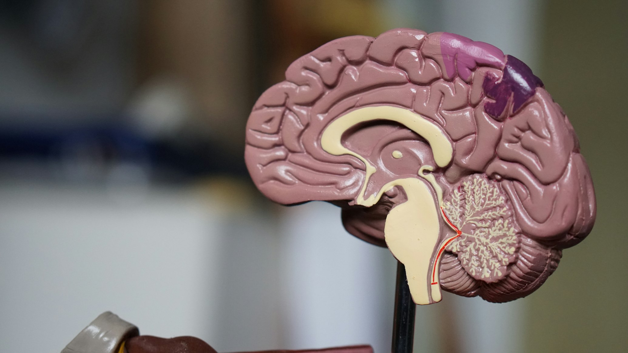
Imagine being able to watch the human brain think—to see which regions spark into activity as a mathematician solves an equation, a musician composes a melody, or a lover recognizes their beloved's face. This seemingly supernatural ability is now scientific reality, thanks to functional Magnetic Resonance Imaging (fMRI), a technology that has fundamentally transformed our understanding of the very organ that defines our humanity 8 .
Since its development in the 1990s, fMRI has become neuroscience's blessed gift, a non-invasive window into the living, functioning brain that has illuminated everything from our most basic sensory processes to the complex neural symphony underlying human consciousness.
"The foundational insight behind fMRI is surprisingly ancient. Over a century ago, Italian scientist Angelo Mosso conducted an experiment where a subject lay on a delicately balanced table, theorizing that blood would rush to the head during mental activity."
Today's fMRI scanners are technological marvels that operationalize this same basic principle, using powerful magnetic fields to track the brain's ever-changing activity patterns, revolutionizing psychology, medicine, and even how we understand ourselves.

The Brain's Secret Language: How fMRI Works
At its core, fMRI doesn't measure brain cells firing directly. Instead, it detects their oxygen consumption—the metabolic aftermath of neural activity. The technology relies on what scientists call the Blood Oxygenation Level-Dependent (BOLD) signal 8 .
Neural Activity
Neurons fire in specific brain regions
Blood Flow
Oxygen-rich blood rushes to active areas
Magnetic Signal
fMRI detects magnetic properties of blood
When neurons in a specific brain region become active, they initially consume more oxygen from the blood. This is quickly followed by a generous—in fact, overcompensating—rush of oxygen-rich blood to the area. Hemoglobin, the iron-rich protein in blood that carries oxygen, has a unique magnetic property: it's diamagnetic when oxygenated but paramagnetic when deoxygenated 8 . An fMRI scanner, typically operating at a magnetic strength 50,000 times that of the Earth's field, can detect this subtle magnetic difference 8 .
Comparison of Major Neuroimaging Techniques
| Technique | What It Measures | Spatial Resolution | Temporal Resolution | Key Advantages |
|---|---|---|---|---|
| fMRI | Blood oxygenation changes (BOLD signal) | Excellent (millimeters) | Good (seconds) | Non-invasive, no radiation, excellent spatial detail |
| EEG | Electrical activity from neurons | Poor (centimeters) | Excellent (milliseconds) | Direct neural measurement, excellent for timing |
| PET | Radioactive tracer distribution | Good (millimeters) | Poor (minutes) | Can track specific neurotransmitters |
| sMRI | Brain structure/anatomy | Excellent (sub-millimeters) | Not applicable | Detailed static pictures of brain anatomy |
This biological chain reaction creates an elegant, indirect map of brain activity. As you perform a task—say, tapping your finger—the part of your brain controlling motor function lights up on researchers' screens. This "activation" is actually a color-coded statistical map superimposed on a structural brain image, showing which voxels (three-dimensional pixels) demonstrated a BOLD signal that closely matched the expected timing of the task 8 .
The greatest advantages of this approach are its non-invasive nature (no injections or radiation), excellent spatial resolution (able to pinpoint activity to regions just millimeters in size), and its ability to capture the entire brain in action simultaneously 8 .
A Window into Brain and Mind: The Expanding Applications of fMRI
Clinical Neuroscience
fMRI has provided unprecedented insights into how brain function alters in neurological and psychiatric conditions. Researchers are using it to identify distinct functional connectivity patterns in conditions like Autism Spectrum Disorder (ASD) and Alzheimer's Disease (AD) 1 7 .
In Age-Related Macular Degeneration (AMD), a leading cause of vision loss, fMRI studies have revealed the brain's remarkable adaptive plasticity, showing how the visual cortex reorganizes itself in response to sensory loss 3 .
Educational Neuroscience
One of the most exciting frontiers is educational neuroscience. Researchers have successfully used fMRI to implement Knowledge Concept Recognition (KCR)—identifying which specific concepts a student is learning or thinking about based on their brain activity patterns 1 .
In one fascinating study, scientists classified fMRI signals from students learning computer science concepts, employing machine learning models to distinguish between different programming knowledge points 1 .
Pharmaceutical Research
The drug development process has been transformed by fMRI's ability to provide objective biomarkers of drug effects on brain function 7 .
Pharmaceutical researchers use what's called pharmacological MRI (phMRI) to measure how experimental compounds alter brain activity and functional connectivity, providing crucial evidence of target engagement and dose-response relationships early in clinical trials 7 .

The Multitasking Brain: A Landmark Experiment
Unveiling the Central Bottleneck
A groundbreaking study published in Nature Communications in 2025 used cutting-edge ultrafast fMRI to solve a long-standing mystery: why can't we effectively perform two cognitively demanding tasks at once? 9
The research team used a 7 Tesla MRI scanner with an unprecedented temporal resolution of 199 milliseconds—far faster than conventional fMRI—to track how the brain processes multiple tasks simultaneously.
Methodology: Isolating the Brain's Processing Stages
The researchers designed two distinct tasks that could be easily distinguished in the brain: an auditory-oculomotor (AO) task (selecting eye movement responses to sounds) and a visual-manual (VM) task (selecting hand movements to colors) 9 . Participants performed these tasks both separately and simultaneously while their brain activity was monitored.
Experimental Steps
- Brain Region Identification
- Temporal Tracking
- Multivariate Pattern Analysis
Task Design
- Auditory-Oculomotor Task
- Visual-Manual Task
- Single and Dual Task Conditions
Key Brain Networks Identified in the Multitasking Study
| Network/Region | Function | Activity Pattern During Multitasking |
|---|---|---|
| Multiple-Demand (MD) Network | General-purpose cognitive operations | Serial processing (one task at a time) |
| Sensory Areas | Processing auditory and visual input | Parallel processing (both tasks simultaneously) |
| Motor Areas | Executing physical responses | Modality-specific parallel processing |
| Fronto-Parietal Regions | Complex task coordination | Central bottleneck location |
Results and Analysis: The Serial Bottleneck Revealed
The findings provided the first direct neural evidence for a "central bottleneck" in human cognition. When participants attempted both tasks simultaneously, the study revealed that:
- Sensory processing occurred in parallel: Both auditory and visual areas processed their respective inputs simultaneously without interference 9 .
- Central cognition operated serially: The fronto-parietal multiple-demand (MD) network processed one task at a time, queuing the second task until the first was completed 9 .
- The bottleneck location was pinpointed: This serial staging occurred specifically during the response selection stage, not during perception or motor execution 9 .
The behavioral data confirmed significant delays in responding to the second task (over half a second), while neural data showed the MD network's activity peaked first for one task, then the other—never both at once 9 .
Behavioral Results from Dual-Task Experiment
| Condition | Task 1 Response Time | Task 2 Response Time | Performance Accuracy |
|---|---|---|---|
| Single Task | ~1000 ms | ~1000 ms | High (similar to baseline) |
| Dual Task (Long Interval) | ~1000 ms | ~1000 ms | High (similar to baseline) |
| Dual Task (Short Interval) | ~1032 ms | ~1500+ ms | Reduced for second task |
This elegant experiment demonstrates how ultrafast fMRI can resolve long-standing questions about human cognition that were previously only addressable through indirect behavioral measures. The findings have implications for everything from interface design to understanding neurological conditions that affect cognitive control.
The Scientist's Toolkit: Essential Equipment in fMRI Research
Conducting cutting-edge fMRI research requires sophisticated technology and carefully controlled materials. Below are key components of the modern fMRI toolkit:
| Item | Function | Example Specifications |
|---|---|---|
| High-Field MRI Scanner | Creates strong magnetic field for signal detection | 3T (clinical), 7T-11.7T (research) 6 9 |
| Gradient Coils | Precisely localize brain signals | 400-1000 mT/m strength, 1000-9000 T/m/s slew rates 6 |
| Radiofrequency Coils | Transmit and receive radio signals | Multi-channel arrays, cryogenically cooled coils 6 |
| Syringe Pump Systems | Precisely deliver liquid rewards in behavioral studies | Computer-controlled with precise timing 4 |
| Response Recording Devices | Capture participant responses | MRI-compatible button boxes, eye-tracking systems 9 |
| Physiological Monitors | Track potential confounds | Respiratory bellows, cardiac pulse oximeters 6 |
| Neutral Solution | Control substance in taste studies | 0.6 mM Sodium Bicarbonate, 6.3 mM Potassium Chloride 4 |
Advanced Equipment
Advanced fMRI research often employs specialized equipment to enhance signal quality or enable novel experiments.
Cryogenic radiofrequency coils cooled to extremely low temperatures can reduce electronic noise, boosting the signal-to-noise ratio by up to three times compared to room-temperature coils 6 .
Animal Studies
In animal studies, where even higher magnetic fields are used, implantable radiofrequency coils can provide dramatic signal improvements—up to 500% increases in some configurations—though these require surgical implantation 6 .
The Future of Brain Imaging: Where fMRI is Headed
As transformative as fMRI has been, neuroscientists continue to push its boundaries. The future direction of fMRI research focuses on several key areas:
Higher Spatial and Temporal Resolution
The move toward ultrahigh magnetic fields (11.7 Tesla and beyond for animal studies) provides stronger signals, enabling researchers to distinguish ever-smaller neural structures 6 . Some researchers are now investigating cortical layers—the thin sheets of cells that form the brain's basic computational units—a spatial scale that has largely been overlooked but bridges cells and brain areas 2 .
Integration Across Scales
Leading neuroscientists argue that fMRI must "break out of its silo" by linking phenomena across levels, from genes and molecules to cells, circuits, networks, and behavior 2 . This integration will require combining fMRI with other techniques like genetics, electrophysiology, and transcriptome analysis.
Improved Accessibility and Analysis
The field is developing more user-friendly software packages, web-based applications, and video tutorials to make neuroimaging accessible to more researchers 2 . Simultaneously, methodological reviews are calling for more transparent reporting of fMRI network estimation methodologies to facilitate the synthesis of findings across studies 5 .
Clinical Translation
Efforts continue to make fMRI more clinically useful by deepening our understanding of the BOLD signal itself and promoting multimodal imaging studies that combine different assessment techniques 2 7 . The regulatory qualification of fMRI as a validated biomarker for specific clinical contexts remains an active pursuit between researchers and agencies like the FDA and EMA 7 .
Conclusion: The Ongoing Blessing of fMRI
From its origins in simple observations about blood flow to its current status as a cornerstone of modern neuroscience, functional Magnetic Resonance Imaging has truly been a benediction to our understanding of the human brain. It has illuminated the neural underpinnings of everything from basic perception to our most complex cognitive capacities, all while remaining safe and non-invasive for human subjects.
"The technology continues to evolve, with ultrafast acquisitions now revealing the brain's information processing in near-real-time and sophisticated analytical techniques decoding the very concepts people are learning."
As fMRI advances, it promises not only to answer fundamental questions about human cognition but also to transform how we diagnose and treat neurological and psychiatric disorders, develop educational strategies, and understand what makes us uniquely human.
While no technology is without limitations—the BOLD signal remains indirect, and the equipment expensive—fMRI's blessings to neuroscience are undeniable. It has given us what previous generations could only dream of: a window into the living, thinking, feeling human mind. As we continue to peer through this window, each discovery brings new questions, ensuring that fMRI will remain neuroscience's blessed tool for exploration for decades to come.