Deep Learning vs. Traditional Methods for EEG Artifact Removal: A Comprehensive Analysis for Biomedical Research
This article provides a systematic comparison of deep learning (DL) and traditional signal processing techniques for electroencephalography (EEG) artifact removal, a critical preprocessing step in neuroscience and clinical diagnostics.
Deep Learning vs. Traditional Methods for EEG Artifact Removal: A Comprehensive Analysis for Biomedical Research
Abstract
This article provides a systematic comparison of deep learning (DL) and traditional signal processing techniques for electroencephalography (EEG) artifact removal, a critical preprocessing step in neuroscience and clinical diagnostics. Tailored for researchers and drug development professionals, it explores the foundational principles, methodological advances, and practical challenges in the field. We detail how DL models like CNNs, GANs, and LSTMs overcome limitations of traditional methods such as ICA and wavelet transforms, particularly for complex artifacts in mobile and wearable EEG. The content covers performance validation metrics, optimization strategies for real-world applications, and a forward-looking perspective on integrating these technologies to enhance the reliability of brain data in biomedical research.
The EEG Artifact Problem: From Classic Challenges to Modern Deep Learning Solutions
Electroencephalography (EEG) is a fundamental tool in neuroscience and clinical diagnostics, providing unparalleled temporal resolution for monitoring brain activity. However, the utility of EEG is critically dependent on signal quality, which is perpetually threatened by various artifacts that can obscure neural information. Artifacts are unwanted signals originating from multiple sources, broadly categorized as physiological (from the subject's body), motion-related (from movement), and technical (from equipment or environment). The expansion of EEG into real-world applications through wearable devices has further exacerbated artifact challenges, as uncontrolled environments and subject mobility introduce additional noise sources. Effective artifact management is therefore not merely a preprocessing step but a pivotal determinant of data validity, influencing applications ranging from clinical diagnosis to brain-computer interfaces (BCIs). This guide systematically defines the EEG artifact landscape, objectively compares the performance of traditional and deep learning removal methods, and provides experimental protocols to inform researcher selection for specific applications.
Defining the Artifact Landscape: A Taxonomy of EEG Contaminants
EEG artifacts can be classified based on their origin, characteristics, and impact on the signal. Understanding this taxonomy is essential for selecting appropriate removal strategies. The table below catalogs the primary artifact types, their sources, and key identifying features.
Table 1: Taxonomy of Common EEG Artifacts
| Artifact Category | Specific Type | Origin/Source | Key Characteristics | Impact on EEG Signal |
|---|---|---|---|---|
| Physiological | Ocular Artifacts [1] [2] | Eye blinks and movements (corneo-retinal dipole, eyelid movement) | High-amplitude, slow deflections (3-15 Hz); Frontally dominant | Overwhelms theta/alpha bands; obscures event-related potentials |
| Muscle Artifacts (EMG) [3] [1] | Head, neck, jaw muscle contractions (talking, swallowing) | High-frequency bursts (>13 Hz to >200 Hz); broad spectral distribution | Masks beta/gamma activity; mimics epileptic spikes | |
| Cardiac Artifacts [1] | Heartbeat (pulse, ECG) | Periodic waveform (~1.2 Hz for pulse); characteristic spike pattern | Can be mistaken for pathological slow waves or spike-wave complexes | |
| Motion | Head Movement [4] | Vertical head movements during walking/gait | Baseline shifts; periodic oscillations | Alters signal baseline and amplitude, corrupting steady-state potentials |
| Electrode Motion [4] | Displacement from scalp due to cable sway, heel strike | Sudden, high-amplitude transients (gait-related amplitude bursts) | Creates spike-like artifacts that mimic neural activity | |
| Technical/Environmental | Powerline Interference [1] [5] | Alternating current from mains electricity | 50/60 Hz sinusoidal oscillation and harmonics | Obscures high-frequency neural activity |
| Instrument Artifacts [1] | Faulty electrodes, high impedance, cable movements | Irregular, non-physiological patterns, sharp transients | Can cause signal dropouts or localized noise |
Diagram 1: A hierarchical taxonomy of common EEG artifacts, showing primary categories and their characteristic signal features.
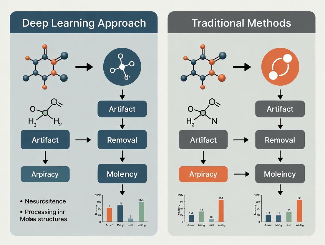
Traditional vs. Deep Learning Approaches: A Comparative Analysis
Artifact removal pipelines generally consist of two phases: detection and removal. Traditional methods often rely on strong statistical or mathematical assumptions about the signal, while deep learning (DL) approaches learn these features directly from data.
Core Methodologies and Workflows
Traditional Workflow: Traditional methods typically involve a sequential process of signal decomposition, component identification, and reconstruction.
Diagram 2: The standard workflow for traditional artifact removal methods, a sequential, component-based process.
Deep Learning Workflow: DL models, particularly those using an end-to-end structure, learn to map contaminated input signals directly to cleaned outputs.
Diagram 3: The end-to-end workflow for deep learning-based artifact removal, showing the direct mapping learned during supervised training.
Quantitative Performance Comparison
The following tables synthesize experimental data from recent studies to compare the performance of various artifact removal methods across different artifact types.
Table 2: Performance Comparison for Ocular and Muscle Artifact Removal
| Method | Category | Artifact Type | Key Metric Results | Experimental Context |
|---|---|---|---|---|
| Regression-based [1] [2] | Traditional | Ocular | N/A | Requires reference EOG channel; performance drops without reference; simple but limited. |
| ICA [3] [1] | Traditional | Ocular, Muscle | N/A | Effective with high-channel counts (>40); requires manual inspection; struggles with low-density wearable EEG. |
| Wavelet Transform [3] | Traditional | Ocular, Muscle | N/A | Commonly used with thresholding; integrated in many pipelines. |
| AnEEG (LSTM-GAN) [6] | Deep Learning | Muscle, Ocular, Environmental | Lower NMSE, RMSE; Higher CC, SNR, SAR vs. wavelet | Outperformed wavelet decomposition; strong agreement with ground truth. |
| CLEnet (CNN-LSTM) [5] | Deep Learning | EMG, EOG, Mixed | SNR: 11.50 dB, CC: 0.93 (Mixed Artifacts) | Excelled in removing mixed artifacts; effective on multi-channel data with unknown artifacts. |
Table 3: Performance Comparison for Motion and Complex Artifacts
| Method | Category | Artifact Type | Key Metric Results | Experimental Context |
|---|---|---|---|---|
| ASR [3] [2] | Traditional | Ocular, Movement, Instrumental | N/A | Widely applied; detects and reconstructs artifact subspaces; suitable for real-time. |
| Motion-Net (CNN) [4] | Deep Learning | Motion | Artifact Reduction (η): 86% ±4.13; SNR Improvement: 20 ±4.47 dB | Subject-specific model; used Visibility Graph features for accuracy on smaller datasets. |
| M4 (State Space Models) [7] | Deep Learning | tACS, tRNS | Best RRMSE, CC for complex tACS/tRNS | Multi-modular network excelled at removing complex transcranial stimulation artifacts. |
| Complex CNN [7] | Deep Learning | tDCS | Best RRMSE, CC for tDCS | Convolutional network performed best on direct current stimulation artifacts. |
Experimental Protocols and Benchmarking
To ensure fair and reproducible comparisons, researchers rely on standardized experimental protocols and benchmark datasets.
Common Experimental Protocols
- Semi-Synthetic Data Generation [6] [7] [5]: This widespread protocol involves clean EEG segments that are artificially contaminated with recorded artifact signals (e.g., EOG, EMG). This provides a known ground truth for quantitative evaluation. Key steps include:
- Acquiring clean EEG and separate artifact signals.
- Linearly or non-linearly mixing them at controlled signal-to-noise ratios (SNRs).
- Using the original clean EEG as the ground truth for training and validation.
- Real Data with Expert Annotation [4]: In this protocol, real EEG data is collected while subjects perform artifact-inducing actions (e.g., walking, blinking). The ground truth is established using advanced methods like artifact-free intervals or high-fidelity artifact removal techniques (e.g., ICA) verified by expert annotation.
- Performance Metrics: Standard quantitative metrics include:
Table 4: Essential Resources for EEG Artifact Removal Research
| Resource/Solution | Function/Role in Research | Example Instances |
|---|---|---|
| Public Benchmark Datasets | Provides standardized data for training and fair comparison of algorithms. | EEGdenoiseNet [5], MIT-BIH Arrhythmia Database [5], PhysioNet Motor/Imagery Dataset [6] |
| Deep Learning Frameworks | Provides the software environment to build, train, and test complex neural network models. | TensorFlow, PyTorch |
| Signal Processing Toolkits | Offers implementations of traditional algorithms for preprocessing and baseline comparisons. | EEGLAB (ICA), ASR, FieldTrip |
| Semi-Synthetic Data Pipelines | Enables controlled validation of removal techniques by providing a known ground truth. | Custom pipelines using clean EEG + artifact libraries (EOG, EMG, ECG) [5] |
The artifact landscape in EEG is diverse, necessitating a nuanced approach to removal. Traditional methods like ICA, regression, and wavelet transforms remain effective for specific, well-defined artifacts, particularly in high-channel-count lab settings. However, their reliance on manual intervention, specific assumptions, and high channel counts limits their efficacy for the low-density, dynamic environments of wearable EEG.
Deep learning approaches represent a paradigm shift, demonstrating superior performance in handling complex artifacts like motion and mixed noise, adapting to unknown artifacts, and operating automatically in an end-to-end fashion. Models like CLEnet [5] and Motion-Net [4] show remarkable versatility and accuracy. The current consensus indicates that while traditional methods are sufficient for controlled settings, deep learning is increasingly critical for real-world, mobile applications. Future research will likely focus on developing more efficient, explainable DL models that require less data and are robust across diverse populations and recording conditions, further bridging the gap between laboratory research and real-world brain monitoring.
In both clinical diagnostics and brain-computer interface (BCI) applications, the presence of artifacts—unwanted signals that obscure data of interest—poses significant challenges to accuracy, reliability, and safety. Artifacts stem from diverse sources, including patient movement, equipment limitations, and environmental interference, and they can severely degrade the performance of medical imaging and neural signal processing systems. The pursuit of robust artifact removal methods has emerged as a critical research frontier, characterized by a fundamental tension between traditional signal processing techniques and modern deep learning approaches. This article examines the impact of artifacts across medical domains and provides a comparative analysis of removal methodologies, emphasizing experimental data and protocols to guide researchers and developers in selecting optimal strategies for their specific applications.
The Impact of Artifacts in Medical Imaging and Neural Data
Artifacts in Medical Imaging
In medical imaging, artifacts can lead to misdiagnosis, false positives, and false negatives. A recent systematic evaluation of Vision-Language Models (VLMs) on medical images containing artifacts revealed significant performance degradation. On original, unaltered images, VLMs achieved moderate accuracy: 0.645 for brain MRI, 0.602 for optical coherence tomography (OCT), and 0.604 for chest X-ray applications. However, the introduction of weak artifacts (images that are partially obscured but still interpretable) caused accuracy to decline by -3.34% (MRI), -9.06% (OCT), and -10.46% (X-ray) [8]. Furthermore, these models demonstrated a poor ability to identify strong artifacts (severely distorted, ungradable images), with detection rates as low as 0.128 for OCT and 0.115 for X-ray images [8]. This indicates that advanced AI models are not yet robust enough to perform reliably on artifact-laden medical images, a critical requirement for clinical deployment.
Artifacts in Brain-Computer Interfaces and EEG
For Brain-Computer Interfaces (BCIs) and electroencephalography (EEG), artifacts are a central problem. BCIs, which enable communication between the brain and external devices, are particularly vulnerable. Artifacts can lead to erroneous interpretations, poor model fitting, and subsequently reduced online performance [9]. This is especially critical for communication BCIs (cBCIs) designed for individuals with severe motor disabilities, where accuracy is paramount [9].
The amplitude and nature of EEG signals make them highly susceptible to contamination. Ocular artifacts from eye blinks can have amplitudes up to ten times greater than the underlying neural signal, while muscle activity introduces noise across a wide frequency range (0–200 Hz), distorting critical beta and gamma brain waves [10]. This contamination hinders the accurate analysis of brain activity, potentially leading to false alarms in clinical settings or errors in BCI control [10].
Comparative Analysis: Traditional vs. Deep Learning Artifact Removal
The evolution of artifact removal techniques has progressed from traditional model-based methods to data-driven deep learning approaches. The table below summarizes the core characteristics of these two paradigms.
| Feature | Traditional Methods | Deep Learning Methods |
|---|---|---|
| Core Principle | Relies on statistical assumptions and linear signal processing [10]. | Learns complex, non-linear mappings from noisy to clean data [10]. |
| Example Techniques | Regression, Independent Component Analysis (ICA), Wavelet Transform, Adaptive Filtering [10] [6]. | Convolutional Neural Networks (CNNs), Transformers, Autoencoders, Generative Adversarial Networks (GANs) [7] [11] [10]. |
| Key Strengths | Well-understood; computationally efficient for some applications [10]. | Superior at handling complex, non-stationary artifacts; end-to-end learning without manual feature engineering [7] [10]. |
| Major Limitations | Linear assumptions may not hold for real data; often requires manual parameter tuning and intervention [10]. | High computational cost; requires large datasets; "black box" nature can reduce interpretability [10]. |
| Generalizability | Often limited, as performance is tied to specific statistical assumptions [10]. | Higher potential, but can be dataset-specific; a key research challenge [10]. |
Performance Comparison of Specific Methods
Experimental data from recent studies allows for a direct comparison of the quantitative performance of various artifact removal methods. The following tables consolidate results from key experiments in EEG and MRI domains.
Table 1: Comparative Performance of Deep Learning Models on EEG Denoising (Semi-Synthetic Data)
| Model | Stimulation Type | Key Metric | Performance | Reference / Context |
|---|---|---|---|---|
| Complex CNN | tDCS | RRMSE (Temporal) | Best Performance | [7] |
| M4 (SSM-based) | tACS | RRMSE (Temporal) | Best Performance | [7] |
| M4 (SSM-based) | tRNS | RRMSE (Temporal) | Best Performance | [7] |
| ART (Transformer) | Multi-Artifact | MSE / SNR | Surpassed other DL models | [11] |
| AnEEG (GAN-LSTM) | Multi-Artifact | NMSE / RMSE / CC | Lower error, higher correlation than wavelet methods | [6] |
| GCTNet (GAN-CNN-Transformer) | Multi-Artifact | RRMSE / SNR | 11.15% reduction in RRMSE, 9.81 improvement in SNR | [6] |
Table 2: Performance of a Deep Learning Model on Knee MRI Motion Artifact Removal
| Model | Input Type | RMSE | PSNR | SSIM | Reference |
|---|---|---|---|---|---|
| De-Artifact Diffusion Model | Motion-corrupted Knee MRI | 11.44 ± 0.12 | 33.23 ± 0.22 | 0.968 ± 0.002 | [12] |
| ESR (Comparison Algorithm) | Motion-corrupted Knee MRI | 14.87 ± 0.11 | 30.85 ± 0.19 | 0.943 ± 0.003 | [12] |
The data shows that the performance of deep learning models can be highly dependent on the specific application and artifact type. For instance, in Transcranial Electrical Stimulation (tES) artifact removal, no single model was best for all stimulation types: Complex CNN excelled for tDCS, while an M4 model based on State Space Models was superior for tACS and tRNS [7]. Furthermore, models like the De-Artifact Diffusion Model for knee MRI demonstrate that deep learning can significantly outperform other advanced algorithms on real-world clinical data [12].
Experimental Protocols and Workflows
To ensure reproducible and valid results, rigorous experimental protocols are essential in artifact removal research. Below are detailed methodologies from key studies.
Protocol for Benchmarking EEG Denoising Models
This protocol, used to evaluate models for removing tES artifacts, highlights the use of semi-synthetic data for controlled evaluation [7].
- Step 1: Dataset Creation. A semi-synthetic dataset is created by combining clean, artifact-free EEG recordings with synthetically generated tES artifacts (simulating tDCS, tACS, and tRNS). This provides a known ground truth for rigorous evaluation.
- Step 2: Model Training. Multiple artifact removal models (eleven in the cited study) are trained on the generated dataset. The model's task is to learn the mapping from the noisy (EEG + artifact) signal to the clean EEG signal.
- Step 3: Quantitative Evaluation. Model performance is evaluated using metrics calculated against the known ground truth. Key metrics include:
- Root Relative Mean Squared Error (RRMSE): Assesses accuracy in both temporal and spectral domains.
- Correlation Coefficient (CC): Measures the linear relationship between the cleaned signal and the ground truth.
Protocol for Knee MRI Motion Artifact Removal
This protocol outlines a supervised deep learning approach for a clinically relevant problem [12].
- Step 1: Paired Data Collection. The model is trained using a dataset of 90 patients, each with two sets of images: one with motion artifacts and a corresponding "ground truth" image without artifacts, acquired via immediate rescanning.
- Step 2: Model Construction and Training. A supervised conditional diffusion model is constructed. This model is trained to learn the conditional distribution of clean images given the artifact-corrupted input.
- Step 3: Validation and Testing. The model is tested on internal and external test datasets from different time periods and hospitals to assess generalizability. Performance is evaluated with:
- Objective Metrics: Root Mean Square Error (RMSE), Peak Signal-to-Noise Ratio (PSNR), and Structural Similarity Index (SSIM).
- Subjective Assessment: Clinical experts rate the quality of the output images.
Workflow Visualization
The following diagram illustrates a generalized supervised deep learning workflow for artifact removal, as applied in both EEG and MRI contexts.
Supervised Deep Learning for Artifact Removal
The Scientist's Toolkit: Key Research Reagents and Materials
Successful experimentation in this field relies on specific data, software, and hardware. The following table details essential "research reagents" for developing and benchmarking deep learning-based artifact removal methods.
Table 3: Essential Research Reagents for Deep Learning-Based Artifact Removal
| Item Name | Function / Application | Specific Examples / Notes |
|---|---|---|
| Semi-Synthetic Datasets | Provides a known ground truth for controlled model training and evaluation. | Clean EEG + synthetic tES artifacts [7]; Clean EEG mixed with EOG/EMG artifacts [6]. |
| Paired Real-World Datasets | Enables supervised training on real artifacts where ground truth is available. | Paired motion-corrupted and immediately rescanned knee MRI [12]. |
| Public EEG Datasets | Serves as benchmark for training and evaluating generalizability. | PhysioNet motor/imaging dataset [6]; EEG Eye Artefact Dataset [6]. |
| Independent Component Analysis (ICA) | Used for generating pseudo clean-noisy training data pairs or for pre-processing. | Enhanced training data generation for the ART transformer model [11]. |
| Deep Learning Frameworks | Provides the programming environment for building and training complex models. | TensorFlow, PyTorch. |
| High-Performance Computing (HPC) | Essential for training large models like transformers and diffusion models. | GPU clusters for reducing training time of deep neural networks. |
| Quantitative Evaluation Metrics | Standardized for objective performance comparison across studies. | RRMSE, CC, SNR, SAR (for EEG) [7] [6]; RMSE, PSNR, SSIM (for MRI) [12]. |
Artifacts present a profound and multi-faceted problem that directly impacts the accuracy of clinical diagnoses and the reliability of emerging technologies like BCIs. The research community's response has been a rapid transition from traditional, assumption-heavy methods toward powerful, data-driven deep learning models. Evidence shows that while no single solution is universally optimal, deep learning approaches—from CNNs and GANs to the latest transformers and diffusion models—consistently push the performance frontier, offering superior artifact suppression and signal restoration. However, challenges remain in computational efficiency, model interpretability, and real-world generalizability. The future of artifact removal lies in addressing these challenges through hybrid architectures, self-supervised learning, and a steadfast commitment to developing solutions with high online parity and clinical translatability.
The analysis of Electroencephalography (EEG) signals is fundamental to both neuroscience research and clinical diagnostics, providing unparalleled insights into brain function with millisecond-level temporal resolution [6]. However, the journey from raw EEG recordings to interpretable neural data is fraught with a major obstacle: artifacts. These unwanted signals, originating from biological sources like eye movements (EOG) and muscle activity (EMG), or environmental sources such as powerline interference, can severely obscure genuine brain activity, leading to misinterpretation and flawed conclusions [6] [13]. For decades, the field has relied on a suite of established traditional methods to combat this problem. Techniques based on regression, filtering, and Blind Source Separation (BSS)—including Independent Component Analysis (ICA), Principal Component Analysis (PCA), and Wavelet Transform—have formed the cornerstone of EEG preprocessing pipelines [14] [15].
These traditional methods are characterized by their strong mathematical foundations, relative computational simplicity, and well-understood behavior. Before the advent of deep learning, they were the definitive tools for enhancing EEG signal quality. This guide provides an objective comparison of these established techniques, detailing their operational principles, experimental performance, and suitability for different artifact types. This establishes a crucial baseline for understanding their enduring value and limitations within the broader thesis of deep learning versus traditional approaches in artifact removal research.
Methodologies and Experimental Protocols
A rigorous evaluation of artifact removal methods requires standardized experimental protocols and performance metrics. The following section outlines the core methodologies of traditional techniques and describes how they are benchmarked in experimental settings.
Core Methodological Principles
- Regression-Based Methods: These techniques employ a reference signal, typically from dedicated EOG or EMG channels, to model and subtract the artifact's contribution from the EEG data. The protocol involves calculating a transfer function between the reference artifact and the contaminated EEG channels, which is then used to estimate and remove the artifact component [14].
- Filtering Techniques (Bandpass/Adaptive):
- Bandpass Filtering: A straightforward protocol where specific frequency bands associated with artifacts are attenuated. For example, a high-pass filter at 0.5 Hz can suppress slow drifts, while a notch filter at 50/60 Hz removes powerline interference [15].
- Adaptive Filtering: This method uses a reference noise signal and an adaptive algorithm (e.g., Recursive Least Squares - RLS) to dynamically adjust filter coefficients, optimizing noise cancellation even when signal statistics change over time [16] [15].
- Blind Source Separation (BSS):
- Independent Component Analysis (ICA): ICA is a statistical technique that decomposes multi-channel EEG data into independent components (ICs). The experimental protocol involves a human expert or automated algorithm visually inspecting and classifying these ICs based on their temporal, spectral, and spatial patterns to identify and remove those corresponding to artifacts [14] [15].
- Principal Component Analysis (PCA): PCA decomposes the EEG signal into orthogonal components ordered by variance. The protocol assumes that high-variance components are likely artifacts. These components are then discarded before reconstructing the signal, preserving the lower-variance neural data [14].
- Wavelet Transform: This method uses a protocol of signal decomposition and thresholding. The EEG signal is decomposed into different frequency sub-bands using a mother wavelet. A threshold is then applied to the wavelet coefficients to suppress those associated with noise, after which the signal is reconstructed [14] [16].
Benchmarking and Evaluation Protocols
To quantitatively assess the performance of these methods, researchers use semi-synthetic or real EEG datasets with known ground truths. Common evaluation metrics include [6] [7] [15]:
- Temporal Similarity: Correlation Coefficient (CC) measures the linear relationship between the cleaned signal and the original clean signal. Root Mean Square Error (RMSE) and Relative RMSE (RRMSE) quantify the magnitude of error introduced by the cleaning process.
- Quality Metrics: Signal-to-Noise Ratio (SNR) and Signal-to-Artifact Ratio (SAR) gauge the improvement in signal quality post-processing.
- Classification Accuracy: In applied settings, the ultimate test is whether denoising improves the accuracy of downstream tasks, such as disease diagnosis using classifiers like Support Vector Machines (SVM) and Random Forests [14].
The diagram below illustrates a generalized experimental workflow for benchmarking artifact removal methods.
Performance Data and Comparative Analysis
The following tables summarize the experimental performance of traditional methods against deep learning approaches and each other, based on published benchmarks.
Table 1: Comparative Performance of Traditional vs. Deep Learning Methods
| Method Category | Method Name | Artifact Type | Key Performance Metrics (vs. Traditional Baseline) | Reference |
|---|---|---|---|---|
| Traditional (Wavelet) | Wavelet Decomposition | Various | Used as a baseline for comparison. | [6] |
| Deep Learning | AnEEG (LSTM-GAN) | Various | Lower NMSE & RMSE, Higher CC, improvements in SNR and SAR. | [6] |
| Deep Learning | GCTNet | Ocular, EMG | 11.15% reduction in RRMSE, 9.81 improvement in SNR. | [6] |
| Traditional (Hybrid) | EMD-DFA-WPD | Ocular, EMG | Achieved classification accuracy of 98.51% for depression diagnosis. | [14] |
| Traditional (Adaptive) | Deep WVFLN (RLS) | Ocular | MSE: 0.18 μV², RMSE: 0.4242 μV. | [16] |
Table 2: Performance of Methods Specific to tES Artifact Removal
| Method | Stimulation Type | Temporal RRMSE | Spectral RRMSE | Correlation Coefficient (CC) |
|---|---|---|---|---|
| Complex CNN | tDCS | Best Performance | - | - |
| M4 Network (SSM) | tACS | Best Performance | - | - |
| M4 Network (SSM) | tRNS | Best Performance | - | - |
| Traditional Methods (e.g., ICA, Filtering) | tDCS/tACS/tRNS | Variable, generally outperformed by the best DL models. | - | - |
Table 3: Performance of Single-Channel EOG Removal Techniques
| Method | Artifact Type | Relative RMSE (RRMSE) | Correlation Coefficient (CC) |
|---|---|---|---|
| FF-EWT + GMETV | EOG (Eye Blink) | Lower | Higher |
| EMD | EOG (Eye Blink) | Higher | Lower |
| SSA | EOG (Eye Blink) | Higher | Lower |
Analysis of Comparative Performance
- Deep Learning Advantages: As shown in Table 1, modern deep learning models like AnEEG and GCTNet consistently demonstrate superior quantitative performance compared to traditional wavelet techniques, achieving lower error (NMSE, RMSE) and higher signal fidelity (CC, SNR) [6]. Their strength lies in learning complex, non-linear artifact patterns directly from data without requiring manual feature engineering.
- Enduring Strengths of Traditional Methods:
- Hybrid Models: Traditional hybrid approaches remain highly competitive. The EMD-DFA-WPD model achieved a remarkable 98.51% accuracy in a clinical depression diagnosis task, proving that well-designed traditional pipelines can deliver state-of-the-art results in applied settings [14].
- Specialized Scenarios: The performance of traditional methods is highly dependent on the artifact type and recording context (Tables 2 & 3). For example, adaptive filters like the WVFLN-RLS are very effective for specific, well-defined noise like ocular artifacts [16].
- Computational Efficiency: Traditional methods like filtering and wavelet transforms are generally less computationally intensive than deep learning models, making them more suitable for real-time applications on devices with limited processing power [13].
Successful experimentation in EEG artifact removal relies on a suite of key resources, from datasets to software tools.
Table 4: Essential Research Resources for Artifact Removal Studies
| Resource Category | Specific Example | Function and Application |
|---|---|---|
| Public EEG Datasets | PhysioNet Motor/Imaging Dataset | Provides real EEG data for training and benchmarking artifact removal algorithms [6]. |
| Mendeley EEG Database | Source of clean and artifactual EEG data, used for validating specific methods like ocular artifact removal [16]. | |
| EEG Eye Artefact Dataset | A dedicated dataset for developing and testing methods against ocular artifacts [6]. | |
| Benchmarking Tools | ABOT (Artefact removal Benchmarking Online Tool) | An online tool that allows researchers to compare over 120 machine learning and traditional methods for artifact removal from various neuronal signals, complying with FAIR principles [13]. |
| Algorithmic Toolboxes | ICA (e.g., in EEGLAB) | Standard software implementation for running Independent Component Analysis on EEG data [14]. |
| Wavelet Toolbox (e.g., in MATLAB) | Provides functions for performing wavelet decomposition and thresholding for denoising [14]. |
The established traditional methods of regression, filtering, and blind source separation form a robust, well-understood foundation for EEG artifact removal. While deep learning approaches are emerging as powerful alternatives, often outperforming traditional methods on quantitative metrics, traditional techniques are far from obsolete.
The future of EEG artifact removal lies not in a single victorious approach, but in the strategic combination of traditional and deep learning methods. Hybrid models that leverage the interpretability and efficiency of traditional techniques with the representational power of deep learning hold significant promise. Furthermore, the development of resources like the ABOT benchmarking tool is critical for providing objective, standardized comparisons that guide researchers in selecting the most appropriate method for their specific signal type, artifact, and application context [13]. As the field moves towards more mobile and real-world applications with wearable EEG, the development of efficient, robust, and hybrid denoising pipelines will be more important than ever.
The analysis of electrophysiological and medical imaging data is fundamental to neuroscience research and clinical diagnostics. However, the accurate interpretation of this data is consistently challenged by the presence of artifacts—unwanted signals that obscure genuine biological activity. Traditional approaches to artifact removal have primarily relied on statistical decompositions and linear filtering techniques. While useful in controlled environments, these methods contain inherent limitations rooted in their requirement for manual intervention and their foundational linear assumptions. The emergence of deep learning represents a paradigm shift, offering data-driven approaches that automatically learn to separate artifact from signal without relying on rigid linear models. This guide provides an objective comparison of these methodological approaches, supported by experimental data quantifying their performance across multiple domains.
Defining the Traditional Paradigm and Its Core Limitations
Traditional artifact removal techniques form the historical foundation of signal processing in biomedical data. These methods can be broadly categorized into several classes, each with specific operational principles and applications.
Regression-based methods primarily rely on setting a reference channel and using linear transformation to subtract the estimated artifact from the contaminated EEG. However, their performance significantly decreases in the absence of a reference signal, which additionally increases operational difficulty and cost [5]. Filtering methods, including notch filters for powerline interference, have relatively limited applicability due to the significant spectral overlap between physiological artifacts (like EMG and EOG) and the effective components of biological signals [5].
Blind Source Separation (BSS) techniques, including Independent Component Analysis (ICA), Principal Component Analysis (PCA), and Canonical Correlation Analysis (CCA), map artifact-contaminated signals into a new data space through mathematical transformations [3] [10]. These methods then remove components corresponding to artifacts using established criteria or manual intervention, reconstructing the remaining components to obtain clean data [5]. Decomposition techniques like Wavelet Transform, Empirical Mode Decomposition (EMD), and Singular Spectrum Analysis (SSA) break signals into constituent elements for selective artifact removal [15] [10].
The core limitations of these traditional approaches emerge from two fundamental constraints: their reliance on manual intervention and their foundation in linear assumptions. ICA and other BSS methods frequently require visual inspection and manual selection of components for rejection, introducing subjectivity and hindering automated processing pipelines [5]. Similarly, techniques like EMD and SSA often depend on manual parameter tuning or threshold-based heuristics, limiting their scalability and generalizability across diverse datasets [10].
The mathematical foundation of many traditional methods rests on the assumption that artifacts and biological signals combine in a linear, stationary manner [10]. This linear assumption fails to account for the complex, nonlinear interactions that characterize real-world biological systems, particularly in mobile acquisition environments [10].
Quantitative Performance Comparison: Traditional vs. Deep Learning Approaches
Experimental data from controlled studies provides objective evidence of the performance disparities between traditional and deep learning methods for artifact removal. The following tables summarize key findings across multiple data modalities and artifact types.
Table 1: Performance Comparison of Methods on EEG Artifact Removal Tasks (RRMSE: Relative Root Mean Square Error; CC: Correlation Coefficient)
| Method Category | Specific Method | Artifact Type | Performance Metrics | Key Limitations |
|---|---|---|---|---|
| Traditional BSS | ICA | Ocular, Muscle | Requires manual component selection [5] | Subjective, non-automated, struggles with low-channel counts [3] |
| Traditional Decomposition | Wavelet Transform | EOG | Depends on threshold parameters [10] | May not perfectly reconstruct original signal [15] |
| Traditional Decomposition | EMD | EOG | Suffers from mode mixing [15] | Can lead to loss of important signal components [15] |
| Deep Learning | Complex CNN | tDCS artifacts | Best performance for tDCS [7] | Specialized for specific artifact types [7] |
| Deep Learning | M4 (SSM-based) | tACS, tRNS artifacts | Best for tACS and tRNS [7] [17] | Higher computational complexity [7] |
| Deep Learning | CLEnet | Multi-artifact (EMG, EOG, unknown) | CC: 0.925, RRMSEt: 0.300 [5] | Complex architecture requiring significant data [5] |
| Deep Learning | AnEEG (GAN-LSTM) | Multiple biological artifacts | Lower NMSE/RMSE, higher CC vs. wavelet [6] | Training instability common in GAN architectures [6] |
Table 2: Performance Metrics for Single-Channel EOG Removal Techniques
| Method | Synthetic Data Performance | Real EEG Performance | Applicability to Single-Channel |
|---|---|---|---|
| FF-EWT + GMETV (Proposed) | Lower RRMSE, Higher CC [15] | Improved SAR and MAE [15] | Specifically designed for SCL [15] |
| ICA | Not evaluated | Effective for MCL [15] | Limited effectiveness for SCL [15] |
| Adaptive Filtering | Requires reference signal [15] | Dependent on reference quality [15] | Applicable but needs reference [15] |
| EMD | Component separation [15] | Mode mixing issues [15] | Applicable but imperfect reconstruction [15] |
Experimental Protocols and Methodologies
To ensure reproducibility and proper interpretation of the comparative data, this section outlines the standard experimental protocols used for evaluating artifact removal methods in the cited studies.
Semi-Synthetic Dataset Creation
A validated approach for rigorous benchmarking involves creating semi-synthetic datasets where clean signals are artificially contaminated with known artifacts, enabling precise ground truth comparisons [7] [5]. For EEG artifact removal studies, researchers typically:
- Source clean EEG segments from publicly available databases like EEGdenoiseNet [5]
- Record or simulate artifact signals (EOG, EMG, ECG, or tES artifacts) [7] [5]
- Linearly mix artifacts with clean EEG at controlled signal-to-noise ratios [5]
- Validate the realism of synthetic datasets against real contaminated recordings [7]
Performance Evaluation Metrics
Standardized metrics enable objective comparison across methods. The most commonly employed metrics include:
- Temporal Domain Accuracy: Relative Root Mean Square Error in temporal domain (RRMSEt) and Correlation Coefficient (CC) between processed and ground truth signals [7] [5]
- Spectral Domain Accuracy: Relative Root Mean Square Error in frequency domain (RRMSEf) assesses preservation of spectral content [7] [5]
- Signal Quality Indices: Signal-to-Noise Ratio (SNR) and Signal-to-Artifact Ratio (SAR) quantify the effectiveness of artifact suppression [6] [5]
- Clinical Validation: For medical imaging, diagnostic performance compared to ground truth using expert ratings and clinical accuracy measures [12] [18]
Implementation Details for Deep Learning Methods
Training deep learning models for artifact removal follows a supervised learning paradigm:
- Network Architecture Selection: Choosing appropriate architectures (CNN, LSTM, GAN, Transformer, or hybrid) based on the signal characteristics [10]
- Loss Function Optimization: Typically using Mean Squared Error (MSE) to minimize differences between denoised output and ground truth [10]
- Parameter Optimization: Utilizing optimizers like Adam, RMSProp, or Stochastic Gradient Descent to update network weights [10]
- Validation Strategy: Employing k-fold cross-validation or hold-out validation sets to prevent overfitting and ensure generalizability [19]
Figure 1: Conceptual workflow comparing the fundamental approaches of traditional and deep learning methods for artifact removal, highlighting their core characteristics and resulting limitations or strengths.
The Scientist's Toolkit: Key Research Reagents and Computational Solutions
Table 3: Essential Resources for Advanced Artifact Removal Research
| Resource Category | Specific Tool/Solution | Function/Purpose | Example Applications |
|---|---|---|---|
| Benchmark Datasets | EEGdenoiseNet | Provides standardized clean/artifact pairs for training & evaluation [5] | Method comparison, model validation |
| Computational Frameworks | TensorFlow/PyTorch | Deep learning framework for model development & training [10] | Implementing CNN, LSTM, GAN architectures |
| Traditional Algorithms | ICA, Wavelet Transform, EMD | Baseline traditional methods for performance comparison [15] [10] | Establishing performance benchmarks |
| Evaluation Metrics | RRMSE, CC, SNR, SAR | Quantitative performance assessment [7] [6] [5] | Objective method comparison |
| Specialized Architectures | CNN-LSTM hybrids (CLEnet) | Capture both spatial and temporal features [5] | Multi-channel EEG artifact removal |
| Specialized Architectures | GAN with LSTM (AnEEG) | Adversarial training for artifact generation/removal [6] | Biological artifact removal |
| Specialized Architectures | State Space Models (M4) | Modeling sequential dependencies [7] [17] | tACS and tRNS artifact removal |
Figure 2: Standardized experimental workflow for developing and evaluating deep learning-based artifact removal methods, showing key stages from data preparation through performance validation.
The experimental evidence consistently demonstrates that deep learning approaches outperform traditional methods across multiple metrics and artifact types. The superiority is particularly evident in handling complex, real-world artifacts where nonlinear relationships and overlapping spectral characteristics prevail. While traditional methods maintain utility in specific, controlled scenarios with clear linear separability, their fundamental limitations of manual intervention and linear assumptions restrict their effectiveness in advanced research and clinical applications.
Deep learning models excel through their capacity for automated operation, preservation of signal integrity, and adaptability to diverse artifact types. However, researchers should consider that this enhanced performance comes with increased computational complexity and data requirements. The choice between methodological approaches should be guided by the specific application constraints, including available computational resources, the necessity for real-time processing, and the diversity of artifacts encountered. Future research directions likely involve developing more efficient architectures, incorporating self-supervised learning to reduce labeled data dependence, and enhancing model interpretability for clinical adoption.
The analysis of electroencephalography (EEG) signals is fundamental to neuroscience research, clinical diagnosis, and brain-computer interface (BCI) applications. However, the inherent vulnerability of EEG signals to contamination by various artifacts—from physiological sources like ocular and muscle activity to environmental interference—has long posed a significant challenge. Traditional artifact removal techniques have primarily operated on linear assumptions and often required manual parameter tuning, limiting their effectiveness and generalizability. The emergence of deep learning represents a paradigm shift in this domain, moving from linear filtering and heuristic-based approaches to data-driven models that learn complex, nonlinear mappings directly from noisy to clean EEG signals. This guide objectively compares the performance of this new deep learning paradigm against traditional methods, providing researchers with the experimental data and methodological insights needed to inform their artifact removal strategies.
Performance Benchmarking: Deep Learning vs. Traditional Methods
Deep learning models have demonstrated superior performance across multiple quantitative metrics compared to traditional artifact removal techniques. The table below summarizes key experimental findings from recent studies.
Table 1: Performance Comparison of EEG Denoising Methods
| Model/Method | Model Type | Key Performance Metrics | Artifact Types Targeted |
|---|---|---|---|
| AnEEG (LSTM-based GAN) [6] | Deep Learning (Generative) | Lower NMSE & RMSE; Higher CC, SNR & SAR values than wavelet techniques | General artifacts (muscle, ocular, environmental) |
| DHCT-GAN [20] | Deep Learning (Hybrid) | Outperforms state-of-the-art networks on 6 metrics for EMG, EOG, ECG, and mixed artifacts | EMG, EOG, ECG, Mixed |
| Deep Lightweight CNN [21] | Deep Learning (Discriminative) | F1-score improvements of +11.2% to +44.9% over rule-based methods | Eye movement, muscle, non-physiological |
| Complex CNN [7] | Deep Learning (Discriminative) | Best performance for tDCS artifact removal (RRMSE, CC) | tES artifacts (tDCS) |
| Multi-modular SSM (M4) [7] | Deep Learning (State Space) | Best performance for tACS and tRNS artifact removal (RRMSE, CC) | tES artifacts (tACS, tRNS) |
| Wavelet Decomposition [6] | Traditional (Thresholding) | Higher NMSE and RMSE, lower CC compared to AnEEG | General artifacts |
| Rule-Based Methods [21] | Traditional (Heuristic) | Lower F1-scores across all artifact categories | Eye movement, muscle, non-physiological |
The data consistently shows that deep learning models achieve a higher degree of signal fidelity. The lower Normalized Mean Square Error (NMSE) and Root Mean Square Error (RMSE) values indicate a better agreement with the original, clean signal, while higher Correlation Coefficient (CC) values reflect a stronger linear relationship with the ground truth [6]. Improvements in Signal-to-Noise Ratio (SNR) and Signal-to-Artifact Ratio (SAR) further confirm the effectiveness of deep learning in isolating neural activity from contamination [6].
Experimental Protocols in Deep Learning EEG Denoising
Data Preprocessing and Model Training
A critical factor in the success of deep learning models is a robust and standardized data preprocessing pipeline. A typical protocol, as used in studies like the lightweight CNN for artifact detection, involves several key stages [21]:
- Signal Standardization: Raw EEG signals are resampled to a uniform sampling rate (e.g., 250 Hz) and filtered with a bandpass (e.g., 1-40 Hz) and a notch filter (50/60 Hz) to remove line noise and out-of-band artifacts.
- Referencing and Normalization: Average referencing is applied to reduce common-mode noise, followed by global normalization (e.g., using RobustScaler) across all channels to standardize the input for stable model training.
- Adaptive Segmentation: The continuous EEG is segmented into non-overlapping windows. Research indicates that optimal window lengths are artifact-specific: 1s for non-physiological, 5s for muscle, and 20s for eye movement artifacts [21].
- Model Optimization: Models are typically trained using loss functions like Mean Square Error (MSE) and optimized with algorithms such as ADAM or RMSProp to minimize the divergence between the denoised output and the ground truth clean signal [10].
Architectures of State-of-the-Art Models
The deep learning paradigm encompasses a diverse set of neural network architectures, each with distinct strengths:
- Generative Adversarial Networks (GANs): Models like AnEEG and DHCT-GAN employ a generator network to produce denoised signals and a discriminator network to distinguish them from real clean EEG. This adversarial training encourages the generator to output highly realistic, artifact-free signals [6] [20].
- Convolutional Neural Networks (CNNs): These models, including the Deep Lightweight CNN, excel at extracting local spatial and temporal features from EEG data. Their hierarchical structure is well-suited for identifying pattern-based artifacts like muscle activity and electrode pops [21] [22].
- Hybrid Architectures: The current state-of-the-art trend involves combining architectural components. DHCT-GAN, for instance, uses a dual-branch hybrid CNN-Transformer network. The CNN branch captures local features, while the Transformer branch models long-range temporal dependencies, allowing the model to handle a wider variety of complex artifacts [20]. Another study combined CNNs for EEG with LSTMs for EDA in a multimodal approach to noise annoyance detection [22].
The following diagram illustrates the typical workflow and architecture of an advanced hybrid denoising model like DHCT-GAN:
Diagram 1: Workflow of a Hybrid Deep Learning Model for EEG Denoising
For researchers embarking on deep learning-based EEG denoising, the following tools, datasets, and models are essential components of the modern toolkit.
Table 2: Research Reagent Solutions for EEG Denoising
| Resource Type | Name/Example | Function & Application |
|---|---|---|
| Public Datasets | Temple University Hospital (TUH) EEG Artifact Corpus [21] | Provides expert-annotated artifact labels for training and validating detection models in realistic clinical scenarios. |
| EEG DenoiseNet [6] | Contains clean EEG segments mixed with EMG and EOG artifacts, useful for semi-simulated validation. | |
| Software & Libraries | EEGLAB/ MATLAB [23] | Standard toolboxes for foundational EEG preprocessing, including filtering and re-referencing. |
| Python Deep Learning Frameworks (TensorFlow, PyTorch) | Essential for building, training, and deploying complex models like CNNs, GANs, and Transformers. | |
| Model Architectures | Lightweight CNNs [21] | Ideal for real-time or resource-constrained applications requiring efficient artifact detection. |
| Hybrid CNN-Transformers (e.g., DHCT-GAN) [20] | State-of-the-art for high-performance denoising, balancing local feature extraction with global context. | |
| Evaluation Metrics | NMSE, RMSE, CC [6] [7] | Quantifies signal fidelity and similarity to ground truth. |
| SNR, SAR [6] | Measures the effectiveness of artifact suppression and neural signal preservation. | |
| Hardware | Portable EEG Systems (e.g., BrainVision LiveAmp) [23] | Enables community-based and naturalistic data collection, expanding data diversity. |
| Dry Electrode Headsets [24] | Facilitates comfortable, long-term monitoring, though may introduce unique artifact profiles. |
The evidence from recent studies solidifies the position of deep learning as a transformative paradigm in EEG artifact removal. By learning complex, nonlinear mappings from noisy to clean signals, models such as hybrid CNN-Transformers and GANs consistently surpass the capabilities of traditional linear methods and heuristic-based rules. The key advantages of this shift are superior denoising performance across a range of quantitative metrics, reduced reliance on manual expertise for parameter tuning, and enhanced adaptability to diverse and complex artifact types. For the research community, adopting this paradigm requires familiarity with new tools and datasets but offers the reward of more accurate, automated, and reliable EEG analysis, thereby strengthening findings in neuroscience, clinical diagnostics, and drug development.
A Deep Dive into Architectures: CNNs, GANs, and Hybrid Models in Action
Convolutional Neural Networks (CNNs) for Spatial and Morphological Feature Extraction
In the field of deep learning, particularly in research focused on artifact removal, a central conflict exists between traditional, often linear, methods and modern deep learning approaches. Convolutional Neural Networks (CNNs) have emerged as a powerful tool for spatial and morphological feature extraction, directly addressing the limitations of traditional techniques. This capability is crucial for distinguishing subtle features in complex datasets, from medical images to biological shapes. The core strength of CNNs lies in their hierarchical architecture, which uses convolution layers with filters to automatically learn and extract relevant spatial features—such as edges, textures, and shapes—from input data [25]. This review objectively compares the performance of CNN-based feature extraction methods against traditional alternatives across various scientific domains, providing researchers and drug development professionals with validated experimental data to guide their methodological choices.
Performance Comparison: CNNs vs. Traditional Methods
Quantitative comparisons reveal that CNN-based methods consistently outperform traditional techniques in extracting discriminative spatial and morphological features. The following tables summarize experimental results from multiple independent studies.
Table 1: Performance Comparison in Morphological Feature Extraction
| Method | Application Domain | Key Performance Metric | Result | Traditional Method Result |
|---|---|---|---|---|
| Morpho-VAE [26] | Primate Mandible Shape Analysis | Cluster Separation Index (CSI) | Well-separated clusters (CSI <1) | PCA: Overlapping clusters (CSI >1) |
| Morphological-Convolutional Neural Network (MCNN) [27] | Melanoma Classification | Area Under Curve (AUC) | 0.94 (95% CI: 0.91 to 0.97) | Popular CNNs (ResNet-18, etc.): Lower AUC |
| Classification CNN [28] | Pharmaceutical Excipient Morphology | Classification Accuracy | High Accuracy | N/A (Traditional classification not used) |
| Morphological Feature Extractor [29] | Dog Breed Identification | Qualitative Recognition Capability | Structured, interpretable feature analysis | Traditional CNN: Confused by background |
Table 2: Performance in Signal & Image Artifact Handling
| Method | Application | Comparison Baseline | Performance Advantage |
|---|---|---|---|
| Specialized Lightweight CNNs [30] | EEG Artifact Detection | Rule-Based Methods | F1-score improvement: +11.2% to +44.9% |
| Complex CNN [7] | tDCS Artifact Removal | Various ML Methods | Best performance for tDCS artifacts (RRMSE, Correlation Coefficient) |
| M4 Network (SSMs) [7] | tACS & tRNS Artifact Removal | Various ML Methods | Best performance for tACS/tRNS artifacts |
| PISC (Physics-Informed + CNN) [31] | CT Metal Artifact Reduction | NMAR, O-MAR, CNN-MAR, DuDoNet | Best qualitative scores, least new artifacts |
| DuDoNet [31] | CT Metal Artifact Reduction | FBP, NMAR, O-MAR, CNN-MAR | Best quantitative Artifact Index (AI) |
Experimental Protocols and Methodologies
Morphological Feature Extraction with Morpho-VAE
The Morpho-VAE framework represents a hybrid approach combining unsupervised and supervised learning for landmark-free morphological analysis [26].
- Data Preparation: The study used 147 mandible samples from seven families. Three-dimensional mandible data were projected from three directions to create two-dimensional input images for analysis [26].
- Architecture: The model integrates a Variational Autoencoder (VAE) with a classifier module. The encoder compresses input images into a low-dimensional latent space, and the decoder reconstructs images. The classifier ensures the extracted features are discriminative between classes [26].
- Loss Function: A weighted total loss function guides the training:
E_total = (1 - α)E_VAE + αE_C, whereE_VAEis the VAE reconstruction and regularization loss, andE_Cis the classification loss. The hyperparameterαwas optimized to 0.1 via cross-validation [26]. - Evaluation: Cluster separation in the latent space was quantified using a custom Cluster Separation Index (CSI) and compared against traditional Principal Component Analysis (PCA) and a standard VAE [26].
Specialized CNNs for EEG Artifact Detection
This protocol outlines the development of specialized CNNs for detecting specific classes of artifacts in continuous Electroencephalography (EEG) signals [30].
- Data Source: The Temple University Hospital (TUH) EEG Corpus was used, containing expert-annotated artifacts. The dataset includes 158,884 annotations across 19 categories, with muscle, eye movement, and electrode artifacts being most common [30].
- Preprocessing:
- Signal Standardization: Recordings were resampled to 250 Hz and converted to a standardized 22-channel bipolar montage.
- Filtering: A bandpass filter (1-40 Hz) and notch filter (50/60 Hz) were applied to remove line noise.
- Referencing & Normalization: Average referencing was applied, followed by global normalization using RobustScaler.
- Segmentation: Data were segmented into non-overlapping windows of varying lengths (1s to 30s) to identify the optimal temporal context for each artifact type [30].
- Model Design and Training: Three distinct CNN systems were developed, each optimized for a specific artifact class (eye movement, muscle, non-physiological). Each system was tailored with an ideal temporal window size identified through experimentation: 20s for eye movements, 5s for muscle activity, and 1s for non-physiological artifacts [30].
- Comparison: The CNN systems were evaluated against standard rule-based clinical detection methods on a held-out test set using metrics like F1-score and ROC AUC [30].
CNN-based Metal Artifact Reduction (MAR) in CT
This protocol describes a comparative benchmark of MAR methods, including deep learning approaches, for CT images with neurovascular coils [31].
- Patient Cohort: 40 patients with intracranial aneurysms treated with endovascular coil embolization were included. Non-contrast brain CT scans were acquired using specific clinical protocols [31].
- Compared Methods: The study compared several algorithms:
- Traditional MAR: Normalized MAR (NMAR) and Metal Artifact Reduction for Orthopedic Implants (O-MAR).
- Deep Learning MAR: CNN-MAR and a Dual Domain Network (DuDoNet).
- Hybrid Method: Physics-Informed Sinogram Completion (PISC), which combines NMAR with physical correction [31].
- Evaluation:
- Quantitative Analysis: The Artifact Index (AI) was calculated by measuring the standard deviation (SD) of ROIs placed near metal coils and in artifact-free regions
(AI = SD_coil / SD_background). - Qualitative Analysis: Two blinded neuroradiologists independently evaluated the images using a five-point Likert scale, assessing metal artifact severity, diagnostic confidence, resolution, new artifacts, and soft tissue interfaces [31].
- Quantitative Analysis: The Artifact Index (AI) was calculated by measuring the standard deviation (SD) of ROIs placed near metal coils and in artifact-free regions
Workflow and Architectural Diagrams
Workflow for Landmark-Free Morphological Analysis
The following diagram illustrates the integrated workflow of the Morpho-VAE model for extracting morphological features without manual landmark annotation.
Specialized CNN Pipeline for EEG Artifact Detection
This diagram outlines the specialized pipeline for detecting different classes of artifacts in EEG signals, highlighting the artifact-specific optimization.
For researchers aiming to implement CNN-based feature extraction or artifact removal, the following tools and datasets are essential.
Table 3: Essential Research Resources for CNN-Based Feature Extraction
| Resource Name | Type | Primary Function in Research | Example Use-Case |
|---|---|---|---|
| TUH EEG Corpus [30] | Datasets | Provides expert-annotated, real-world EEG data for training and validating artifact detection models. | Benchmarking EEG artifact detection algorithms. |
| Primate Mandible Image Data [26] | Datasets | Enables landmark-free morphological analysis of biological shapes. | Studying evolutionary and developmental biology. |
| SEM Images of Excipients [28] | Datasets | Allows quantitative evaluation of particle morphology for pharmaceutical materials. | Clustering and classifying excipients by particle shape. |
| Pre-trained Models (VGG16, ResNet) [25] | Software/Tools | Provides a feature extraction backbone via transfer learning, saving training time and computational resources. | Rapid prototyping for custom image classification tasks. |
| Morphological Regulated VAE (Morpho-VAE) [26] | Algorithm | A hybrid architecture for extracting discriminative morphological features in an unsupervised manner. | Analyzing shapes where homologous landmarks are difficult to define. |
| Physics-Informed Sinogram Completion (PISC) [31] | Algorithm | A hybrid method combining traditional physical correction with deep learning for superior artifact reduction. | Correcting metal artifacts in clinical CT images. |
| RobustScaler [30] | Preprocessing Tool | Global normalization method that preserves relative amplitude relationships, crucial for stable CNN training. | Preprocessing EEG signals before input into a CNN. |
Generative Adversarial Networks (GANs) and LSTM Integration for Temporal Dynamics
The removal of artifacts from biological signals represents a significant challenge in data analysis, particularly within neuroscience and clinical diagnostics. Traditional methods often rely on statistical assumptions and linear models, which struggle to capture the complex, non-linear, and temporal nature of artifacts contaminating signals like electroencephalography (EEG) or Heart Rate Variability (HRV). The integration of Generative Adversarial Networks (GANs) and Long Short-Term Memory (LSTM) networks has emerged as a powerful, model-free approach for this task. This hybrid architecture leverages the adversarial training of GANs to learn the underlying distribution of clean data, while the LSTM components excel at modeling long-range temporal dependencies inherent in time-series data. This synergy creates a robust framework for generating high-fidelity, artifact-free signals, moving beyond the limitations of conventional techniques and offering a new paradigm for data preprocessing in critical applications such as brain-computer interfaces (BCIs) and personalized healthcare monitoring [6] [11] [32].
Performance Comparison: GAN-LSTM vs. Alternative Methods
Experimental results across various domains demonstrate the superior performance of the GAN-LSTM fusion model compared to both traditional methods and other deep learning architectures. The following tables summarize quantitative findings from key studies in artifact removal and sequential data prediction.
Table 1: Performance Comparison in EEG Artifact Removal
| Model | Dataset | Key Metric 1 | Key Metric 2 | Key Metric 3 |
|---|---|---|---|---|
| AnEEG (LSTM-based GAN) [6] | Various EEG datasets | Lower NMSE & RMSE vs. wavelet methods | Higher Correlation Coefficient (CC) | Improved SNR and SAR |
| ART (Transformer) [11] | Multiple open BCI datasets | Superior Mean Squared Error (MSE) | Better Signal-to-Noise Ratio (SNR) | Sets new benchmark in EEG processing |
| GCTNet (GAN+CNN+Transformer) [6] | Semi-simulated & real datasets | 11.15% reduction in Relative RMSE | 9.81 improvement in SNR | Outperforms existing approaches |
| Wavelet Decomposition [6] | Various EEG datasets | Higher NMSE & RMSE | Lower CC | Lower SNR and SAR |
Table 2: Performance in Sequential Data Prediction Tasks
| Model | Application | Performance Metrics |
|---|---|---|
| GAN-LSTM [33] | Coiled Tubing Drilling Parameter Prediction | ~90% accuracy (≈17% higher than standalone GAN or LSTM) |
| LSTM [34] | Daily Streamflow Forecasting | Lowest NRMSE; Highest Nash-Sutcliff efficiency (E, EH, EL) and correlation |
| Standalone GAN [33] | Coiled Tubing Drilling Parameter Prediction | Lower accuracy compared to the fused GAN-LSTM model |
| SVR (Support Vector Regression) [35] | Traffic Flow Prediction | Outperformed by hybrid deep learning models |
Table 3: Performance in Other Domains
| Model | Application | Key Findings |
|---|---|---|
| TL-LSTM-GAN [35] | Adaptive Traffic Signal Control | Reduces congestion and energy usage; superior to state-of-the-art methods |
| LSTM [36] | Schottky Diode Behavior Prediction | R² > 0.98; RMSE as low as 6.22 mA; reliable, cost-effective alternative to experiments |
| Adaptive LSTM [32] | HRV-based Psychosis Prediction | Mean F1 score of 0.9817 without artifact preprocessing |
Experimental Protocols and Methodologies
GAN-LSTM for Artifact Removal in EEG
The AnEEG model presents a typical protocol for using an LSTM-based GAN to remove artifacts from EEG signals. The generator is typically composed of a two-layer LSTM network, which processes the noisy EEG input sequence. This architecture is chosen specifically to capture the temporal dependencies and contextual information in the signal. The generator's role is to transform the noisy input into a cleaned version of the signal. The discriminator, often a one-dimensional convolutional neural network (1D-CNN), then judges whether its input is a real, clean signal from the training set or a generated one from the generator. This adversarial process, guided by a loss function that may include terms for temporal-spatial-frequency consistency, trains the generator to produce artifact-free EEG that preserves the underlying neural information [6]. The model is trained on datasets containing pairs or sets of EEG recordings with various artifacts (e.g., from the HaLT dataset or PhysioNet), allowing it to learn a robust mapping from noisy to clean signals [6].
GAN-LSTM for Parameter Prediction in Engineering
In a different application, a GAN-LSTM fusion network was employed to predict multiple drilling parameters (e.g., circulation pressure, ROP, wellhead pressure) in coiled tubing operations. Here, the LSTM network serves as the core predictive model, processing the historical time-series data of drilling parameters. Its ability to capture long-term dependencies in sequential data is crucial for accurate forecasting. To overcome the challenge of multi-variable output and maintain high accuracy, the powerful generative model of a GAN is used to refine the LSTM's outputs. The low-dimensional data from the LSTM is fed into the GAN's generator, which produces the final, high-dimensional predictions. The discriminator then evaluates these predictions against real data. This combined approach mitigates the accuracy drop typically seen when LSTMs output multiple variables and leads to more reliable predictions for complex engineering systems [33].
The Scientist's Toolkit: Essential Research Reagents and Materials
Table 4: Key Research Reagents and Computational Tools
| Item / Solution | Function / Explanation |
|---|---|
| EEG Datasets (e.g., PhysioNet, HaLT) | Provide the essential raw, often artifact-laden, biological signals required for training and validating deep learning models. [6] |
| Independent Component Analysis (ICA) | A blind source separation technique used as a preprocessing step to generate pseudo clean-noisy data pairs for supervised training of models like ART. [11] |
| Graph Data & Spatial Embeddings | Provide structural and relational context (e.g., sensor locations in a network) which can be fused with time-series data in advanced spatio-temporal models. [37] |
| Wearable Device HRV Recordings | Source long-term, real-world physiological time-series data for personalized health prediction models, often containing inherent artifacts. [32] |
| Pre-trained Models (e.g., ResNet-50 on ImageNet) | Used in Transfer Learning (TL) to initialize discriminators, leveraging learned feature representations to improve convergence and performance on new tasks. [35] |
Architectural Workflows and Signaling Pathways
The following diagram illustrates the typical workflow of a GAN-LSTM model for signal denoising, integrating the components and processes described in the experimental protocols.
GAN-LSTM Denoising Workflow
The logical flow begins with a Noisy Signal Input (e.g., artifact-contaminated EEG) fed into the LSTM Generator. The generator's role is to capture the temporal context and produce a Generated "Clean" Signal. This generated signal, along with a Real Clean Signal from the training dataset (ground truth), is presented to the Discriminator. The discriminator's task is to distinguish between the two. The result of this discrimination generates Adversarial Feedback (a gradient signal), which is propagated back to the LSTM Generator. This feedback loop forces the generator to continuously improve its output until it can produce a Clean Output that the discriminator can no longer distinguish from the real clean data. Throughout this process, the LSTM components are critical for understanding the temporal evolution of the signal, ensuring that the denoising is context-aware and not just a point-wise operation [6] [33].
Emerging Transformer Networks and Attention Mechanisms for Global Context
The quest to effectively capture global context—the complex, long-range dependencies within data—represents a central challenge in deep learning. For years, traditional architectures like Convolutional Neural Networks (CNNs) and Recurrent Neural Networks (RNNs) have been constrained in their ability to model these relationships, primarily due to their inductive biases toward local connectivity and sequential processing [38]. The introduction of the Transformer architecture in 2017, with its core self-attention mechanism, marked a paradigm shift by enabling direct, parallelized interactions between all elements in a sequence, regardless of their positional distance [39]. This foundational innovation unlocked unprecedented capabilities in modeling global context, establishing Transformers as the backbone of modern large language models and vision systems [40] [41].
However, the standard self-attention mechanism's computational and memory requirements scale quadratically with sequence length (O(L²)), creating a significant bottleneck for processing very long sequences [42]. This review explores the cutting-edge landscape of emerging Transformer networks and attention mechanisms specifically designed to overcome this limitation while preserving or even enhancing the model's capacity for global context understanding. We will objectively compare the performance of these novel architectures, analyze supporting experimental data, and frame their development within the applied context of artifact removal in biomedical signal processing, a domain where distinguishing global signal from local noise is paramount.
Foundational Principles and the Need for Evolution
Core Components of the Original Transformer
The original Transformer architecture, introduced in "Attention Is All You Need," relies on three key components to process information. The Self-Attention Mechanism allows the model to weigh the importance of all other tokens when encoding a specific token. It dynamically computes a weighted sum of value (V) vectors based on compatibility scores between a query (Q) and a set of keys (K), formally expressed as Attention(Q, K, V) = softmax(QKᵀ/√dₖ)V [39]. Multi-Head Attention extends this by running multiple self-attention operations in parallel, each with linearly projected versions of Q, K, and V. This allows the model to jointly attend to information from different representation subspaces at different positions, capturing diverse contextual relationships [40] [39]. Finally, Positional Encodings are added to the input embeddings to inject information about the order of the sequence, as the self-attention mechanism itself is permutation invariant. The original model used fixed sine and cosine functions for this purpose [38] [39].
The Quadratic Bottleneck and Its Implications
The fundamental constraint of the standard Transformer is its quadratic complexity. Calculating the attention scores for every token against every other token in a sequence of length L requires computing and storing an L×L matrix. This O(L²) complexity makes it computationally prohibitive and memory-intensive to process very long sequences, such as extended documents, high-resolution images, or lengthy biomedical time-series recordings [42]. This limitation has spurred a major research direction focused on developing more efficient architectures that can maintain strong performance on global context modeling while being scalable for long-context applications.
Emerging Architectures and Mechanisms
The field has evolved along several innovative paths to address the quadratic bottleneck. The following table summarizes the core approaches and their representative models.
Table 1: Categories of Emerging Efficient Transformer Architectures
| Architecture Category | Core Innovation | Key Representative Models | Primary Advantage |
|---|---|---|---|
| Linearized Attention | Reformulates attention as linear operations via kernel approximation or recurrence. | Linear Transformer [42], Performer [42], RetNet [40], Mamba [40] | Linear O(L) time and memory complexity. |
| State Space Models (SSMs) | Replaces attention with structured state-space layers for sequence modeling. | Mamba [40] [7], S4 [7] | Efficient long-sequence handling; data-dependent reasoning (Mamba). |
| Hybrid & Alternative Models | Combines elements from different architectures or introduces novel mechanisms. | Hyena [40], RWKV [40], Multi-Range Attention [43] | Leverages strengths of multiple paradigms (e.g., convolution + attention). |
| Sparse Attention | Restricts attention computation to a selected subset of tokens. | Fixed-Pattern, Clustering-Based, Block-Sparse [42] | Reduces computation by ignoring presumably irrelevant token pairs. |
Linear Attention and Recurrent Formulations
Linear Attention methods aim to break the quadratic barrier by re-parameterizing the softmax attention. The core idea is to find a feature map, φ(·), that allows the attention operation to be expressed as a linear function, often leveraging the associativity property of matrix multiplication to compute the outputs in reverse order. For example, the Linear Transformer uses a positive feature map like ELU(x)+1, while the Performer leverages random Fourier features to unbiasedly approximate the softmax kernel [42]. A significant development in this space is the Retentive Network (RetNet), which employs a retention mechanism that mimics recurrence within Transformer blocks, enabling constant-time (O(1)) inference while maintaining compatibility with Transformer-like APIs [40].
State Space Models (SSMs)
State Space Models (SSMs) have emerged as a powerful alternative to attention, particularly for long sequences. Inspired by classical control theory, SSMs map a one-dimensional input sequence to an output via a hidden state, showing linear scalability. Mamba is a leading SSM architecture that enhances traditional linear SSMs by introducing data-dependent weights, allowing it to selectively propagate or forget information based on the current input. This makes Mamba capable of context-dependent reasoning, a capability previously dominated by attention-based models. Mamba has demonstrated high performance in domains like genomics, audio, and long-text modeling, offering 100x faster inference on sequences of 64k tokens compared to standard Transformers in some benchmarks [40] [7].
Multi-Range and Hybrid Attention Mechanisms
Beyond fully replacing attention, other approaches enhance it with more efficient or flexible patterns. Multi-Range Attention Mechanisms are a class of architectures that enable a single model to dynamically integrate features across multiple spatial, temporal, or semantic scales. They overcome the limitation of fixed-context attention by employing techniques like variable window sizes, hierarchical compression, and parallel multi-scale heads. For instance, Multi-Scale Window Attention (MSWA) assigns different window sizes to each attention head and layer, allowing fine-grained local and broad global contexts to be processed concurrently. Empirical results show that such multi-range approaches deliver better performance on language modeling perplexity and image super-resolution tasks than single-scale models at a comparable computational cost [43].
Hyena is another notable hybrid model that replaces attention with long convolutions and gated activations, reportedly delivering 100x faster inference on long sequences (64k tokens) while maintaining competitive accuracy on NLP benchmarks [40].
Performance Comparison and Experimental Data
To objectively evaluate these emerging architectures, we summarize their performance based on reported benchmarks. The following table synthesizes quantitative data from the literature.
Table 2: Comparative Performance of Emerging Architectures on Key Benchmarks
| Model / Architecture | Complexity (Seq. Length L) | Reported Performance Highlights | Key Experimental Findings |
|---|---|---|---|
| Standard Transformer | O(L²) | Baseline for tasks (e.g., LM, BV) | Becomes computationally infeasible for very long sequences (L > 50k). |
| Mamba (SSM) | O(L) | Language Modeling: Matches Transformer quality on Pile (800GB text) [40].Long Context: 100x faster inference at 64k tokens [40].Artifact Removal: SOTA on tACS/tRNS artifact removal (RRMSE, CC) [7] [17]. | Excels in long-context and data-dependent reasoning tasks; efficient on hardware. |
| Hyena | O(L) | Long Context: 100x faster inference at 64k tokens [40].Benchmarks: Comparable accuracy to Transformers on NLP benchmarks [40]. | Proves attention is not strictly necessary for high-level reasoning on long sequences. |
| RetNet (Retentive) | O(1) Inference | Language Modeling: Competitive with LLMs (e.g., LLaMA) [40].Inference: Constant-time inference, ideal for low-power deployment [40]. | A strong candidate for replacing Transformers in production systems due to O(1) inference. |
| Multi-Range Attention | O(L·Σkᵢ) | Image SR: +0.16 dB PSNR, 3.3x speedup vs. SRFormer [43].Language Modeling: Lower perplexity vs. sliding-window attention [43]. | Provides an effective efficiency-performance trade-off by fusing multi-scale context. |
| RWKV (RNN+Transformer) | O(L) | Language Modeling: Rivals LLaMA across 100+ languages [40].Inference: Stateful, efficient for streaming [40]. | Merges training parallelism of Transformers with efficient inference of RNNs. |
Experimental Protocols in Benchmarking
The performance claims for architectural models like Mamba and Hyena are typically validated through standardized benchmarks. For language modeling, models are trained on large-scale corpora like the Pile (800GB of text) and evaluated on held-out test data using metrics such as perplexity (PPL), where a lower score indicates better performance [40] [43]. For long-context efficiency, a common protocol involves measuring training and inference speed (e.g., tokens/second) and memory usage on sequences of varying lengths (e.g., 8k to 64k tokens), comparing against a standard Transformer baseline [40]. On specialized tasks like image super-resolution, models are evaluated on benchmark datasets (e.g., Urban100) using pixel-level accuracy metrics like Peak Signal-to-Noise Ratio (PSNR) and structural similarity metrics [43].
Application in Biomedical Artifact Removal: A Case Study
The theoretical advantages of these new architectures are being tested in critical real-world applications, notably in the removal of artifacts from biomedical signals like electroencephalography (EEG). This domain perfectly illustrates the clash between deep learning and traditional methods, framed within the need to preserve global neural context while removing local noise.
Traditional vs. Deep Learning Approaches
Traditional methods for EEG artifact removal include regression-based techniques, Blind Source Separation (BSS) like Independent Component Analysis (ICA), and wavelet transforms [6]. These are often mathematically elegant and interpretable but can struggle with non-linear and complex artifacts, sometimes requiring manual intervention and leading to the unwanted loss of neural information [6].
Deep learning approaches, particularly CNNs and RNNs, offered an improvement by learning complex, non-linear filters directly from data. For example, Generative Adversarial Networks (GANs) have been successfully used to generate artifact-free EEG signals [6]. However, their ability to capture long-range dependencies in the signal can be limited.
The Emergence of State Space Models and Attention
A 2025 benchmark study on Transcranial Electrical Stimulation (tES) artifact removal directly compared 11 different methods, providing a clear snapshot of the current landscape [7] [17]. The study's experimental protocol involved creating a semi-synthetic dataset by combining clean EEG data with synthetic tES artifacts (tDCS, tACS, tRNS), allowing for controlled evaluation using Root Relative Mean Squared Error (RRMSE) and Correlation Coefficient (CC) as key metrics [7] [17].
The results were telling: while a Complex CNN performed best for the simpler tDCS artifacts, a multi-modular network (M4) based on State Space Models (SSMs) yielded the best results for the more complex tACS and tRNS artifacts [7] [17]. This suggests that SSMs, with their superior ability to model long-range temporal dependencies, are better equipped to handle oscillatory and noisy artifacts that require a more global understanding of the signal context. This positions SSMs like Mamba as a powerful tool for robust analysis of neural dynamics in clinical applications [7].
Diagram 1: A simplified workflow for EEG artifact removal using an SSM-based model, which leverages global context to separate neural signal from noise.
The Scientist's Toolkit: Essential Research Reagents
For researchers seeking to implement or experiment with these emerging architectures, the following "toolkit" outlines key components and their functions.
Table 3: Research Reagent Solutions for Model Development and Evaluation
| Research Reagent | Function / Purpose | Examples / Notes |
|---|---|---|
| Efficient Attention Core | The fundamental algorithm for linear or sparse attention. | Linear Attention [42], FlashAttention [40], Multi-Range Attention [43]. |
| State Space Model Layer | The core building block for SSM-based architectures. | Mamba Block [40] [7], S4 Layer. Often available in open-source repositories. |
| Long-Range Benchmarks | Datasets and tasks to evaluate global context understanding. | Long-Range Arena [43], PG-19 (long text), Long-context EEG datasets [7]. |
| Artifact Removal Datasets | Semi-synthetic or real data for biomedical validation. | tES-EEG datasets [7] [17], EEG DenoiseNet [6], MIT-BIH [6]. |
| Optimization Libraries | Hardware-optimized kernels for efficient training/inference. | FlashAttention [40], CUDA-optimized SSM kernels (for Mamba etc.) [40]. |
| Evaluation Metrics | Quantitative measures for model performance. | Perplexity (PPL) [43], RRMSE [7] [17], Correlation Coefficient (CC) [7] [17], PSNR [43]. |
The landscape of global context modeling is undergoing a rapid and exciting transformation. While the canonical Transformer architecture unveiled the critical importance of attention, its computational limits have catalyzed a new wave of innovation. Emerging architectures like Mamba, RetNet, and Hyena, along with advanced mechanisms like Multi-Range Attention, are demonstrating that it is possible to achieve—and in some cases surpass—the global context modeling capabilities of Transformers with dramatically improved efficiency.
The application of these models in challenging domains like biomedical artifact removal provides a compelling validation of their potential. The success of State Space Models in particular highlights a broader trend: moving beyond a one-size-fits-all attention mechanism toward a diverse ecosystem of specialized, efficient, and powerful architectures for understanding context. For researchers and practitioners, this evolution offers a new set of powerful tools to decode increasingly complex data, pushing the boundaries of what is possible in fields ranging from drug development to clinical neuroscience.
Electroencephalography (EEG) is a crucial non-invasive tool for capturing neural activity in fields ranging from clinical diagnostics to cognitive neuroscience and Brain-Computer Interfaces (BCI) [6] [10]. However, the utility of EEG is severely compromised by its vulnerability to various artifacts—unwanted signals that do not originate from neural activity [44]. These artifacts, which can be physiological (e.g., from eye movements, muscle activity, cardiac rhythms) or non-physiological (e.g., power line interference, electrode pops), often possess amplitudes much larger than genuine brain signals and exhibit overlapping spectral characteristics, making their separation a formidable challenge [6] [10] [44].
The inability to effectively address these artifacts not complicates the interpretation of brain dynamics but also has profound implications in clinical practice, where their presence can lead to misdiagnosis [6]. This challenge frames a critical thesis in biomedical signal processing: Can advanced deep learning (DL) methodologies surpass the capabilities of traditional artifact removal techniques? This case study investigates this question by examining "AnEEG," a novel LSTM-based Generative Adversarial Network (GAN) designed for multi-artifact removal. We will evaluate its performance against other contemporary deep learning models and established traditional methods, using quantitative metrics and detailed experimental protocols to provide a comprehensive comparison.
Methodological Deep Dive: From Traditional Techniques to Deep Learning
The Landscape of Traditional EEG Artifact Removal
Traditional approaches to artifact removal are primarily divided into two categories: those that estimate and remove artifacts using reference channels, and those that decompose EEG signals into alternative domains for artifact extraction [6]. Key traditional methods include:
- Regression-based Methods: These use reference signals (e.g., EOG for ocular artifacts) to estimate and subtract the artifact component from the EEG recording [6].
- Blind Source Separation (BSS): Techniques like Independent Component Analysis (ICA) separate recorded signals into statistically independent components, allowing for the manual or semi-automated identification and removal of artifact-related components before reconstruction [10].
- Wavelet Transform: This method decomposes a signal into time-frequency representations, enabling the selective filtering of coefficients associated with artifacts [6].
- Empirical Mode Decomposition (EMD): EMD adaptively decomposes non-stationary signals like EEG into intrinsic mode functions, which can be filtered or thresholded to remove noise [6].
While these methods have been widely used, they often rely on linear assumptions, manual intervention, or pre-defined thresholds, limiting their effectiveness and generalizability across diverse artifacts and recording conditions [10].
The Rise of Deep Learning and the AnEEG Framework
Deep learning models have demonstrated remarkable potential in overcoming the limitations of traditional techniques by automatically learning complex, non-linear mappings from noisy to clean EEG signals without relying on rigid statistical assumptions or reference channels [10]. The "AnEEG" model represents an innovative approach within this paradigm.
Core Architecture of AnEEG: An LSTM-based GAN AnEEG is built on a Generative Adversarial Network framework, which consists of two neural networks engaged in an adversarial contest [6]:
- Generator (
G): Its role is to take a noisy EEG signal as input and generate a clean, artifact-free version. The key innovation in AnEEG is the integration of Long Short-Term Memory (LSTM) layers within the generator. LSTMs are a type of Recurrent Neural Network (RNN) exceptionally adept at capturing temporal dependencies and contextual information in time-series data, making them ideally suited for modeling the dynamic nature of EEG signals [6]. - Discriminator (
D): Its task is to distinguish between the clean EEG signals produced by the generator and the ground-truth clean signals. The discriminator's feedback is used to train the generator to produce increasingly realistic outputs.
The model is trained on datasets containing EEG recordings contaminated with various artifacts, with the objective of enabling the generator to produce pure EEG signals that preserve the essential information of the original neural activity [6]. The adversarial training process can be summarized as a minimax game with the following objective function: ( \minG \maxD V(D, G) = \mathbb{E}{x \sim p{data}(x)}[\log D(x)] + \mathbb{E}{z \sim pz(z)}[\log(1 - D(G(z)))] ) where ( x ) is a real clean signal and ( z ) is a noisy input signal.
Experimental Workflow for AnEEG Validation
The validation of AnEEG, and similar deep learning models, typically follows a structured pipeline. The workflow below illustrates the key stages from data preparation to model training and evaluation.
To replicate and validate advanced EEG denoising studies like AnEEG, researchers require access to specific datasets, computational tools, and evaluation frameworks. The following table details key resources in this field.
| Resource Name/Type | Primary Function in Research | Example Sources / Libraries |
|---|---|---|
| Public EEG Datasets | Provide standardized, often annotated data for training and benchmarking models. | EEGdenoiseNet [45], TUH EEG Corpus [21], UC San Diego RS EEG [19] |
| Simulated Artifact Data | Allow for controlled creation of noisy-clean EEG pairs for supervised learning. | Semi-synthetic mixtures of clean EEG w/ EOG/EMG [6] [20] |
| Deep Learning Frameworks | Offer libraries and tools for building and training complex neural network architectures. | Python-based: TensorFlow, PyTorch |
| Signal Processing Tools | Used for data preprocessing, filtering, and implementing traditional baselines. | MATLAB, SciPy, MNE-Python |
| Quantitative Metrics | Provide objective, numerical assessment of denoising performance. | NMSE, RMSE, CC, SNR, SAR [6] [45] |
Performance Benchmarking: AnEEG vs. The State-of-the-Art
Quantitative Performance Comparison
The effectiveness of AnEEG was confirmed through a battery of quantitative metrics, which were also used to compare it against a traditional method, Wavelet Decomposition [6]. The table below summarizes a comparative analysis based on reported results for AnEEG and other contemporary deep learning models.
Table 1: Comparative Performance of Deep Learning Models for EEG Artifact Removal
| Model / Architecture | Key Artifacts Targeted | Reported Performance Metrics | Key Advantages / Focus |
|---|---|---|---|
| AnEEG (LSTM-GAN) [6] | Multiple (Muscle, Ocular, Environmental) | Lower NMSE & RMSE; Higher CC, SNR & SAR vs. Wavelet | Captures temporal dependencies via LSTM; Adversarial training. |
| LK-DARTS (NAS w/ Large Kernels) [45] | EMG, EOG, ECG, Motion | Avg. CC >0.95; RRMSE <0.3; SNR >12 dB | Automated architecture search; Large receptive field. |
| ART (Transformer) [11] | Multiple | Outperforms other DL models in MSE, SNR & component classification. | Captures transient, millisecond-scale dynamics; End-to-end. |
| DHCT-GAN (Dual-Branch Hybrid) [20] | EMG, EOG, ECG, Mixed | Outperforms recent SOTA networks across 6 metrics. | Learns artifact features explicitly; Stable multi-discriminator GAN. |
| Complex CNN / M4 (SSM) [7] | tES (tDCS, tACS, tRNS) | Best for tDCS (Complex CNN) & tACS/tRNS (M4). | Specialized for tES artifacts; Stimulation-type dependent performance. |
| Lightweight CNN [21] | Eye, Muscle, Non-Physio | ROC AUC: 0.975 (Eye); Accuracy: 93.2% (Muscle); F1: 77.4% (Non-Physio) | Artifact-specific detection; Optimized for clinical deployment. |
Interpreting the Metrics
The performance of these models is gauged using a set of standardized metrics, each providing a different lens on quality [6] [45]:
- NMSE (Normalized Mean Squared Error) & RMSE (Root Mean Squared Error): Measure the difference between the denoised and ground-truth clean signal. Lower values are better, indicating higher fidelity reconstruction.
- CC (Correlation Coefficient): Quantifies the linear relationship between the denoised and clean signal. Higher values (closer to 1) indicate the denoised signal better preserves the original signal's morphology.
- SNR (Signal-to-Noise Ratio) & SAR (Signal-to-Artifact Ratio): Assess the enhancement of the desired signal relative to the background noise and artifacts. Higher values signify more effective denoising.
Logical Workflow for Model Selection and Application
Choosing the right model depends on the specific research goals, artifact types, and operational constraints. The decision pathway below outlines a logical process for selecting an appropriate denoising strategy.
Discussion and Future Directions
Performance Analysis and the Deep Learning Advantage
The quantitative evidence strongly supports the thesis that deep learning models, including AnEEG, offer a significant performance advantage over traditional artifact removal methods. AnEEG's demonstrated lower NMSE and RMSE, coupled with higher CC, SNR, and SAR compared to wavelet decomposition, confirms its superior capability in producing a denoised signal that more closely aligns with the original neural data [6]. This advantage stems from the ability of DL models to learn complex, non-linear representations directly from data, without relying on the linear assumptions or manual parameter tuning that limit many traditional techniques [10].
Furthermore, the success of architectures like the Transformer-based ART [11] and the dual-branch DHCT-GAN [20] highlights specific architectural innovations. ART excels at capturing long-range, millisecond-scale dependencies crucial for EEG dynamics, while DHCT-GAN's approach of explicitly learning artifact features demonstrates the benefit of modeling the noise itself to better separate it from the signal.
Challenges and Emerging Trends
Despite their promise, deep learning approaches for EEG denoising face several challenges:
- Generalizability: Models often struggle to perform consistently across diverse subject populations, recording equipment, and artifact profiles not seen during training [10] [21].
- Computational Cost: High-performing models like transformers can be computationally intensive, making them less suitable for low-latency or resource-constrained real-time applications [10].
- Data Availability: The scarcity of large, high-quality datasets with ground-truth clean EEG signals remains a significant bottleneck for robust supervised learning [10] [11].
Future research is poised to address these challenges through several emerging trends:
- Hybrid Architectures: Combining the strengths of different models, such as CNNs for local feature extraction and Transformers for global context, will likely yield more powerful and efficient denoisers [10] [20].
- Self-Supervised and Unsupervised Learning: These paradigms aim to reduce dependency on scarce clean data by learning effective representations directly from noisy recordings [10].
- Neural Architecture Search (NAS): As demonstrated by LK-DARTS [45], automating the design of network architectures can discover highly effective, task-specific models that might be overlooked by human experts.
- Real-Time and Lightweight Models: There is a growing focus on developing optimized models like LTDNet-EEG [20] and artifact-specific CNNs [21] for clinical deployment and mobile health applications.
This case study demonstrates that AnEEG represents a meaningful advancement in the domain of EEG artifact removal, effectively leveraging the synergy of LSTM networks and GANs to tackle multiple artifacts with notable efficacy. The broader comparison firmly establishes deep learning as a superior paradigm compared to traditional methods, offering enhanced reconstruction fidelity and adaptability to complex, non-linear noise.
However, the field is rapidly evolving beyond single-model solutions. The future of EEG artifact removal lies in a diversified toolkit of specialized models—from lightweight detectors for clinical triage to high-power hybrids for offline analysis—all driven by automated design and efficient learning techniques. As these technologies mature and become more accessible, they promise to unlock the full potential of EEG, leading to more reliable brain-computer interfaces, more accurate clinical diagnoses, and a deeper understanding of brain function in real-world environments.
Electroencephalography (EEG) is a fundamental tool in clinical neurology and neuroscience research, providing non-invasive, high-temporal-resolution monitoring of brain electrical activity. However, a persistent challenge compromising EEG data quality is the presence of non-neural artifacts originating from both physiological sources (such as eye movements [EOG], muscle activity [EMG], and cardiac rhythms [ECG]) and non-physiological sources (like environmental electromagnetic noise) [46] [3]. These artifacts can obscure genuine neural signals, leading to misinterpretation in both clinical diagnosis and scientific research. Traditional artifact removal methods, including regression, filtering, and blind source separation techniques like Independent Component Analysis (ICA), often require manual intervention, perform poorly without reference signals, or struggle with the frequency overlap between artifacts and neural signals [46] [47]. Furthermore, these methods can inadvertently remove neural activity alongside artifacts, sometimes artificially inflating effect sizes and biasing source localization estimates [47]. The emergence of wearable EEG systems with fewer channels and dry electrodes has further exacerbated these challenges, limiting the effectiveness of traditional approaches like ICA that rely on high channel counts [3]. This landscape has catalyzed a significant shift toward deep learning-based methods, which promise automated, end-to-end artifact removal without the need for manual component inspection. This case study examines CLEnet, a novel dual-branch deep learning model, and evaluates its performance against both traditional methods and contemporary deep learning alternatives within the broader thesis of deep learning versus traditional approaches in EEG artifact removal research.
CLEnet Architecture: An Advanced Dual-Branch Design
CLEnet is a sophisticated deep learning architecture specifically engineered to overcome the limitations of existing artifact removal methods. Its design integrates three powerful components to simultaneously address the morphological and temporal characteristics of multi-channel EEG data [46].
Core Architectural Components
- Dual-Scale CNN Branch: The model employs two convolutional kernels of different scales to identify and extract morphological features from the EEG signal at multiple resolutions. This allows the network to capture both fine-grained and broader pattern characteristics associated with either neural activity or artifacts [46].
- LSTM Branch for Temporal Dynamics: The features extracted by the CNN are then processed by a Long Short-Term Memory (LSTM) network. This component is crucial for capturing the temporal dependencies and long-range contextual information inherent in genuine EEG signals, which often exhibit strong sequential patterns [46].
- EMA-1D Attention Mechanism: An improved one-dimensional Efficient Multi-Scale Attention (EMA-1D) module is embedded within the CNN. This mechanism enhances the network's capability to focus on the most relevant features across different scales and time points, thereby maximizing the extraction of genuine EEG morphological features while preserving temporal integrity [46].
The following diagram illustrates the flow of information through these core components and their integration:
Comparative Performance Analysis
Experimental Protocol and Benchmarking
The performance evaluation of CLEnet was conducted through comprehensive experiments on three distinct datasets to ensure robustness and generalizability [46]:
- Dataset I: A semi-synthetic dataset created by combining single-channel clean EEG from EEGdenoiseNet with recorded EMG and EOG artifacts.
- Dataset II: A semi-synthetic dataset formed by adding ECG artifacts from the MIT-BIH Arrhythmia Database to clean EEG signals.
- Dataset III: A real 32-channel EEG dataset collected from healthy participants performing a 2-back task, containing unknown physiological artifacts.
CLEnet was benchmarked against several mainstream deep learning models, including 1D-ResCNN, NovelCNN, and DuoCL, using four standard quantitative metrics: Signal-to-Noise Ratio (SNR), Correlation Coefficient (CC), Relative Root Mean Square Error in temporal domain (RRMSEt), and Relative Root Mean Square Error in frequency domain (RRMSEf) [46].
Quantitative Performance Results
Table 1: Performance Comparison on Mixed (EMG + EOG) Artifact Removal
| Model | SNR (dB) | CC | RRMSEt | RRMSEf |
|---|---|---|---|---|
| CLEnet | 11.498 | 0.925 | 0.300 | 0.319 |
| DuoCL | 10.923 | 0.901 | 0.322 | 0.330 |
| NovelCNN | 10.215 | 0.885 | 0.351 | 0.345 |
| 1D-ResCNN | 9.874 | 0.872 | 0.363 | 0.356 |
Table 2: Performance on ECG Artifact Removal and Multi-Channel EEG with Unknown Artifacts
| Model | Artifact Type | SNR Improvement | CC Improvement | RRMSEt Reduction | RRMSEf Reduction |
|---|---|---|---|---|---|
| CLEnet | ECG | +5.13% vs DuoCL | +0.75% vs DuoCL | -8.08% vs DuoCL | -5.76% vs DuoCL |
| CLEnet | Unknown (Multi-channel) | +2.45% vs DuoCL | +2.65% vs DuoCL | -6.94% vs DuoCL | -3.30% vs DuoCL |
The experimental data demonstrates CLEnet's consistent superiority across all artifact types and evaluation metrics. Particularly noteworthy is its robust performance on the challenging multi-channel EEG dataset with unknown artifacts, where it achieved a 2.45% SNR improvement and 2.65% CC improvement over the next-best model (DuoCL), while simultaneously reducing temporal and frequency domain errors by 6.94% and 3.30%, respectively [46]. This performance advantage stems from CLEnet's ability to effectively separate EEG from artifacts by leveraging both morphological and temporal features while preserving the neural signal integrity through its attention mechanism.
The Broader Research Context: Deep Learning vs. Traditional Methods
The development of CLEnet occurs within a broader paradigm shift from traditional artifact removal methods to deep learning approaches. Understanding this context is essential for appreciating its significance.
Limitations of Traditional Methods
Traditional EEG artifact removal techniques face several fundamental limitations:
- Regression Methods: Performance significantly decreases without reference signals and requires additional recording channels [46].
- Filtering Techniques: Struggle with the substantial frequency overlap between physiological artifacts (like EMG/EOG) and neural EEG rhythms [46].
- Blind Source Separation (BSS): Methods like ICA and PCA require numerous channels, sufficient prior knowledge, and often manual component selection, making them unsuitable for low-channel wearable EEG systems [46] [3].
- Non-Targeted Removal: ICA-based approaches typically subtract entire components, potentially removing neural signals alongside artifacts and introducing false positive effects in event-related potential analyses and source localization [47].
The Deep Learning Advantage
Deep learning models like CLEnet overcome these limitations through:
- End-to-End Automation: Eliminating the need for manual intervention or component selection [46].
- Adaptability to Various Artifacts: Capable of handling multiple artifact types without structural changes [46].
- Multi-Channel Compatibility: Effectively processing multi-channel EEG data while considering inter-channel correlations [46].
- Preservation of Neural Signals: Advanced architectures can remove artifacts while minimizing impact on underlying neural activity [47].
Table 3: Key Experimental Resources for EEG Artifact Removal Research
| Resource | Type | Function/Application | Example Sources |
|---|---|---|---|
| EEGdenoiseNet | Benchmark Dataset | Provides semi-synthetic EEG data with recorded EMG/EOG artifacts for controlled algorithm validation [46]. | Publicly available dataset |
| MIT-BIH Arrhythmia Database | Reference Data | Source of ECG artifacts for creating semi-synthetic datasets evaluating cardiac artifact removal [46]. | PhysioNet |
| RELAX Pipeline | Software Tool | ICA-based artifact removal with targeted cleaning; useful for comparative performance benchmarking [47]. | EEGLAB Plugin (GitHub) |
| Wavelet Transform Toolboxes | Signal Processing | For time-frequency analysis and feature extraction from non-stationary EEG signals [48] [3]. | MATLAB, Python (PyWT) |
| BCI Competition IV 2b | Benchmark Dataset | Standardized dataset for evaluating algorithm performance on motor imagery tasks with few-channel EEG [49]. | Public competition dataset |
CLEnet represents a significant advancement in the field of EEG artifact removal, demonstrating the clear potential of sophisticated deep learning architectures to outperform both traditional methods and simpler deep learning models. Its dual-branch CNN-LSTM design with integrated attention mechanisms achieves superior performance across diverse artifact types, including the challenging scenario of unknown artifacts in multi-channel EEG data. While traditional methods like ICA continue to have value, particularly with improvements such as targeted cleaning approaches [47], the comprehensive quantitative evidence shows that deep learning models like CLEnet offer tangible improvements in output quality, adaptability, and automation. For researchers and clinicians working with EEG data, particularly in contexts involving wearable systems, low channel counts, or complex artifact profiles, CLEnet and similar advanced deep learning approaches present a compelling solution for enhancing data quality and analytical reliability. Future developments in this field will likely focus on further improving model interpretability, computational efficiency for real-time processing, and adaptability to individual patient characteristics.
Motion artifacts present a significant obstacle in biomedical signal acquisition, particularly in neuroimaging techniques such as electroencephalography (EEG), functional near-infrared spectroscopy (fNIRS), and magnetic resonance imaging (MRI). These artifacts arise from both physiological sources (e.g., muscle activity, eye blinks) and technical sources (e.g., electrode displacement, cable sway), often obscuring genuine neural signals and compromising data integrity [4] [3]. The expansion of wearable EEG systems for real-world brain monitoring has intensified this challenge, as the relaxed constraints of acquisition setups often compromise signal quality [3]. Traditional artifact removal methods, including signal processing-based filters and blind source separation techniques like Independent Component Analysis (ICA), have shown limitations when dealing with the complex, non-stationary nature of motion artifacts, especially in mobile recording environments [4] [50].
This landscape has prompted the exploration of deep learning approaches, which can learn complex, non-linear mappings from artifact-corrupted signals to clean counterparts. Within this context, Motion-Net emerges as a novel, subject-specific convolutional neural network framework specifically designed for motion artifact removal from EEG signals [4]. This case study examines Motion-Net's architectural innovations, performance metrics, and experimental protocols, positioning it within the broader methodological debate between deep learning and traditional approaches for artifact removal in neuroimaging research.
Motion-Net Deep Learning Architecture and Workflow
Core Architectural Framework
Motion-Net employs a 1D U-Net architecture, a convolutional neural network (CNN) specifically adapted for one-dimensional signal processing [4]. This design is particularly suited for EEG time-series data. The model follows an encoder-decoder structure with skip connections, enabling it to capture features at multiple temporal scales while preserving high-frequency details essential for neural signal analysis.
A key innovation in Motion-Net is its incorporation of visibility graph (VG) features. This novel approach converts EEG time series into graph structures, providing structural information that enhances the model's learning capability and stability, particularly when working with smaller datasets [4]. Unlike generalized models, Motion-Net implements a subject-specific training paradigm, where the model is trained and tested separately for each individual subject using their own EEG recordings with ground-truth references [4].
End-to-End Experimental Workflow
The following diagram illustrates the comprehensive experimental workflow for Motion-Net, from data acquisition through to performance validation:
The workflow begins with synchronized acquisition of EEG and accelerometer data, followed by comprehensive preprocessing to ensure temporal alignment. The core processing phase involves feature extraction combining raw EEG with visibility graph transformation, processing through the 1D U-Net architecture, and subject-specific model training. The final phase produces artifact-reduced signals with quantitative validation against multiple performance metrics.
The Scientist's Toolkit: Essential Research Reagents and Materials
Table 1: Essential Materials and Experimental Components for Motion-Net Research
| Component Category | Specific Examples | Research Function |
|---|---|---|
| Mobile EEG System | Dry or semi-dry electrode systems with ≤16 channels [3] | Enables EEG acquisition in naturalistic, moving scenarios outside controlled labs. |
| Motion Tracking Sensors | Accelerometers, IMUs (Inertial Measurement Units) [4] [3] | Provides reference motion data for artifact correlation and ground-truth validation. |
| Computational Framework | U-Net CNN, 1D convolutional architectures [4] [51] | Core deep learning structure for learning artifact-to-clean signal mapping. |
| Signal Features | Visibility Graph (VG) transformation of time series [4] | Enhances model accuracy by providing structural information from EEG signals. |
| Validation Metrics | Artifact Reduction Percentage (η), SNR improvement, MAE [4] | Quantifies performance against ground-truth signals and benchmark methods. |
Performance Comparison: Motion-Net vs. Alternative Approaches
Quantitative Performance Metrics Across Methods
Table 2: Performance Comparison of Motion-Net Against Alternative Artifact Removal Methods
| Method Category | Specific Method | Application Domain | Key Performance Metrics | Reported Performance |
|---|---|---|---|---|
| Subject-Specific Deep Learning | Motion-Net (Proposed) | Mobile EEG | Artifact Reduction (η): 86% ±4.13 [4]SNR Improvement: 20 ±4.47 dB [4]Mean Absolute Error: 0.20 ±0.16 [4] | Best |
| Traditional Blind Source Separation | Independent Component Analysis (ICA) | General EEG | Component Dipolarity [50]Quality of ERP components [50] | Variable, degrades with high motion [50] |
| Online Univariate Processing | 1D CNN with Penalty (1DCNNwP) | fNIRS | SNR Improvement: >11.08 dB [51]Processing Speed: 0.53 ms/sample [51] | Moderate |
| Multivariate Motion Correction | Artifact Subspace Reconstruction (ASR) | Mobile EEG | Power reduction at gait frequency [50]ICA component dipolarity [50] | Good |
| Reference-Based Noise Cancellation | iCanClean (with pseudo-reference) | Mobile EEG | Power reduction at gait frequency [50]ICA component dipolarity [50]P300 congruency effect recovery [50] | Better |
Comparative Analysis Across Methodological Categories
Traditional vs. Deep Learning Approaches Traditional artifact removal methods include signal processing techniques (low-pass/high-pass filters, Wiener filtering) and blind source separation methods (ICA, PCA). While these approaches are computationally efficient and well-understood, they struggle when motion artifacts overlap with neural signals in frequency or temporal domains [4]. ICA, a widely used method, particularly suffers from degraded decomposition quality when dealing with high-amplitude motion artifacts common during locomotion [50].
Contemporary Motion-Specific Algorithms More recently developed methods like Artifact Subspace Reconstruction (ASR) and iCanClean specifically target motion artifacts. ASR uses a sliding-window principal component analysis to identify and remove high-variance artifacts [50]. iCanClean leverages canonical correlation analysis with reference noise signals (either physical or pseudo-references derived from EEG) to detect and subtract noise subspaces [50]. These methods show improved performance for mobile EEG during activities like walking and running, with iCanClean particularly effective in recovering expected event-related potential components like the P300 during a Flanker task [50].
Deep Learning Innovations Motion-Net represents a different paradigm by employing a subject-specific deep learning approach. Unlike generalized models, Motion-Net is trained separately for each individual, allowing it to adapt to unique artifact characteristics and neural signatures [4]. The incorporation of visibility graph features provides an additional advantage by capturing structural information that enhances performance with smaller datasets. The reported artifact reduction of 86% and SNR improvement of 20dB demonstrate its effectiveness [4].
Other deep learning architectures show promise in related domains. For fNIRS signal processing, a 1D convolutional network with a penalty network (1DCNNwP) achieved >11.08 dB SNR improvement with exceptional processing speed (0.53 ms per sample), enabling real-time application [51]. In MRI, CNN-based approaches have been successfully applied to remove motion artifacts from dynamic contrast-enhanced liver images [52] and detect corrupted k-space lines for improved reconstruction [53].
Detailed Experimental Protocols and Methodologies
Motion-Net Experimental Protocol
Data Acquisition and Preprocessing: The Motion-Net study utilized real EEG recordings with ground-truth references, employing a subject-specific approach where models were trained and tested separately for each individual [4]. Data preprocessing involved cutting data according to experiment triggers, resampling to synchronize EEG and accelerometer data, and baseline correction using polynomial fitting. This synchronization was validated by comparing motion artifact amplitude peak locations in EEG and accelerometer signals [4].
Training Methodology and Feature Engineering: The model was trained using three distinct experimental approaches incorporating visibility graph features. These graph-based features provide structural information about EEG signals that enhances learning efficiency and stability [4]. The training leveraged a U-Net architecture adapted for 1D signal processing, with separate encoding of visibility graph features to improve artifact removal consistency and signal integrity [4].
Validation Framework: Performance was quantified using three primary metrics: (1) Artifact Reduction Percentage (η), calculated as the percentage reduction in motion artifact power; (2) Signal-to-Noise Ratio (SNR) improvement in dB; and (3) Mean Absolute Error (MAE) between processed and ground-truth signals [4].
Comparative Method Protocols
iCanClean and ASR Protocol for Mobile EEG: A recent comparison study evaluated these methods during overground running using a Flanker task [50]. Performance was assessed based on: (1) ICA component dipolarity, measuring how well independent components represent true brain sources; (2) power reduction at gait frequency and harmonics; and (3) ability to recover expected event-related potential components (P300) compared to a static standing condition [50]. iCanClean was implemented with pseudo-reference noise signals created by applying a notch filter to raw EEG, then using canonical correlation analysis to identify and subtract noise subspaces [50].
fNIRS Processing with 1DCNNwP: This approach employed a specialized architecture combining a 1D CNN with a penalty network [51]. The CNN component included seven convolutional layers with max-pooling and up-sampling, while the penalty network operated in parallel to enhance robustness [51]. Training used simulated data derived from the balloon model and semi-simulated data for experimental validation [51].
MRI Deep Learning Approaches: For MRI motion artifact reduction, researchers simulated motion-corrupted k-spaces using pseudo-random sampling orders and random motion tracks [53]. A CNN was trained to filter motion-corrupted images, followed by k-space analysis to detect unaffected phase-encoding lines [53]. Compressed sensing reconstruction using unaffected data points then produced the final artifact-reduced image [53].
The development of Motion-Net and comparable deep learning architectures represents a significant advancement in motion artifact removal for neuroimaging. The subject-specific approach, combined with innovative feature engineering using visibility graphs, addresses fundamental limitations of traditional one-size-fits-all solutions. The reported performance metrics—86% artifact reduction and 20dB SNR improvement—demonstrate the potential of tailored deep learning solutions for mobile brain imaging applications [4].
When positioned within the broader methodological landscape, deep learning approaches like Motion-Net offer distinct advantages for handling complex, non-stationary artifacts during natural movement, while traditional methods like ICA and ASR maintain utility for less challenging scenarios. The emergence of real-time capable networks like 1DCNNwP for fNIRS further expands the practical applications for real-world neuroimaging [51].
For researchers in neuroscience and drug development, these advancements enable more reliable data acquisition in ecological settings and patient populations where motion cannot be fully constrained. This promises to enhance the validity of neural signatures as biomarkers for therapeutic development and clinical assessment. As deep learning methodologies continue evolving, their integration with multimodal data (e.g., accelerometers, reference sensors) and adaptive learning paradigms will likely further bridge the gap between laboratory-controlled recordings and real-world brain activity monitoring.
Navigating Practical Challenges: From Data Scarcity to Real-Time Processing
In the field of artifact removal, particularly for electrophysiological data like electroencephalography (EEG), the ability of a model to maintain high performance across diverse, unseen datasets—known as generalization—remains a critical benchmark for real-world utility. The fundamental challenge stems from the vast variability in data characteristics across different recording environments, subject populations, and artifact types. While deep learning (DL) models have demonstrated superior artifact removal capabilities in controlled settings, their performance often degrades when applied to data that differs statistically from their training sets. Traditional machine learning (ML) methods, though sometimes less powerful on specific datasets, frequently exhibit more consistent performance across varied conditions due to their simpler architectures and stronger inductive biases. This comparison guide examines the generalization performance of deep learning versus traditional methods for artifact removal, providing researchers with experimental data and methodological frameworks to guide model selection for their specific applications.
Traditional vs. Deep Learning Approaches: A Generalization Perspective
Traditional Machine Learning Methods
Traditional artifact removal approaches encompass a range of signal processing and machine learning techniques, including regression methods, blind source separation (BSS) algorithms like Independent Component Analysis (ICA), wavelet transforms, and adaptive filtering [1]. These methods typically rely on explicit statistical assumptions or physiological principles to separate artifacts from neural signals. For instance, regression methods require reference channels to estimate and subtract artifacts, while ICA separates signals based on statistical independence [1].
The principal strength of traditional methods regarding generalization lies in their interpretability and stability. Because they incorporate domain knowledge through their mathematical formulations, they tend to perform consistently across datasets with similar artifact characteristics. However, their performance is often limited by their fixed assumptions, which may not hold across diverse recording conditions or artifact types. The need for manual parameter tuning and their limited capacity to model complex, non-linear relationships further constrains their adaptability to new data environments [10].
Deep Learning Approaches
Deep learning models for artifact removal include convolutional neural networks (CNNs), recurrent neural networks (RNNs), autoencoders (AEs), generative adversarial networks (GANs), and more recently, transformer architectures and state space models (SSMs) [7] [6] [10]. These models learn complex, non-linear mappings from noisy to clean signals directly from data, without relying on pre-defined features or strong statistical assumptions.
The primary generalization challenge for DL models stems from their data hunger and capacity for overfitting. Models with millions of parameters can potentially memorize training set peculiarities rather than learning generalizable artifact features. However, when trained on sufficiently diverse and representative datasets, DL models can develop robust representations that transfer effectively to new data environments. Studies have shown that carefully designed DL architectures can adapt to variable artifact characteristics across subjects and recording sessions better than traditional methods [6].
Comparative Performance Analysis: Experimental Evidence
Performance Metrics for Artifact Removal
Evaluating generalization requires metrics that assess both artifact removal efficacy and neural signal preservation. Common quantitative metrics include:
- Root Relative Mean Squared Error (RRMSE): Measures reconstruction error in temporal and spectral domains [7]
- Correlation Coefficient (CC): Quantifies waveform similarity with ground truth [7]
- Signal-to-Noise Ratio (SNR): Measures noise reduction [6]
- Signal-to-Artifact Ratio (SAR): Quantifies artifact suppression [6]
- Normalized Mean Squared Error (NMSE): Evaluates overall denoising performance [6]
Comparative Performance Across Modalities
Table 1: Performance Comparison of Artifact Removal Methods Across Different Stimulation Types
| Method Category | Specific Model | tDCS Performance | tACS Performance | tRNS Performance | Generalization Score |
|---|---|---|---|---|---|
| Traditional ML | Regression | Moderate | Low | Low | Medium |
| Traditional ML | ICA | Moderate | Moderate | Low | Medium |
| Deep Learning | Complex CNN | Best | Moderate | Moderate | Medium-High |
| Deep Learning | M4 (SSM) | High | Best | Best | High |
| Deep Learning | AnEEG (LSTM-GAN) | High | High | High | Medium-High |
Table 2: Quantitative Performance Metrics for EEG Denoising Models
| Model | NMSE | RMSE | CC | SNR Improvement | Computational Cost |
|---|---|---|---|---|---|
| AnEEG (LSTM-GAN) | Low | Low | 0.95 | High | High |
| Complex CNN | Low | Low | 0.93 | High | Medium |
| M4 (SSM) | Low | Low | 0.94 | High | Medium-High |
| Wavelet-based | Medium | Medium | 0.87 | Medium | Low |
| ICA | Medium-High | Medium | 0.82 | Medium | Low |
Experimental evidence demonstrates that the optimal model varies significantly by artifact type. For transcranial electrical stimulation (tES) artifacts, studies show Complex CNN performs best for tDCS artifacts, while multi-modular State Space Models (M4) excel for tACS and tRNS artifacts [7]. This specialization highlights the generalization challenge—models optimized for one artifact type may not transfer effectively to others.
The AnEEG model, which combines LSTM networks with GANs, demonstrates particularly strong generalization capabilities for ocular and muscle artifacts, achieving correlation coefficients up to 0.95 with ground truth signals [6]. This performance stems from its ability to capture temporal dependencies in EEG data while leveraging adversarial training to produce physiologically plausible clean signals.
Methodological Framework for Assessing Generalization
Robust Experimental Design
Evaluating generalization requires rigorous experimental protocols that test models under conditions beyond their training data:
- Cross-Dataset Validation: Training and testing on different datasets with varying recording parameters, subject populations, and artifact characteristics [54]
- Leave-One-Subject-Out Validation: Assessing performance on completely unseen subjects to evaluate person-independent generalization [6]
- Semi-Synthetic Data Generation: Creating controlled datasets by adding artificial artifacts to clean EEG, enabling precise ground truth comparison [7] [10]
Table 3: Essential Research Reagents and Datasets for Generalization Studies
| Resource Type | Specific Name | Key Characteristics | Utility in Generalization Research |
|---|---|---|---|
| Benchmark Dataset | BOT-IOT | Large, simulated network forensics data | Tests model scalability [54] |
| Benchmark Dataset | CICIOT2023 | Real-time IoT topology | Evaluates real-world performance [54] |
| EEG Dataset | EEG Eye Artefact Dataset | 50 subjects, pre-processed with notch filter | Standardized ocular artifact testing [6] |
| Evaluation Metric | RRMSE | Temporal and spectral error measurement | Comprehensive performance assessment [7] |
| Evaluation Metric | SAR | Specific artifact quantification | Targeted artifact removal evaluation [6] |
Diagram 1: Generalization Assessment Workflow. The critical transition from single-dataset to multi-dataset validation is highlighted in red, representing the essential step for proper generalization evaluation.
Strategies for Enhancing Model Generalization
Data-Centric Approaches
Multi-Dataset Training: Incorporating diverse datasets during training exposes models to wider data distributions. Studies show models trained on multiple datasets (BOT-IOT, CICIOT2023, IOT23) outperform those trained on single datasets by up to 6.2% in accuracy on unseen data [54].
Advanced Data Augmentation: Creating synthetic variations of training data through techniques like Quantile Uniform transformation helps models learn invariant features, with one study achieving near-zero skewness (0.0003 vs. 1.8642 for log transformation) while preserving critical signal characteristics [54].
Feature Selection Optimization: Multi-layered feature selection combining correlation analysis, Chi-square statistics with p-value validation, and distribution analysis enhances discriminative power while reducing overfitting to dataset-specific noise [54].
Model-Centric Approaches
Hybrid Architectures: Combining traditional signal processing with deep learning leverages the strengths of both approaches. For instance, integrating State Space Models (SSMs) with convolutional networks has shown excellent results for complex tACS and tRNS artifacts [7].
Ensemble Methods: Weighted voting ensembles that combine deep learning and traditional models demonstrate superior performance on complex datasets, achieving up to 100% accuracy on standardized tests while maintaining robustness across data variations [54].
Transfer Learning: Pre-training on large, diverse datasets followed by fine-tuning on specific domains helps models learn generalizable features while adapting to specialized requirements.
The generalization problem remains a significant challenge in artifact removal, with no single approach universally superior. Deep learning models typically achieve higher performance on specific datasets and artifact types but may require extensive customization and diverse training data to generalize effectively. Traditional methods offer more consistent performance across datasets but often lag in maximum achievable accuracy.
For researchers and practitioners, selection criteria should include:
- Data similarity: Traditional methods often suffice for consistent, well-characterized artifacts
- Performance requirements: DL approaches offer superior results for complex artifacts when sufficient training data exists
- Computational constraints: Traditional methods remain preferable for resource-limited environments
- Interpretability needs: Traditional methods provide more transparent operation for clinical validation
Future research directions should focus on self-supervised learning to reduce data dependencies, federated learning to access diverse datasets while preserving privacy, neuromorphic computing for efficient deployment, and hybrid architectures that combine the generalization strengths of both traditional and deep learning approaches [10]. The development of standardized, large-scale benchmark datasets will remain crucial for advancing generalization capabilities across the field.
The pursuit of high-fidelity signal and image reconstruction is a cornerstone of modern scientific research, directly impacting the quality of data in fields ranging from medical diagnostics to drug development. A fundamental challenge persists: the inherent trade-off between computational complexity and denoising performance. As researchers strive for more accurate artifact removal, the computational cost of achieving these gains often increases exponentially, creating a critical decision point in experimental design. This guide provides an objective comparison of contemporary denoising methods, from traditional algorithms to advanced deep learning models, framing their performance within this essential trade-off. We summarize quantitative experimental data and detail methodological protocols to offer scientists a clear framework for selecting the optimal denoising strategy for their specific computational constraints and performance requirements.
Methodological Approaches and Comparative Performance
Denoising methods can be broadly categorized into traditional model-based approaches and modern deep learning techniques, each with distinct computational profiles and performance characteristics.
Traditional Denoising Methods
Traditional methods typically rely on mathematical models of signal or image priors. Spatial and transform domain filters, such as Gaussian filtering, median filtering, and Wiener filtering, operate by processing pixels based on neighboring information. These methods offer high computational efficiency but often struggle with preserving fine details, as they can blur edges and small features [55]. The Gaussian Pyramid (GP) framework represents a more advanced multi-scale strategy that attenuates noise at coarser levels while preserving details at higher resolutions. One study reported that a GP approach achieved a PSNR of 36.80 dB and SSIM of 0.94 with a computational complexity of just 0.0046 seconds, outperforming wavelet transforms like Coiflet4, Haar, and Daubechies in both performance and efficiency [56]. Block-matching and 3D filtering (BM3D) stands as a benchmark in traditional denoising, leveraging non-local self-similarity and sparse representation in transform domains to effectively remove noise while retaining structural information [55].
Deep Learning-Based Methods
Deep learning approaches use trained neural networks to learn a direct mapping from noisy to clean data. Convolutional Neural Networks (CNNs) form the backbone of many modern denoisers. For instance, a ResNet model with 16 residual blocks demonstrated superior denoising performance for Gaussian and speckle noise in ultrasound images compared to median, Wiener, and median-modified Wiener filters [57]. Generative Adversarial Networks (GANs) have shown remarkable success in artifact removal. In EEG data, an LSTM-based GAN model ("AnEEG") achieved lower NMSE and RMSE values and higher correlation coefficients with ground truth signals compared to wavelet decomposition techniques [6]. Similarly, a conditional diffusion model for knee MRI motion artifact removal outperformed other deep learning algorithms, achieving an RMSE of 11.44, PSNR of 33.05 dB, and SSIM of 0.92 [12]. Hybrid architectures seek to balance performance and cost. The Feature-Enhanced Denoising Network (FEDNet) combines CNN efficiency with the long-range dependency capture of Transformers, but strategically places Transformer blocks only at the minimum-scale layers of the network to significantly reduce multiplier-accumulator operations (MACs) while maintaining state-of-the-art performance on standard datasets [58].
Table 1: Quantitative Performance Comparison Across Denoising Methods
| Method | Domain | Reported PSNR (dB) | Reported SSIM | Computational Complexity | Key Strengths |
|---|---|---|---|---|---|
| Gaussian Pyramid (GP) [56] | Image (X-ray, MRI) | 36.80 | 0.94 | 0.0046 s | High speed, good detail preservation |
| ResNet (16 block) [57] | Medical Ultrasound | Not Specified | Not Specified | High (300 epoch training) | Superior noise reduction vs. traditional filters |
| Conditional Diffusion Model [12] | Knee MRI | 33.05 | 0.92 | High (Model training) | Effective motion artifact removal |
| FEDNet [58] | Natural Images | State-of-the-art on SIDD | State-of-the-art on SIDD | Lower MACs than pure Transformers | Balances performance and cost |
| Denoising Autoencoder (DAR) [59] | EEG/fMRI | N/A (Signal) | 0.89 (Signal SSIM) | N/A | Effective for physiological artifacts |
Experimental Protocols and Workflows
Understanding the experimental methodology is crucial for interpreting performance data and replicating results. Below is a generalized workflow for developing and evaluating a deep learning-based denoiser, synthesized from multiple studies.
Diagram Title: Deep Learning Denoising Experimental Workflow
Data Preparation and Ground Truth
A foundational step involves curating a high-quality dataset. For supervised learning, this requires pairs of noisy data and their corresponding clean "ground truth" references. Protocols vary:
- Paired Data Acquisition: In image denoising, this often involves capturing a noisy image (e.g., low-light, high-ISO) and a clean reference of the exact same scene, typically via long-exposure shots at base ISO, ensuring pixel-wise alignment [60].
- Synthetic Noise Addition: For simulated experiments, a clean image is corrupted with known noise models (e.g., Additive White Gaussian Noise (AWGN), Poisson noise, or more complex mixtures) to create the noisy input [57] [61].
- Clinical Ground Truth: In medical contexts, ground truth may be established using immediately re-scanned images after motion artifacts are resolved [12].
Model Architecture and Training
The choice of architecture defines the performance-complexity frontier.
- CNN/ResNet Training: As in ultrasound denoising, a model is trained using optimized hyperparameters (e.g., Adam optimizer, MSE loss function) over hundreds of epochs. The dataset is typically split into training, validation, and test sets (e.g., 8:1:1 ratio) [57].
- GAN Training: For EEG artifact removal, a generator network learns to produce clean signals from noisy inputs, while a discriminator network tries to distinguish the generated signals from the ground truth. This adversarial process guides the generator to produce increasingly realistic, artifact-free outputs [6].
- Cross-Validation: To ensure generalizability, especially with limited clinical data, techniques like Leave-One-Subject-Out (LOSO) cross-validation are employed, where the model is trained on all subjects but one, which is held out for testing [59].
Analysis of the Trade-off: Performance vs. Computational Cost
The relationship between computational complexity and denoising quality is not linear, and different methods occupy different points on this Pareto frontier. The following diagram conceptualizes this relationship and the positioning of various methods.
Diagram Title: Performance vs Complexity Trade-off Concept
The Law of Diminishing Returns
As model capacity and complexity increase, performance gains become progressively smaller and more costly. Research in speech denoising has empirically shown that as computational complexity (measured in Multiply-Accumulate Operations or MACs) is scaled over a wide range, metrics like PESQ-WB and SI-SNR increase linearly with the logarithm of MACs [62]. This log-linear relationship highlights the inherent inefficiency of simply scaling up model size.
Strategic Choices for Efficiency
- Multi-Scale Processing: Methods like the Gaussian Pyramid and FEDNet exploit multi-scale representations. By processing data at lower resolutions where computations are cheaper, they achieve significant noise attenuation before refining details at higher resolutions, leading to an excellent performance-to-complexity ratio [56] [58].
- Architectural Optimization: Replacing standard convolutions with asymmetric convolutions [58] or removing non-linear activation functions [58] can reduce computational overhead without significant performance loss. The strategic placement of complex components (e.g., Transformers only at minimum-scale layers in FEDNet) is another effective strategy [58].
- Semi-Supervised Learning: To reduce dependency on costly paired clean-noisy data, semi-supervised models like N2N75 have been developed. These models can be trained with only a fraction of clean reference images, reducing data acquisition costs while still delivering accurate petrophysical estimates in applications like micro-CT imaging [61].
The Scientist's Toolkit: Essential Research Reagents
Selecting the right tools is critical for designing a denoising pipeline. The following table details key solutions and their functions as derived from the cited experimental protocols.
Table 2: Key Research Reagent Solutions for Denoising Experiments
| Reagent / Solution | Function in Denoising Research | Exemplary Use Case |
|---|---|---|
| SIDD Benchmark Dataset [58] | Provides real-world noisy images from smartphones with ground truths for training and fair benchmarking of image denoising algorithms. | Used to evaluate the FEDNet model's PSNR and SSIM performance against state-of-the-art methods [58]. |
| CWL EEG-fMRI Dataset [59] | Contains paired artifact-contaminated and clean EEG signals, essential for supervised training of models to remove MR-induced artifacts. | Served as the primary data source for training and evaluating the Denoising Autoencoder (DAR) for EEG artifact removal [59]. |
| Adaptive Moment Estimation (Adam) Optimizer [57] | A stochastic optimization algorithm that adapts learning rates for each parameter, enabling efficient and effective neural network training. | Used to train the ResNet model for denoising Gaussian and speckle noise in ultrasound phantom images [57]. |
| Mean Squared Error (MSE) Loss Function [57] | A common loss function that measures the average squared difference between the denoised output and the ground truth, driving the model to minimize pixel-wise error. | Employed as the objective function to optimize the ResNet model during training [57]. |
| Peak Signal-to-Noise Ratio (PSNR) [56] | An objective, full-reference metric for evaluating the fidelity of a denoised image by measuring the ratio between the maximum possible signal power and corrupting noise power. | Used as a primary quantitative metric to compare the performance of the Gaussian Pyramid method against wavelet transforms [56]. |
| Structural Similarity Index (SSIM) [56] | A perceptual metric that assesses image quality based on structural information, often correlating better with human visual perception than PSNR. | Reported alongside PSNR to validate the structural preservation capability of the Gaussian Pyramid denoiser [56]. |
Strategies for Effective Training with Limited Labeled EEG Data
The following table summarizes the performance of various machine learning and deep learning methods on EEG data, highlighting their effectiveness in scenarios with limited labeled data.
| Method | Architecture/Model | Task | Key Strategy for Limited Data | Performance (Accuracy) | Dataset Details |
|---|---|---|---|---|---|
| Spectral Feature + SVM [19] | SVM with power spectral features from 5 bands | Parkinson's Disease (PD) Detection | Standardized spectral feature engineering from pre-defined frequency bands [19] | 82% - 94% (Subject-Dependent); 68.09% (Subject-Independent) [19] | UC San Diego & IOWA EEG Datasets [19] |
| Deep Learning (CNN) [19] | Convolutional Neural Network (CNN) | Parkinson's Disease (PD) Detection | Capturing complex, cross-frequency patterns from multi-dimensional feature sets [19] | 96% - 99% (Subject-Dependent) [19] | UC San Diego & IOWA EEG Datasets [19] |
| Topological Deep Learning [63] | Neural Network (NN) with Topological Descriptors | Alzheimer's Disease (AD) vs. FTD vs. CN Classification | Integrating persistent homology to extract robust topological features from EEG data [63] | Up to 90% [63] | OpenNeuro Dataset (88 subjects) [63] |
| Artifact Removal (CLEnet) [5] | Dual-scale CNN + LSTM with EMA-1D | Multi-channel EEG Artifact Removal | End-to-end model extracting morphological and temporal features to separate clean EEG from artifacts [5] | SNR: 11.498 dB; CC: 0.925 [5] | Semi-synthetic and real 32-channel datasets [5] |
| Artifact Removal (M4 Model) [7] | Multi-modular State Space Model (SSM) | tES Artifact Removal | State space models excelling at removing complex, oscillatory artifacts like tACS and tRNS [7] | Best results for tACS/tRNS (RRMSE, CC) [7] | Synthetic dataset (clean EEG + tES artifacts) [7] |
Detailed Experimental Protocols and Methodologies
Protocol: PD Detection with Spectral Features and CNN
This study established a robust protocol for PD detection, comparing a traditional machine learning baseline with a more powerful deep learning model [19].
- Data Preparation: Utilized two publicly available EEG datasets: the UC San Diego Resting State EEG dataset and the IOWA dataset. The raw EEG data underwent standard preprocessing, including artifact removal and segmentation [19].
- Feature Extraction (for SVM): A key step involved spectral feature engineering. Power spectral density values were extracted from five key frequency bands: delta, theta, alpha, beta, and gamma. This created a standardized set of features for the SVM classifier [19].
- Model Training & Evaluation: An SVM classifier was used as a baseline, trained on the feature matrix from each frequency band individually. The deep learning approach used a CNN designed to process a complex, multi-dimensional feature set combining power values from all frequency bands. Models were evaluated using five-fold cross-validation in both subject-dependent and subject-independent scenarios [19].
Protocol: Advanced Artifact Removal with CLEnet
The CLEnet model demonstrates a modern deep-learning approach to a critical preprocessing step—artifact removal—which is vital for improving downstream analysis, especially when labeled data is scarce [5].
- Network Architecture: CLEnet is a dual-branch network integrating:
- Dual-scale CNN: Uses convolutional kernels of different scales to extract robust morphological features from the EEG signal.
- LSTM: Follows the CNN to capture the temporal dependencies in the data.
- EMA-1D Module: An improved attention mechanism that enhances the extraction of genuine EEG features across dimensions [5].
- Training Process: The model is trained in a supervised, end-to-end manner using a Mean Squared Error (MSE) loss function. It learns to map artifact-contaminated EEG input to clean EEG output [5].
- Validation: The model was tested on three datasets, including a semi-synthetic dataset with known ground truth and a real 32-channel dataset collected by the authors containing "unknown" artifacts, proving its generalizability [5].
Protocol: Alzheimer's Classification with Topological Deep Learning
This protocol showcases an innovative way to generate informative features from complex EEG data by leveraging its underlying topological structure [63].
- Data Preprocessing: The EEG preprocessing pipeline included discarding initial seconds of recording to remove transient artifacts, followed by digital filtering (high-pass and low-pass) to isolate the frequency range of interest. Independent Component Analysis (ICA) was employed to separate and remove artifacts from non-neural sources (e.g., eye movements, muscle contractions) [63].
- Topological Feature Extraction: The method's novelty lies in using persistent homology to create stable topological descriptors known as persistence images. These images quantify the shape and connectivity patterns of the brain's functional networks as captured by EEG, capturing features often overlooked by conventional methods [63].
- Model Integration and Classification: The extracted persistence images were integrated into a neural network framework. These topological features provided a rich, compressed representation of the EEG data, enabling the model to achieve high accuracy in distinguishing between Alzheimer's disease, frontotemporal dementia, and cognitively normal subjects [63].
Workflow Visualization of Key Strategies
EEG Data Analysis Pipeline
Strategies for Limited Labeled Data
The following table details key computational tools and data resources that are essential for conducting effective EEG research with limited labeled data.
| Tool/Resource | Type | Primary Function in Research |
|---|---|---|
| Public EEG Datasets [19] [64] [63] | Data | Provide foundational data for training and benchmarking models, mitigating the challenge of collecting large, proprietary datasets. Examples include the UC San Diego EEG dataset [19], the WBCIC-MI dataset [64], and OpenNeuro datasets [63]. |
| Independent Component Analysis (ICA) [63] | Algorithm | A blind source separation technique critical for preprocessing; it identifies and removes artifacts from recordings without requiring labeled artifact data. |
| Power Spectral Density (PSD) [19] | Feature Extraction | A traditional signal processing method that quantifies signal power across frequency bands (e.g., alpha, beta), creating well-defined features for model input. |
| Topological Data Analysis (TDA) [63] | Feature Extraction | A mathematical framework that computes topological descriptors (e.g., persistence images) from data, capturing robust shape and connectivity features invariant to noise. |
| Convolutional Neural Network (CNN) [19] [5] | Deep Learning Model | Excellently extracts spatial and morphological patterns from EEG data, either from raw signals or engineered features. |
| Long Short-Term Memory (LSTM) [5] | Deep Learning Model | Captures long-range temporal dependencies in time-series EEG data, often used in conjunction with CNNs. |
| State Space Models (SSMs) [7] | Deep Learning Model | Particularly effective at modeling and removing complex, structured noise like transcranial electrical stimulation (tES) artifacts from EEG recordings. |
The application of electroencephalography (EEG) is rapidly expanding beyond controlled clinical settings into real-world environments, driven by advancements in wearable technology. These portable systems are revolutionizing domains from community medicine and personalized neurotherapy to cognitive monitoring in professional sports and industrial safety [65] [3]. This shift is fueled by the development of dry electrodes and systems with a low number of channels (typically sixteen or fewer), which prioritize user comfort and long-term usability [65]. However, this transition presents a significant signal processing challenge: the relaxed constraints of the acquisition setup often compromise signal quality. The core obstacles for wearable EEG are low channel counts, which limit spatial resolution and the effectiveness of traditional source separation techniques, and prominent motion artifacts, which are inherent to monitoring active, mobile subjects [65] [4]. Artifacts from ocular, muscular, and motion sources introduce noise that can obscure neural signals of interest, leading to potential misinterpretation [66].
Consequently, optimizing artifact handling is paramount for the reliability of wearable EEG. This guide provides a comparative analysis of methodological approaches for tackling these challenges, framing the discussion within the broader research thesis of deep learning versus traditional methods for artifact management. We will objectively compare the performance of various pipelines, supported by experimental data and detailed protocols, to inform researchers and developers in selecting and refining strategies for robust wearable brain-computer interfaces and neuroscience research.
Methodological Comparison: Deep Learning vs. Traditional Techniques
The selection of an optimal artifact management strategy involves navigating key trade-offs between denoising performance, computational cost, and applicability to low-channel-count systems. The following table provides a high-level comparison of the dominant approaches.
Table 1: High-Level Comparison of Artifact Management Approaches for Wearable EEG
| Method Category | Key Techniques | Primary Strengths | Primary Limitations | Suitability for Low-Channel Systems |
|---|---|---|---|---|
| Traditional Blind Source Separation | Independent Component Analysis (ICA), Principal Component Analysis (PCA) | Well-established, effective for ocular and other physiological artifacts with enough channels [65] | Requires multiple channels; performance degrades significantly with low channel count [65] [66] | Low |
| Traditional Signal Processing | Wavelet Transform, Adaptive Filtering, ASR-based Pipelines [65] | Lower computational demand, applicable to single-channel data (Wavelets) [65] | Often requires manual parameter tuning; may not fully capture complex, non-linear artifact dynamics [10] | Medium to High |
| Modern Deep Learning (DL) | CNNs (e.g., U-Net, Motion-Net), Autoencoders, Hybrid Networks [4] [10] | End-to-end learning; superior at modeling complex, non-linear artifacts (e.g., motion); potential for single-channel application [4] [10] | High computational cost for training; requires large, labeled datasets; risk of overfitting and neural signal loss [10] | Medium (evolving) |
Quantitative Performance Comparison
Reported performance varies significantly based on the dataset, artifact type, and evaluation metrics. The table below summarizes quantitative results from recent studies to enable a direct comparison.
Table 2: Quantitative Performance Comparison of Selected Artifact Management Techniques
| Study & Method | Artifact Focus | Key Performance Metrics | Reported Result | Computational Note |
|---|---|---|---|---|
| Motion-Net (CNN) [4] | Motion Artifacts | Artifact Reduction (η)Signal-to-Noise Ratio (SNR) ImprovementMean Absolute Error (MAE) | 86% ± 4.1320 ± 4.47 dB0.20 ± 0.16 | Subject-specific training; incorporates Visibility Graph features for stability with smaller datasets [4] |
| Gradient Boosted Trees [66] | Seizure Detection (Artifact-Induced False Alarms) | Sensitivity (CHB-MIT)Sensitivity (Private)Artifact Detection Accuracy | 65.27% (182 seizures)57.26% (25 seizures)93.95% | Integrated artifact detection reduced false alarms by up to 96%. Optimized for a low-power platform with a battery life of 300 hours [66] |
| Multi-modular SSM (M4) [7] | tES Artifacts (tACS, tRNS) | Root Relative Mean Squared Error (RRMSE) & Correlation Coefficient (CC) | Best for tACS/tRNS | Benchmarking study; performance is stimulation-type dependent [7] |
| Complex CNN [7] | tES Artifacts (tDCS) | Root Relative Mean Squared Error (RRMSE) & Correlation Coefficient (CC) | Best for tDCS | Benchmarking study; performance is stimulation-type dependent [7] |
| Wavelet Transform + ICA [65] | Ocular & Muscular | Accuracy (when clean signal is reference) | ~71% (reported as most common metric) | Among the most frequently used traditional techniques in wearable EEG; often uses thresholding [65] |
Deep Dive into Experimental Protocols
To ensure reproducibility and provide a clear understanding of the experimental groundwork behind these methods, this section details the protocols for two representative studies: a deep learning model for motion artifact removal and a machine learning approach for reducing false alarms in seizure detection.
Protocol 1: Motion-Net for Motion Artifact Removal
Motion-Net is a subject-specific, 1D convolutional neural network (CNN) based on a U-Net architecture, designed to remove motion artifacts from EEG signals in mobile settings [4].
- Data Acquisition and Preprocessing: The model is trained and tested on data from individual subjects separately. The protocol uses real EEG recordings with ground-truth (GT) references, often obtained during stationary baselines or via artifact-free intervals. Data is preprocessed by synchronizing EEG and accelerometer (Acc) signals using experiment triggers and resampling. A baseline correction is applied by deducting a fitted polynomial. This process increased the Pearson correlation coefficient between motion artifact (MA) and GT signals from 0.52 to 0.80 in clean signal parts [4].
- Feature Engineering: A key innovation is the incorporation of Visibility Graph (VG) features. VG transforms a 1D EEG time series into a graph network, capturing the structural properties of the signal. This provides supplementary information to the raw EEG data, enhancing the model's learning accuracy and stability, particularly with smaller datasets [4].
- Model Architecture and Training: The 1D U-Net architecture consists of an encoder path to capture context and a decoder path for precise localization. The model was trained using three experimental approaches, all operating on a subject-specific basis. The loss function was Mean Absolute Error (MAE), and the model was optimized to directly reconstruct the clean EEG signal from its motion-corrupted counterpart [4].
The following diagram illustrates the Motion-Net experimental workflow.
Protocol 2: Gradient-Boosted Trees for Seizure and Artifact Detection
This protocol outlines a framework combining seizure and artifact detection using Gradient-Boosted Trees (GBT), specifically targeting the reduction of false alarms in wearable epilepsy monitoring devices [66].
- Data and Preprocessing: The study utilized two main datasets: the CHB-MIT dataset (182 seizures) and a private dataset (25 seizures) for seizure detection, and the TUH-EEG Artifact Corpus for artifact detection. The preprocessing likely involved segmenting the continuous EEG data into windows and extracting relevant features.
- Feature Extraction and Model Training: Instead of raw waveforms, the GBT model relies on a set of discriminative features extracted from the EEG signals. While the specific features were not fully detailed, they are chosen to differentiate between seizure activity, brain activity, and various artifacts (ocular, cardiac, muscular). The GBT model, an ensemble of decision trees, is then trained on these features. For the artifact detector, the model was trained to classify data segments as artifact or clean.
- Integration and Optimization: The key to this protocol is the integration of the seizure detector and artifact detector. When the seizure detector identifies a potential seizure, the artifact detector analyzes the same epoch. If the segment is also flagged as containing significant artifacts, the potential seizure alarm can be suppressed, drastically reducing false positives. The entire pipeline was optimized for a Parallel Ultra-Low Power (PULP) platform, demonstrating feasibility for long-term wearable use with a projected battery life of 300 hours [66].
The logical relationship and workflow of this protocol is shown below.
Successfully developing and benchmarking artifact removal methods requires a suite of datasets, software, and hardware tools. The following table details essential "research reagents" for this field.
Table 3: Essential Research Reagents for Wearable EEG Artifact Research
| Tool Name | Type | Primary Function in Research | Example Use Case |
|---|---|---|---|
| TUH-EEG Artifact Corpus (TUAR) [66] | Public Dataset | Benchmarking and training artifact detection algorithms. Provides labeled examples of various artifact types. | Used to validate artifact detectors, achieving state-of-the-art accuracy (e.g., 93.95% with GBT [66]). |
| CHB-MIT Scalp EEG Database [66] | Public Dataset | Evaluating seizure detection performance in the presence of artifacts. | Served as a primary benchmark for seizure detection sensitivity and false alarm rate [66]. |
| Visibility Graph (VG) Transform [4] | Computational Feature | Extracts topological and structural features from a 1D time-series signal. | Enhanced deep learning model accuracy and stability for motion artifact removal with smaller datasets [4]. |
| Independent Component Analysis (ICA) [65] [10] | Algorithm / Software Library | Blind source separation for isolating artifact components in multi-channel EEG. | A traditional baseline method for managing ocular and muscular artifacts; often used in hybrid pipelines [65]. |
| Parallel Ultra-Low Power (PULP) Platform [66] | Hardware Platform | A low-power microcontroller for implementing and profiling algorithms for wearable devices. | Used to demonstrate the feasibility of GBT models for long-term (300h) monitoring on wearable hardware [66]. |
| Semi-synthetic Datasets [7] | Constructed Dataset | Evaluating denoising methods where the ground-truth clean signal is known. Created by adding synthetic artifacts to clean EEG. | Enabled controlled and rigorous benchmarking of 11 different artifact removal techniques for tES artifacts [7]. |
The optimization of wearable EEG systems hinges on effectively managing the dual constraints of low channel counts and motion artifacts. The experimental data and comparative analysis presented in this guide demonstrate that there is no one-size-fits-all solution. The choice between traditional signal processing methods like wavelet transforms and ICA, which offer lower computational costs, and modern deep learning approaches, which excel at handling complex non-linear artifacts, is highly application-dependent.
Emerging trends point toward a hybrid future. Key research directions include the development of more subject-specific models that adapt to individual users [4], the integration of auxiliary sensors (like IMUs) to improve artifact detection under real-world conditions [65], and the creation of modular, adaptive pipelines that can selectively apply the best denoising strategy based on the identified artifact category [65] [10]. Furthermore, the critical importance of benchmarking on public datasets and optimizing for computational efficiency to enable real-time processing on wearable hardware is clearer than ever [66] [10]. As the field progresses, the synergy between deep learning's power and the interpretability of traditional methods, all constrained by the realities of wearable system design, will continue to drive innovations in reliable, real-world brain monitoring.
In the field of artifact removal from biomedical signals, the core challenge lies in isolating noise from physiologically relevant data. While traditional algorithms and modern deep learning models form the methodological backbone, their performance is often constrained by the quality and richness of their input. The integration of auxiliary sensors and the strategic creation of pseudo-references represent a significant evolution in this domain, providing supplemental information that dramatically enhances the ability to identify and remove contaminants. These approaches are particularly valuable in mobile and real-world settings, such as those using wearable Electroencephalography (EEG), where uncontrolled environments and subject movement introduce complex artifacts that are difficult to parse using standard signal processing techniques alone [3].
The debate between deep learning and traditional methods often centers on architectural differences, yet the availability of high-quality, information-rich input data is an equally critical factor. This review objectively compares the performance of artifact removal strategies that leverage these enhanced inputs against conventional single-source methods. By examining experimental data across EEG, motion, and other biomedical signal processing applications, we demonstrate how auxiliary sensors and pseudo-references provide a tangible performance advantage, irrespective of the underlying classification or regression algorithm used.
Core Concepts and Definitions
Auxiliary Sensors
Auxiliary sensors are physical hardware components separate from the primary data acquisition system, dedicated to measuring noise sources. They provide a direct, concurrent record of contaminating signals. Common examples include:
- Inertial Measurement Units (IMUs): Measure head acceleration and movement, providing a direct correlate of motion artifacts [3].
- Electrooculography (EOG) Electrodes: Placed around the eyes to specifically capture ocular movements and blinks, which are major sources of EEG contamination.
- Electromyography (EMG) Sensors: Monitor muscle activity from the jaw, neck, or forehead, identifying myogenic artifacts.
- Dual-Layer EEG Electrodes: A specialized setup where a second layer of electrodes is mechanically coupled to the primary scalp electrodes but is not in contact with the scalp, thus recording only environmental and motion-based noise [67].
Pseudo-references (or synthetic references) are computationally derived noise signals generated from the primary data stream itself, rather than from a separate physical sensor. They are engineered to approximate the noise contaminating the signal of interest.
- Creation Method: This often involves applying targeted filters or transformations to the raw primary signal to isolate components dominated by noise. For example, a notch filter below 3 Hz can be temporarily applied to an EEG signal to create a pseudo-reference highly correlated with motion artifacts [67].
- Utility: They offer a practical solution when adding physical hardware is infeasible, too costly, or would compromise the usability of a wearable system.
Performance Comparison: Quantitative Data
The integration of auxiliary sensors and pseudo-references leads to measurable improvements in standard signal quality metrics. The table below summarizes key experimental results from recent studies.
Table 1: Performance Comparison of Artifact Removal Methods Using Different Reference Inputs
| Method | Reference Type | Primary Signal | Key Performance Metrics | Reported Performance |
|---|---|---|---|---|
| iCanClean [67] | Pseudo-Reference (from EEG) & Dual-Layer Sensors | EEG during running | • Component Dipolarity• Power at Gait Frequency• P300 ERP Congruency Effect | • Most dipolar brain components (R²=0.65, 4s window)• Significant power reduction at gait frequency• Recovered expected P300 effect |
| ASR [67] | None (Relies on calibration) | EEG during running | • Component Dipolarity• Power at Gait Frequency• P300 ERP Congruency Effect | • Improved dipolarity vs. no cleaning• Significant power reduction• Recovered P300 latency, but not congruency effect |
| AnEEG (LSTM-GAN) [6] | None (Deep Learning) | EEG with mixed artifacts | • NMSE• RMSE• Correlation Coefficient (CC) | • Lower NMSE/RMSE & higher CC than Wavelet Decomposition |
| VMD-BSS [68] | None (Traditional BSS) | EEG with ocular artifacts | • Euclidean Distance (ED)• Spearman Correlation (SCC) | ED: 704.04SCC: 0.82 |
| DWT-BSS [68] | None (Traditional BSS) | EEG with ocular artifacts | • Euclidean Distance (ED)• Spearman Correlation (SCC) | ED: 703.64SCC: 0.82 |
| ICA [3] | None (Traditional BSS) | Wearable EEG | • Accuracy• Selectivity | Assessed in 71% and 63% of studies, respectively |
Detailed Experimental Protocols
To ensure reproducibility and provide a clear basis for comparison, this section outlines the core methodologies from key experiments cited in this guide.
iCanClean Protocol for Motion Artifact Removal
This protocol is designed for removing motion artifacts during locomotion, such as running, from EEG data [67].
- Data Acquisition: Record high-density EEG simultaneously with:
- Primary Signal: Scalp EEG from standard electrodes.
- Auxiliary Data: Either (a) signals from mechanically coupled "dual-layer" noise electrodes, or (b) a pseudo-reference created by applying a temporary notch filter (e.g., <3 Hz) to the raw EEG.
- Signal Processing:
- Apply Canonical Correlation Analysis (CCA) to identify subspaces within the scalp EEG that are highly correlated with subspaces in the auxiliary noise data.
- A user-defined criterion (e.g., R² threshold of 0.65) determines which correlated components are considered noise.
- These noise components are projected back to the channel space and subtracted from the original EEG using a least-squares solution.
- Validation:
- Component Dipolarity: Evaluate the quality of subsequent Independent Component Analysis (ICA) by measuring how many brain-independent components exhibit a dipolar topography.
- Spectral Power: Calculate power spectral density to confirm reduction of power at the fundamental gait frequency and its harmonics.
- Event-Related Potentials (ERPs): Test if expected neural components (e.g., the P300 in a Flanker task) are recovered after artifact removal.
VMD-BSS/DWT-BSS Protocol for Ocular Artifact Removal
This protocol compares two hybrid methods for removing ocular artifacts from multi-channel EEG [68].
- Data Preparation: Use semi-simulated EEG datasets containing clean EEG segments with added real ocular artifacts (EOG).
- Decomposition:
- VMD-BSS Path: Decompose each EEG channel into a set of Band-Limited Intrinsic Mode Functions (BLIMFs) using Variational Mode Decomposition (VMD). The number of modes (K) is a critical tuning parameter.
- DWT-BSS Path: Decompose each EEG channel into wavelet coefficients (approximation and detail) using Discrete Wavelet Transform (DWT).
- Blind Source Separation (BSS):
- Apply a BSS algorithm (e.g., AMICA, SOBI) to the decomposed components (the BLIMFs from VMD or the approximation coefficients from DWT) to separate neural and artifactual sources.
- Reconstruction & Thresholding:
- Identify and remove components correlated with the known artifact.
- Reconstruct the clean EEG signal from the remaining components.
- Validation:
- Euclidean Distance (ED): Measure between the denoised signal and the original clean EEG (pre-artifact addition). A lower ED indicates better reconstruction.
- Spearman Correlation Coefficient (SCC): Measure the rank-based correlation between the denoised and original clean signal. A value closer to 1 indicates better preservation of the neural signal's structure.
Visualizing Workflows and Logical Relationships
Pseudo-Reference Workflow
The following diagram illustrates the step-by-step process for creating and using a pseudo-reference for artifact removal, as implemented in tools like iCanClean.
Multi-Sensor Fusion Logic
This diagram outlines the logical decision process for integrating data from an auxiliary sensor, such as an IMU, to detect and classify artifacts in a primary signal like EEG.
The Scientist's Toolkit: Essential Research Reagents and Materials
For researchers seeking to implement these advanced artifact removal strategies, the following table details key hardware and software solutions used in the featured studies.
Table 2: Key Research Reagents and Materials for Advanced Artifact Removal
| Item Name / Category | Specific Examples / Models | Primary Function in Research |
|---|---|---|
| Auxiliary Motion Sensors | Inertial Measurement Units (IMUs) with accelerometer/gyroscope [3] | Provides a direct, time-synchronized measure of head movement to identify motion artifacts in EEG and other physiological signals. |
| Dual-Layer EEG Systems | Custom research setups [67] | The top layer records only environmental and motion noise, serving as a near-perfect hardware reference for noise cancellation in the scalp-layer EEG. |
| Signal Processing Toolboxes | EEGLAB, Python (MNE, SciPy), MATLAB | Provides built-in and community-developed functions for implementing BSS (ICA, SOBI), wavelet transforms, and other core decomposition algorithms [67] [68]. |
| Specialized Artifact Removal Algorithms | iCanClean, Artifact Subspace Reconstruction (ASR) [67] | Software tools specifically designed to leverage auxiliary or pseudo-reference signals for robust, automated artifact removal in dynamic settings. |
| Public Datasets for Validation | EEG Eye Artefact Dataset, PhysioNet Motor/Imagery Dataset, MASS [6] | Provides standardized, often annotated data for benchmarking the performance of new artifact removal methods against established baselines. |
The experimental data consistently demonstrates that methods incorporating auxiliary sensors or pseudo-references achieve superior artifact removal compared to those relying solely on the primary signal. The key advantage lies in providing the algorithm—whether a traditional BSS method or a complex deep learning network—with a disambiguating signal. This reference allows for a more precise separation of noise from the underlying physiology, which is particularly crucial for non-stationary artifacts like those from motion [3] [67].
Within the broader thesis of deep learning versus traditional methods, this analysis reveals a critical insight: the input data's structure can be as important as the model's architecture. A traditional method like iCanClean, which leverages a well-designed pseudo-reference, can outperform a more complex deep learning model that lacks such targeted input information [67]. However, the most promising future direction lies in hybridization. Deep learning models, particularly GANs and LSTM networks, show immense potential for learning the complex, non-linear relationships between auxiliary references and the primary signal, potentially leading to even more robust and adaptive denoising pipelines [6].
In conclusion, the use of auxiliary sensors and pseudo-references is a powerful paradigm shift in artifact removal, offering a tangible path to cleaner data and more reliable analysis in real-world research and clinical applications.
Benchmarking Performance: Quantitative Metrics and Real-World Efficacy
In the field of artifact removal, particularly from neural signals like electroencephalography (EEG), quantifying the performance of different processing techniques is crucial. Researchers and clinicians rely on a set of key metrics—Signal-to-Noise Ratio (SNR), Root Mean Square Error (RMSE), Correlation Coefficient (CC), and Signal-to-Artifact Ratio (SAR)—to objectively compare the effectiveness of novel deep learning methods against traditional algorithms. This guide provides a structured comparison of these metrics, grounded in recent experimental studies, to inform method selection in clinical and research applications.
Quantitative Comparison of Artifact Removal Methods
The table below summarizes the performance of various artifact removal methods as reported in recent scientific literature. These findings allow for a direct, quantitative comparison between deep learning and traditional approaches.
| Method Category | Specific Method / Model | SNR | RMSE | Correlation Coefficient (CC) | SAR | Key Findings / Context |
|---|---|---|---|---|---|---|
| Deep Learning (State Space Models) | M4 Network (SSM) | - | Lower RRMSE* for tACS & tRNS | Best for tACS & tRNS | - | Excelled at removing complex tACS and tRNS artifacts [7]. |
| Deep Learning (Convolutional) | Complex CNN | - | Lower RRMSE* for tDCS | Best for tDCS | - | Optimal performance for tDCS artifact removal [7]. |
| Deep Learning (GAN-based) | AnEEG (LSTM-GAN) | Improvement reported | Lower values reported | Higher values reported | Improvement reported | Outperformed wavelet decomposition techniques across multiple metrics [6]. |
| Deep Learning (GAN-based) | GCTNet (GAN-CNN-Transformer) | 9.81% Improvement | 11.15% Reduction in RRMSE* | - | - | Showed significant performance improvement on semi-simulated datasets [6]. |
| Traditional Method | Wavelet Decomposition | - | Higher than AnEEG | Lower than AnEEG | - | Used as a baseline for comparison; was outperformed by the deep learning model AnEEG [6]. |
Note: RRMSE stands for Root Relative Mean Squared Error, a normalized version of RMSE that facilitates comparison between different datasets or models [7] [6].
Metric Definitions and Experimental Protocols
A clear understanding of each performance metric and how it is measured is fundamental to interpreting the data in the comparison table.
Signal-to-Noise Ratio (SNR)
- Definition: SNR is a measure that compares the level of a desired signal to the level of background noise. It is defined as the ratio of signal power to noise power and is often expressed in decibels (dB). A higher SNR indicates a clearer, more distinguishable signal [69].
- Calculation:
- ( \mathrm{SNR{dB}} = 10 \log{10}\left(\frac{P{\mathrm{signal}}}{P{\mathrm{noise}}}\right) = P{\mathrm{signal,dB}} - P{\mathrm{noise,dB}} )
- Here, (P) represents average power. For amplitude measures like voltage, the formula adjusts to ( \mathrm{SNR{dB}} = 20 \log{10}\left(\frac{A{\mathrm{signal}}}{A{\mathrm{noise}}}\right) ) [69].
- Experimental Protocol: In artifact removal studies, the clean, ground-truth EEG signal is considered the "signal." The "noise" is the artifact-contaminated signal minus the clean signal. The power of each component is calculated, and the ratio is computed as above. An effective artifact removal method will result in a higher output SNR [6].
Root Mean Square Error (RMSE)
- Definition: RMSE measures the average magnitude of the difference between a model's predicted values and the actual observed values. It is the standard deviation of the residuals (prediction errors). Lower RMSE values indicate a better fit and more precise predictions, as the model's output is closer to the ground truth [70] [71].
- Calculation:
- Experimental Protocol: Researchers calculate RMSE by comparing the artifact-cleaned signal produced by a model against the known, clean ground-truth signal (often from a semi-synthetic dataset). Each point-by-point difference is squared, averaged, and then square-rooted to yield the RMSE [7] [6].
Correlation Coefficient (CC)
- Definition: Typically referring to Pearson's Correlation Coefficient, this metric measures the strength and direction of a linear relationship between two variables. In this context, it assesses how well the cleaned signal linearly correlates with the original, clean ground-truth signal. A value of +1 indicates a perfect positive linear relationship [72] [73].
- Calculation:
- ( r{xy} = \frac{\sum{i=1}^{n}(xi - \bar{x})(yi - \bar{y})}{\sqrt{\sum{i=1}^{n}(xi - \bar{x})^2} \sqrt{\sum{i=1}^{n}(yi - \bar{y})^2}} )
- Where (xi) and (yi) are the individual data points of the two signals, and (\bar{x}) and (\bar{y}) are their means [72].
- Experimental Protocol & Interpretation: After generating a cleaned signal, researchers compute the CC between it and the ground-truth signal.
Signal-to-Artifact Ratio (SAR)
- Definition: SAR is a metric specific to artifact removal that quantifies the success of suppressing unwanted artifacts while preserving the underlying signal of interest. A higher SAR indicates that the method has more effectively reduced the artifact component [6].
- Experimental Protocol: While the exact formula can be context-dependent, SAR is generally calculated by comparing the power of the residual signal (which should contain mostly neural information) to the power of the removed artifact component. This requires a dataset where the artifact and clean signal can be separated or are known, such as in semi-synthetic experiments where artifacts are added to clean EEG recordings [6].
The Scientist's Toolkit: Research Reagent Solutions
The following table details key computational tools and data resources essential for conducting rigorous artifact removal research.
| Resource Name | Type | Primary Function |
|---|---|---|
| Synthetic Datasets | Data | Provides a known ground truth by combining clean EEG signals with synthetic artifacts, enabling controlled and rigorous model evaluation [7]. |
| SNAP (Sentinel-1 Toolbox) | Software | A graphical user interface for processing SAR data; used in other domains but analogous to the need for specialized EEG processing toolboxes [74]. |
| pyroSAR | Software | A Python framework for large-scale SAR data processing; exemplifies the trend towards programmable, scalable processing environments [74]. |
| GMTSAR | Software | An open-source software for Interferometric SAR (InSAR) processing; highlights the availability of specialized tools for complex signal analysis [74]. |
| WInSAR Data Archive | Data Repository | A consortium archive for SAR data; underscores the importance of collaborative, curated data sources for method development and validation [75]. |
Method Selection Workflow and Metric Relationships
The process of selecting and evaluating an artifact removal method involves several key steps and relies on the interdependent relationship between different performance metrics. The following diagram visualizes this workflow and the role of SNR, RMSE, CC, and SAR within it.
Artifact Removal Validation Workflow
The diagram illustrates that evaluation is not a final step but part of an iterative process. For example, a model might achieve a high SAR (effective artifact removal) but if the RMSE is also high, it suggests the method may be distorting the underlying neural signal. Therefore, these metrics must be interpreted collectively, not in isolation, to provide a holistic view of a model's performance.
Key Takeaways for Researchers
- No Single Best Model: The optimal artifact removal method is highly dependent on the stimulation type and artifact characteristics. For instance, Complex CNNs excel for tDCS, while State Space Models (M4) are superior for tACS and tRNS [7].
- Deep Learning's Edge: Advanced deep learning models, particularly GANs and SSMs, consistently demonstrate superior performance across key metrics (SNR, RMSE, CC, SAR) compared to traditional methods like wavelet decomposition [7] [6].
- The Need for Comprehensive Evaluation: Relying on a single metric can be misleading. A robust assessment requires a multi-faceted approach using all four metrics—SNR, RMSE, CC, and SAR—to ensure both effective artifact suppression and faithful preservation of the original neural signal [7] [6] [73].
The removal of artifacts from electroencephalography (EEG) signals is a critical preprocessing step in neuroscience research and clinical applications. For years, traditional signal processing techniques, including Independent Component Analysis (ICA) and Wavelet Transform, have been the cornerstone of EEG denoising. However, with the advent of artificial intelligence, deep learning (DL) models have emerged as powerful alternatives. This guide provides an objective comparison of the performance of these methodologies, drawing on recent benchmark studies to inform researchers and drug development professionals about the current state of the art. The evidence indicates that while traditional methods remain valuable, deep learning approaches consistently achieve superior performance in artifact removal tasks on standard benchmarks, albeit with specific trade-offs regarding computational complexity and interpretability.
Performance Comparison on Standard Benchmarks
Recent comparative studies have quantitatively evaluated a wide range of artifact removal techniques, from traditional algorithms to modern deep learning networks. The table below summarizes key performance metrics from these benchmarks, highlighting the distinct advantages of deep learning models.
Table 1: Performance Comparison of Artifact Removal Methods on Standardized Benchmarks
| Method Category | Specific Model/Method | Artifact Type | Key Performance Metrics | Overall Assessment |
|---|---|---|---|---|
| Deep Learning | Complex CNN [7] [17] | tDCS | Best performance for tDCS artifact removal [7] [17] | Superior for specific artifact types |
| Deep Learning | M4 Network (SSM-based) [7] [17] | tACS, tRNS | Best performance for tACS and tRNS artifact removal [7] [17] | Superior for complex, oscillatory artifacts |
| Deep Learning | CLEnet (CNN-LSTM with EMA-1D) [5] | Mixed (EMG, EOG, ECG) | Highest SNR (11.50 dB) and CC (0.925); Lowest RRMSEt (0.300) and RRMSEf (0.319) on mixed artifacts [5] | Superior generalizability across multiple artifact types |
| Deep Learning | 1D-ResCNN, NovelCNN [5] | EMG, EOG | High performance, but tailored to specific artifacts; may show deviations when applied to other types [5] | Specialized Superiority |
| Wavelet-Based | WT-LSTM-SAE [76] | PM10 (Air Pollution) | Outperforms standalone BiLSTM and LSTM models [76] | Effective, outperforms some DL but is often hybrid |
| ICA & Hybrid | ICA-Wavelet, ICA-ML [10] | General Artifacts | Limited by statistical assumptions and manual thresholding; struggles with nonlinear artifacts [10] | Moderate, with known limitations |
The data reveals a clear trend: deep learning models not only set the new state-of-the-art but also offer greater adaptability. For instance, the M4 network, based on State Space Models (SSMs), excelled at removing complex tACS and tRNS artifacts, while the Complex CNN was optimal for tDCS artifacts [7] [17]. Furthermore, hybrid models like CLEnet, which integrates CNNs for morphological feature extraction and LSTMs for temporal dependencies, demonstrated robust performance across a wide range of artifacts including EMG, EOG, and ECG, showcasing significant improvements in Signal-to-Noise Ratio (SNR) and correlation coefficient (CC) over other mainstream models [5].
In contrast, traditional methods like ICA face challenges with nonlinear and dynamically changing artifacts due to their reliance on linear assumptions [10]. While wavelet-based techniques are powerful for feature extraction and noise removal, they often perform best when coupled with deep learning architectures in a hybrid framework, rather than as standalone solutions [76] [10].
Detailed Experimental Protocols and Methodologies
A critical understanding of the performance data requires a thorough examination of the experimental protocols used to generate it. Standardized benchmarking involves a structured pipeline from data preparation to model evaluation.
Benchmarking Data Preparation
A common and rigorous approach involves the use of semi-synthetic datasets. In this paradigm, clean EEG data is artificially contaminated with well-defined synthetic artifacts [7] [5]. This method provides a crucial ground truth, enabling precise quantitative evaluation of how much neural information is preserved and how much artifact is removed. For example, one benchmark created synthetic datasets by combining clean EEG with synthetic tES artifacts for tDCS, tACS, and tRNS modalities [7]. Similarly, another study used EEGdenoiseNet, a semi-synthetic dataset that mixes single-channel EEG with recorded EMG and EOG signals [5].
Model Training and Evaluation Framework
The training of deep learning models for denoising is typically formulated as a supervised regression task. The model learns a nonlinear function ( f_\theta ) that maps a noisy input signal ( y ) (where ( y = x + z ), with ( x ) being the clean signal and ( z ) being the artifact) to an estimate of the clean signal ( \hat{x} ) [10].
- Loss Function: The Mean Squared Error (MSE) between the denoised output and the ground truth clean signal is the most common loss function, directly punishing large deviations and driving the model to accurately reconstruct the neural data [10] [5]: ( \mathcal{L} = \frac{1}{n} \sum{i=1}^{n} (f\theta(yi) - xi)^2 )
- Optimization: Optimizers like Adam, RMSProp, or Stochastic Gradient Descent (SGD) are used to minimize the loss function and update the model's parameters (weights and biases) [10].
- Evaluation Metrics: A standard set of metrics is used for comprehensive comparison [7] [5]:
- Temporal Domain: Root Relative Mean Squared Error (RRMSEt) and Correlation Coefficient (CC).
- Spectral Domain: Relative Root Mean Squared Error in the frequency domain (RRMSEf).
- Signal Quality: Signal-to-Noise Ratio (SNR).
The following diagram visualizes this standardized benchmarking workflow.
The Scientist's Toolkit: Key Research Reagents and Solutions
For researchers aiming to implement or build upon these methods, familiarity with the following key "research reagents" - in this context, datasets, models, and software solutions - is essential.
Table 2: Essential Research Tools for EEG Artifact Removal
| Tool Name | Type | Primary Function | Relevance to Research |
|---|---|---|---|
| EEGdenoiseNet [5] | Benchmark Dataset | Provides semi-synthetic data with clean EEG and recorded EMG/EOG artifacts. | Serves as a standard benchmark for training and fairly comparing different denoising algorithms. |
| Complex CNN [7] [17] | Deep Learning Model | A convolutional network architecture specialized for removing tDCS artifacts. | Represents a high-performing, task-specific DL solution for a well-defined artifact problem. |
| M4 Network (SSM) [7] [17] | Deep Learning Model | A multi-modular network based on State Space Models for tACS/tRNS artifact removal. | Demonstrates the effectiveness of modern sequential models (SSMs) for oscillatory noise. |
| CLEnet [5] | Deep Learning Model | A hybrid CNN-LSTM network with an attention mechanism for multi-artifact removal. | Exemplifies the trend towards hybrid, general-purpose models that handle multiple artifact types effectively. |
| ICA (e.g., in EEGLAB) | Traditional Algorithm | A blind source separation method for decomposing signals into independent components. | Remains a fundamental baseline and a useful tool for exploratory analysis and component rejection. |
| Wavelet Transform (WT) | Traditional Algorithm | A time-frequency analysis method for decomposing signals into different frequency components. | Useful for feature extraction and as a preprocessing step in hybrid deep learning models [76]. |
The comparative analysis of artifact removal methods on standard benchmarks presents a clear narrative: deep learning models consistently outperform traditional methods like Wavelet Transform and ICA. The superiority of DL is evidenced by superior quantitative metrics across diverse artifact types, from transient muscle activity (EMG) to complex electrical stimulation artifacts (tES). This performance advantage stems from the ability of deep learning to learn complex, nonlinear mappings directly from data, without relying on the restrictive assumptions that limit traditional techniques. While challenges in interpretability and computational cost remain, the trajectory of the field points toward the continued dominance of deep learning, particularly through sophisticated hybrid architectures that leverage the strengths of multiple network types to achieve robust, high-fidelity EEG denoising.
Electroencephalography (EEG) provides a non-invasive window into brain dynamics with millisecond temporal resolution, making it indispensable for cognitive neuroscience research and clinical applications. However, the analysis of neural signals, particularly event-related potentials (ERPs) and the performance of multivariate decoding analyses, is critically dependent on the quality of the preprocessed EEG data. The central challenge lies in removing pervasive artifacts—generated by ocular movements, muscle activity, cardiac signals, and motion—while preserving the neural information essential for understanding brain function. This balance represents a fundamental trade-off in neuroscience research, where over-aggressive cleaning can eliminate genuine neural signals alongside artifacts, while insufficient cleaning allows contaminants to distort analytical outcomes.
The field is currently divided between traditional signal processing approaches and emerging deep learning methods, each with distinct strengths and limitations for preserving neural information. Traditional algorithms, such as Independent Component Analysis (ICA) and artifact subspace reconstruction (ASR), offer transparency and established validation but may struggle with complex or novel artifacts. Meanwhile, deep learning approaches provide adaptive, data-driven artifact removal with increasing sophistication but often operate as "black boxes" with limited interpretability. This comprehensive guide objectively compares the performance of these competing methodologies, focusing specifically on their impact on downstream ERP and decoding analyses—the cornerstone techniques for investigating neural correlates of cognition and developing brain-computer interfaces.
Comparative Performance Analysis of Artifact Removal Methods
Quantitative Performance Metrics Across Methodologies
Table 1: Performance comparison of artifact removal methods across multiple studies
| Method Category | Specific Method | Artifact Type | Performance Metrics | Impact on Downstream Analyses |
|---|---|---|---|---|
| Deep Learning | Complex CNN [7] | tDCS artifacts | Best RRMSE/CC for tDCS | Optimal for temporal domain preservation |
| Deep Learning | M4 (SSM-based) [7] | tACS/tRNS artifacts | Best RRMSE/CC for tACS/tRNS | Superior for complex oscillatory artifacts |
| Deep Learning | CLEnet [5] | Mixed/unknown artifacts | SNR: 11.498dB, CC: 0.925 | Effective for multi-channel EEG with unknown artifacts |
| Deep Learning | 3D-CNN [77] | Movement components | Test accuracy: 79.81-82.00% | Preserves spatiotemporal features for decoding |
| Traditional | ICA + wavelet [78] | EOG artifacts | High time/spectral accuracy | Selective correction minimizes neural loss |
| Traditional | iCanClean [67] | Motion artifacts | Improved dipolarity, reduced gait frequency power | Enables ERP recovery during locomotion |
| Traditional | ASR [67] | Motion artifacts | Moderate dipolarity improvement | Recovers ERP components but with reduced effect strength |
| Traditional | ICA correction [79] | Ocular artifacts | No significant decoding improvement | Avoids artificial inflation of decoding accuracy |
Domain-Specific Method Performance
Table 2: Method recommendations for specific research applications
| Research Application | Optimal Methods | Evidence | Neural Preservation Characteristics |
|---|---|---|---|
| ERP Studies during Motion | iCanClean, ASR [67] | P300 recovery during running | Preserves stimulus-locked components, reduces gait artifacts |
| Motor Imagery Decoding | 3D-CNN [77] | 82.00% direction classification | Maintains spatiotemporal patterns in sensor space |
| tES-EEG Applications | M4 (SSM) for tACS/tRNS; Complex CNN for tDCS [7] | Stimulation-specific performance | Tailored to artifact characteristics of stimulation type |
| EOG Artifact Removal | Wavelet-enhanced ICA [78] | Selective region correction | Minimizes neural information loss in non-artifact regions |
| Multi-channel Unknown Artifacts | CLEnet [5] | 2.45% SNR improvement over alternatives | Dual-scale CNN with LSTM preserves morphological/temporal features |
| Functional Connectivity Analysis | FCNet [80] | Spectral directed connectivity | Interpretable deep learning for network connectivity patterns |
Experimental Protocols and Methodologies
Benchmarking Frameworks for Method Evaluation
Research evaluating artifact removal methods increasingly employs standardized benchmarking approaches to ensure fair comparison. The most robust evaluations utilize semi-synthetic datasets where clean EEG is artificially contaminated with known artifacts, enabling precise calculation of performance metrics against ground truth [7] [5]. These datasets typically include various artifact types—EOG (eye blinks and movements), EMG (muscle activity), ECG (cardiac signals), and motion artifacts—at different signal-to-noise ratios.
Key evaluation metrics include:
- Temporal Domain Accuracy: RRMSEt (relative root mean square error) and CC (correlation coefficient) between cleaned and ground truth signals [7] [5]
- Spectral Domain Accuracy: RRMSEf measuring preservation of spectral content [7]
- Component Dipolarity: Quality of ICA decomposition after cleaning, indicating preservation of neural sources [67]
- Downstream Analysis Impact: ERP component recovery and decoding performance [79] [67]
For real datasets where ground truth is unavailable, evaluation typically employs indirect metrics such as ICLabel component classification, power spectral density analysis, and recovery of expected neurophysiological effects (e.g., P300 congruency effects in Flanker tasks) [67].
Deep Learning Architectures for Neural Signal Preservation
Modern deep learning approaches for EEG artifact removal employ specialized architectures designed to capture the unique characteristics of neural signals:
CLEnet integrates dual-scale convolutional neural networks with Long Short-Term Memory (LSTM) networks and an improved EMA-1D (One-Dimensional Efficient Multi-Scale Attention Mechanism). This architecture simultaneously extracts morphological features through CNN while preserving temporal dependencies via LSTM, addressing a key limitation of methods that focus exclusively on one domain [5]. The model operates in three stages: (1) morphological feature extraction and temporal enhancement using dual convolutional kernels with EMA-1D attention; (2) temporal feature extraction through dimensionality reduction and LSTM processing; (3) EEG reconstruction via fully connected layers.
3D-CNN for movement component decoding employs topography-preserving input representations that maintain the spatial relationships between EEG sensors. The network performs 2D convolution in the sensor space and 1D convolution in the temporal space, explicitly modeling the spatiotemporal dependencies inherent in neural signals [77]. This approach outperformed conventional 2D-CNN and LSTM architectures by 1.1% to 29.84% across different classification tasks, demonstrating the importance of preserving spatial information.
M4 Network based on State Space Models (SSMs) excels particularly for removing complex tACS and tRNS artifacts where traditional methods struggle. SSMs effectively model the temporal dynamics of oscillatory artifacts, enabling more precise separation from neural activity compared to conventional approaches [7].
Traditional Signal Processing Approaches
Traditional methods continue to evolve with sophisticated adaptations:
Wavelet-Enhanced ICA improves upon standard ICA by applying discrete wavelet transform to independent components identified as artifactual. Rather than removing entire components, this method selectively corrects only the artifact-dominated regions within components, significantly reducing neural information loss [78]. The approach identifies high-amplitude transient features characteristic of EOG artifacts in the wavelet domain, thresholds these components, and reconstructs the component with minimal distortion to neural content.
iCanClean leverages reference noise signals—either from dedicated sensors or derived pseudo-references—and canonical correlation analysis (CCA) to identify and subtract noise subspaces from EEG data [67]. When dual-layer sensors are unavailable, pseudo-reference noise signals are created by applying a notch filter to identify noise within specific frequency bands (e.g., below 3 Hz for motion artifacts). The method identifies EEG subspaces correlated with noise subspaces beyond a user-defined R² threshold and subtracts these using a least-squares solution.
Artifact Subspace Reconstruction (ASR) employs a sliding-window principal component analysis (PCA) to identify and remove high-variance components indicative of artifacts [67]. The method first calibrates to a "clean" reference period, then identifies artifact components in new data that exceed a predetermined standard deviation threshold (typically k=20-30). The approach is particularly effective for motion artifacts but requires careful threshold selection to avoid overcleaning.
Diagram 1: Methodological workflows for traditional vs. deep learning approaches
Impact on Downstream Analyses
ERP Analysis Preservation
The impact of artifact removal methods on ERP analysis is particularly crucial given the typically small signal amplitude of cognitive ERPs relative to background noise and artifacts. Studies directly comparing methods have revealed significant differences in ERP recovery:
During Motor Tasks: iCanClean preprocessing enabled recovery of P300 ERP components during running conditions that closely matched those obtained during static standing, with the expected greater P300 amplitude to incongruent flankers successfully identified [67]. ASR also recovered P300 components but with reduced effect strength, suggesting slightly more attenuation of neural signals.
For tES-EEG Studies: The M4 network (SSM-based) demonstrated superior performance for preserving neural activity during transcranial alternating current stimulation (tACS), where artifact characteristics overlap substantially with neural oscillations of interest [7]. This is particularly important for investigating entrainment effects and other tACS-mediated neural changes.
Temporal Domain Preservation: Methods with superior RRMSEt values, such as Complex CNN for tDCS artifacts, better preserve the temporal characteristics of ERPs, ensuring accurate latency and amplitude measurements essential for cognitive neuroscience research [7].
Decoding and Classification Performance
Perhaps counterintuitively, extensive artifact correction does not necessarily translate to improved decoding performance in multivariate pattern analysis:
Minimal Performance Impact: A comprehensive evaluation of artifact correction and rejection on SVM- and LDA-based decoding found that the combination of these approaches did not significantly enhance decoding performance in the vast majority of cases across seven common ERP paradigms [79]. This suggests that decoding algorithms may be inherently robust to certain types of artifacts or that the features used for classification remain relatively intact despite contamination.
Confound Prevention: Despite the minimal performance impact, the same study strongly recommended using artifact correction prior to decoding analyses to reduce artifact-related confounds that might artificially inflate decoding accuracy [79]. This is particularly important for ensuring that decoding reflects neural processing rather than systematic artifact patterns.
Movement Component Decoding: For complex decoding tasks such as classifying movement direction, reaction time, and active vs. passive movements, 3D-CNN artifact removal facilitated significantly higher classification accuracy (79.81-82.00%) compared to modern 2D-CNN and LSTM architectures [77]. The topography-preserving approach specifically benefited from maintaining spatial relationships in the input data.
Functional Connectivity and Network Analysis
Emerging applications in functional connectivity analysis present unique challenges for artifact removal:
Spectral Directed Connectivity: The Functional-Connectivity-Net (FCNet) represents a novel interpretable convolutional neural network specifically designed for analyzing spectral directed functional connectivity [80]. By combining interpretable layers with explanation techniques like DeepLIFT, this approach identifies the most relevant frequency contents and connectivity inflow/outflow patterns while removing artifacts.
Network Centrality Measures: Traditional graph theory measures of inflow and outflow (in-degree, out-degree, authority, hubness) can be enhanced through deep learning approaches that optimally weight both brain interactions and frequency components to maximize separation between experimental conditions [80].
Table 3: Key research reagents and computational tools for artifact removal research
| Tool/Resource | Type | Function | Application Context |
|---|---|---|---|
| EEGdenoiseNet [5] | Dataset | Semi-synthetic EEG with ground truth | Method benchmarking and validation |
| ICLabel [67] | Software | Automated ICA component classification | Component selection for rejection/correction |
| EEGLAB [67] | Software | Interactive MATLAB toolbox | Implementation of ICA, ASR, and plugin methods |
| iCanClean [67] | Algorithm | Motion artifact removal using CCA | Mobile EEG during locomotion |
| Artifact Subspace Reconstruction (ASR) [67] | Algorithm | PCA-based artifact removal | Real-time and offline artifact cleaning |
| CLEnet [5] | Deep Learning Model | Dual-scale CNN with LSTM | Multi-channel EEG with unknown artifacts |
| 3D-CNN [77] | Deep Learning Model | Spatiotemporal feature extraction | Movement component decoding |
| M4 Network [7] | Deep Learning Model | State Space Model architecture | tACS and tRNS artifact removal |
| wICA [78] | Algorithm | Wavelet-enhanced ICA | EOG artifact correction with minimal neural loss |
| FCNet [80] | Deep Learning Model | Spectral connectivity analysis | Directed functional connectivity with artifact removal |
The optimal choice between traditional and deep learning approaches for artifact removal depends critically on the specific research context, artifact type, and analytical goals. Traditional methods—particularly wavelet-enhanced ICA and iCanClean—offer compelling performance for well-characterized artifacts like EOG and motion, with the advantage of methodological transparency and interpretability. Deep learning approaches excel in handling complex, unknown, or overlapping artifacts, with architectures like CLEnet and 3D-CNN demonstrating superior preservation of spatiotemporal neural patterns essential for decoding analyses.
For ERP research, methods that prioritize temporal domain accuracy (low RRMSEt) and component preservation are essential, with iCanClean particularly effective for motion-rich environments. For decoding applications, the minimal impact of extensive artifact correction on performance suggests that conservative approaches that avoid introducing additional variance may be preferable. In cutting-edge applications like functional connectivity analysis, emerging deep learning frameworks like FCNet offer the dual advantage of artifact removal and optimized feature extraction for the specific analytical goal.
The field continues to evolve toward hybrid approaches that combine the interpretability of traditional signal processing with the adaptive power of deep learning. Future developments will likely focus on improving the explainability of deep learning methods while maintaining their performance advantages, ultimately providing neuroscientists with increasingly sophisticated tools for extracting meaningful neural signals from contaminated recordings.
The accurate interpretation of electroencephalography (EEG) and medical imaging data is fundamentally compromised by the presence of biological and motion artifacts. These unwanted signals, originating from ocular movements (EOG), muscle activity (EMG), cardiac rhythms (ECG), and subject motion, obscure underlying physiological data, potentially leading to misdiagnosis in clinical settings or reduced performance in brain-computer interfaces (BCIs) [6] [10]. For decades, researchers relied on traditional signal processing techniques such as Independent Component Analysis (ICA), wavelet transforms, and adaptive filtering to mitigate these artifacts [3] [10]. However, these methods often rely on linear assumptions, require manual intervention, and struggle with artifacts that spectrally overlap with neural signals [10]. The advent of deep learning has ushered in a new paradigm, offering data-driven models capable of learning complex, non-linear mappings from noisy to clean signals. This guide provides a comparative analysis of modern deep learning architectures against traditional methods, evaluating their effectiveness against specific artifact types through experimental data and detailed protocols.
Deep learning models have demonstrated remarkable success in artifact removal, but their performance varies significantly across different artifact types and architectural choices. The following table summarizes the quantitative performance of various state-of-the-art methods, providing a clear basis for comparison.
Table 1: Performance Comparison of Artifact Removal Methods Across Modalities
| Method Category | Specific Method | Artifact Type | Key Performance Metrics | Reference |
|---|---|---|---|---|
| Deep Learning (Supervised) | De-Artifact Diffusion Model (Knee MRI) | Motion | RMSE: 11.44±2.19, PSNR: 33.01±1.86, SSIM: 0.925±0.03 | [12] |
| Deep Learning (GAN-based) | AnEEG (LSTM-GAN for EEG) | Muscle, Ocular, Environmental | Lower NMSE & RMSE, Higher CC, SNR, and SAR vs. wavelet methods | [6] |
| Deep Learning (Hybrid) | FDC-Net (EEG Emotion Recognition) | Physiological (EMG, EOG) | Correlation Coefficient (CC): 96.30% on DEAP, 90.31% on DREAMER | [81] |
| Deep Learning (Multi-Modal) | IMU-Enhanced LaBraM (EEG) | Motion | Outperforms established ASR-ICA benchmark under diverse motion scenarios | [82] |
| Deep Learning (Hybrid) | CNN-LSTM with EMG (EEG) | Muscle (Jaw Clenching) | Effective SSVEP preservation, increased Signal-to-Noise Ratio (SNR) | [83] |
| Traditional Method | ICA / ASR (EEG) | Ocular, Muscle, Motion | Effective separation but requires manual intervention, struggles with overlap | [3] [82] |
| Traditional Method | Wavelet Transform (EEG) | Ocular, Muscle | Depends on threshold parameters, can struggle with non-stationary signals | [6] [10] |
Detailed Experimental Protocols and Methodologies
Understanding the experimental design behind the data is crucial for assessing the validity and applicability of these methods. This section details the protocols for several key studies cited in the comparison.
Protocol: Deep Learning-Based De-Artifact Model for Knee MRI
- Objective: To construct a deep learning model for removing motion artifacts from knee MRI scans using real-world, paired data [12].
- Dataset: The model was trained on a dataset from 90 consecutive patients, comprising 1997 2D slices with motion artifacts paired with immediately rescanned, artifact-free images serving as the ground truth. Internal and external testing involved additional datasets from 25 and 39 patients, respectively [12].
- Model Architecture: A supervised conditional diffusion model was employed. These models generate clean images by iteratively denoising a noisy input, conditioned on the artifact-corrupted image.
- Training & Assessment: The model was trained to minimize the difference between its output and the ground-truth rescanned image. Performance was assessed using objective metrics (RMSE, PSNR, SSIM) and subjective radiologist ratings, comparing against other super-resolution algorithms [12].
Protocol: FDC-Net for EEG Artifact Removal and Emotion Recognition
- Objective: To develop an end-to-end framework that deeply couples EEG denoising with emotion recognition, improving robustness in noisy environments [81].
- Dataset: The study used two popular EEG emotion datasets, DEAP and DREAMER, which contain multi-dimensional emotional labels (e.g., valence and arousal). The signals are contaminated with physiological artifacts like EMG and EOG.
- Model Architecture: FDC-Net introduces a novel, feedback-driven collaborative network. Its core innovation is a bidirectional gradient propagation mechanism between a denoising module (an EEG-specific Transformer) and a classification (emotion recognition) module. This allows the denoiser to adaptively retain features important for classification [81].
- Training & Assessment: The model was trained with a joint optimization strategy, simultaneously minimizing denoising loss (e.g., correlation coefficient with clean signals) and classification loss (emotion recognition accuracy). This ensures the denoised output is not just clean but also retains task-critical neural information [81].
Protocol: Hybrid CNN-LSTM with EMG for Muscle Artifact Removal
- Objective: To remove muscle artifacts from EEG signals while preserving cognitively relevant components like Steady-State Visual Evoked Potentials (SSVEPs) [83].
- Dataset: A custom dataset was collected from 24 participants. EEG and facial/neck EMG were recorded simultaneously while participants were presented with SSVEP stimuli and performed strong jaw clenching to induce artifacts.
- Model Architecture: A hybrid CNN-LSTM model was designed. The CNN extracts spatial and local temporal features from the EEG and reference EMG signals, while the LSTM models long-range temporal dependencies.
- Training & Assessment: The model was trained on augmented EEG-EMG data to map noisy inputs to cleaner outputs. Performance was uniquely evaluated by the increase in SSVEP Signal-to-Noise Ratio (SNR) after cleaning, directly measuring the preservation of neural information alongside artifact reduction [83].
Visualizing Workflows and Logical Relationships
The following diagrams illustrate the core architectures and workflows of the discussed methods, highlighting the logical relationships between traditional and deep learning approaches.
Generalized Deep Learning Artifact Removal Pipeline
FDC-Net's Feedback-Driven Collaborative Architecture
For researchers aiming to implement or benchmark these artifact removal methods, the following tools and datasets are essential.
Table 2: Essential Research Resources for Artifact Removal Studies
| Resource Name | Type | Primary Function in Research | Example Use Case |
|---|---|---|---|
| DEAP Dataset | Dataset | Multimodal dataset for emotion analysis; contains EEG and peripheral physiological signals with artifact contamination. | Benchmarking artifact removal methods in the context of affective computing [81]. |
| DREAMER Dataset | Dataset | EEG and ECG dataset for emotion recognition; useful for evaluating artifact removal impacting cardiac signals. | Comparing denoising performance on ECG artifacts and its effect on downstream tasks [81]. |
| ICLabel | Software Tool | A neural network-based classifier for Independent Components (ICs) derived from ICA. | Automating the identification of artifact-related components (e.g., eye, heart, muscle) in ICA, replacing manual selection [83]. |
| Mobile BCI Dataset | Dataset | Includes scalp EEG and IMU recordings during various movement conditions (standing, walking, running). | Training and evaluating multi-modal deep learning models for motion artifact removal [82]. |
| PyTorch/TensorFlow | Software Framework | Open-source libraries for building and training deep learning models. | Implementing custom architectures like CNN-LSTM hybrids or Transformer models [81] [84]. |
| BrainAmp System | Hardware | A high-performance EEG amplifier system for research-grade data acquisition. | Collecting clean, high-fidelity EEG data for creating ground-truth datasets [82]. |
| IMU (Inertial Measurement Unit) | Hardware Sensor | Measures acceleration, angular velocity, and orientation. | Providing a reference signal for motion artifacts to guide denoising in wearable EEG systems [82]. |
Discussion and Future Directions
The experimental data clearly demonstrates the superiority of deep learning approaches in handling complex, non-stationary artifacts like muscle and motion noise, where traditional methods like ICA and wavelet transforms fall short. The key advantage of deep learning is its ability to learn complex, non-linear mappings without relying on rigid statistical assumptions [10]. Furthermore, architectures that integrate multi-modal data (e.g., EMG or IMU) show a significant performance boost by directly leveraging information about the interference source [82] [83].
A pivotal development is the move from simple cascaded pipelines to deeply coupled, task-aware models like FDC-Net [81]. By providing feedback from a downstream task (e.g., emotion recognition) to the denoising module, these models ensure that artifact removal does not inadvertently discard neurally relevant information, leading to more robust and applicable results in real-world scenarios.
Future research will likely focus on enhancing model generalizability across diverse subjects and recording setups, reducing computational complexity for real-time applications, and improving model interpretability. Emerging trends such as self-supervised learning, which can reduce reliance on scarce, paired clean-and-noisy data, and federated learning, for building models without centralizing sensitive patient data, are poised to address some of the most pressing challenges in the field [10].
Electroencephalography (EEG) has traditionally been confined to controlled laboratory environments, but recent technological advancements have spurred a paradigm shift toward mobile and wearable EEG systems. These portable technologies enable the recording of neural activity in real-world, ecological settings, such as homes, schools, and urban environments, thereby enhancing the scalability and ecological validity of neuroscientific research [23] [85]. However, this transition from the lab to the field introduces significant challenges, primarily due to increased artifacts from motion, environmental interference, and the absence of controlled conditions. A critical component of this validation involves the comparison of methods for artifact removal, where sophisticated deep learning architectures are increasingly competing with established traditional signal processing techniques. This guide provides an objective comparison of the performance of various artifact removal methods, presenting experimental data and protocols to inform researchers and professionals in neuroscience and drug development.
Performance Comparison of Artifact Removal Methods
The effectiveness of artifact removal is typically quantified using metrics that compare the processed signal to a ground-truth, clean signal. The table below summarizes the quantitative performance of various deep learning and traditional methods as reported in recent studies.
Table 1: Performance Comparison of Deep Learning and Traditional Artifact Removal Methods
| Method Category | Specific Method/Model | Key Performance Metrics | Reported Performance | Study Context |
|---|---|---|---|---|
| Deep Learning | AnEEG (LSTM-based GAN) | NMSE, RMSE, CC, SNR, SAR | Lower NMSE/RMSE, higher CC, and improved SNR & SAR compared to wavelet techniques [6]. | General EEG Artifact Removal |
| Deep Learning | GCTNet (GAN with CNN & Transformer) | RRMSE, SNR | 11.15% reduction in RRMSE, 9.81 improvement in SNR [6]. | Semi-simulated and real EEG data |
| Traditional / Other | Artifact Subspace Reconstruction (ASR) | Dipolarity, Power at gait frequency, P300 recovery | Effective power reduction at gait frequency; recovered P300 ERP components during running [67]. | Motion artifact during running |
| Traditional / Other | iCanClean (with pseudo-reference) | Dipolarity, Power at gait frequency, P300 recovery | Most effective in producing dipolar brain components and recovering P300 congruency effect during running [67]. | Motion artifact during running |
| Traditional / Other | Polynomial Fitting | Spike & MUA recovery quality | Outperformed other methods (e.g., Template Subtraction, Linear Interpolation) in recovering spikes and multi-unit activity [86]. | Electrical microstimulation artifact |
| Traditional / Other | Exponential Fitting | Spike & MUA recovery quality | Performance comparable to Polynomial Fitting for spike recovery; good trade-off with computational complexity [86]. | Electrical microstimulation artifact |
Interpretation of Comparative Data
The data indicates a contextual advantage for different methods. Deep learning models, such as AnEEG and GCTNet, demonstrate superior performance in terms of raw signal fidelity metrics (e.g., NMSE, CC) when a clean training target is available [6]. Their ability to learn complex, non-linear artifact patterns makes them highly powerful. In contrast, traditional methods like iCanClean and ASR show exceptional utility in specific challenging scenarios, such as removing motion artifacts during whole-body movement (e.g., running), where they can successfully recover expected neural components like the P300 [67]. For specialized artifacts like those from electrical microstimulation in prostheses, simpler fitting-based methods (Polynomial, Exponential) can provide an optimal balance of performance and computational efficiency [86].
Detailed Experimental Protocols
To evaluate and compare these methods, researchers employ rigorous experimental protocols. Below, we detail two key types of experiments from the literature: one validating ecological portability and another comparing motion artifact removal techniques.
Protocol 1: Direct Comparison of Lab and Community-Based EEG
This protocol is designed to validate the signal quality of portable EEG systems against gold-standard lab-based systems in developmental populations [23].
- Objective: To determine whether portable EEG systems can produce neural data comparable to lab-based systems in terms of signal quality and neural signal integrity [23].
- Participants: A developmentally diverse group of young children (e.g., under four years of age), including those with neurodevelopmental disorders to ensure real-world applicability [23].
- EEG Collection:
- Lab Setting: High-density EEG (e.g., 129-channel HydroCel Geodesic Sensor Net) recorded in a soundproofed, electrically shielded room. Participants sit on a caregiver's lap [23].
- Community Setting: Portable EEG (e.g., 32-channel BrainProducts actiCAP) recorded in homes or community settings chosen by families. Participants also sit on a caregiver's lap [23].
- Procedure: In both settings, the goal is to acquire five minutes of continuous, task-free EEG. Data are sampled at 1000 Hz, with electrode impedances maintained below 100 kΩ [23].
- EEG Processing & Analysis:
- For comparison, the high-density lab data are down-sampled to match the 32 electrode positions of the portable system [23].
- Identical processing pipelines are applied using tools like EEGLAB and custom MATLAB scripts, including high-pass filtering (e.g., below 1 Hz) and assessment of signal-to-noise ratio (SNR) [23].
- Key Metrics: Data retention rates, noise levels, and spectral power measures (e.g., in delta, theta, alpha, beta bands) are compared across settings. Intraclass correlation coefficients (ICCs) are calculated to assess individual-level consistency for spectral power [23].
Protocol 2: Comparing Motion Artifact Removal during Running
This protocol evaluates the efficacy of different preprocessing methods in removing motion artifacts generated during vigorous activities like running [67].
- Objective: To evaluate artifact removal approaches (e.g., ASR, iCanClean) for reducing motion artifact from EEG during overground running and identifying stimulus-locked ERP components [67].
- Participants: Young adult athletes capable of performing a Flanker task while jogging and during static standing [67].
- Experimental Task: A dynamic Flanker task is administered during both jogging and static standing conditions. The standing condition serves as a control with minimal motion artifact [67].
- EEG Collection & Preprocessing:
- Mobile EEG is recorded during the task.
- The raw data are preprocessed using the methods under comparison (e.g., ASR with a specific
kparameter, iCanClean with a specificR²criterion and pseudo-reference signals) [67].
- Analysis & Key Metrics:
- Component Dipolarity: The quality of Independent Component Analysis (ICA) decomposition is assessed by measuring the number and proportion of dipolar brain components. Higher dipolarity indicates better decomposition [67].
- Spectral Power: Power at the fundamental frequency of the gait (step rate) and its harmonics is calculated. Effective artifact removal should significantly reduce power at these frequencies [67].
- Event-Related Potentials (ERPs): The ability to recover the P300 ERP component, and specifically the expected greater P300 amplitude for incongruent versus congruent Flanker trials, is assessed and compared to the standing condition [67].
Diagram 1: Lab vs Community EEG Validation Workflow. This flowchart outlines the protocol for directly comparing EEG recordings across laboratory and community settings, highlighting the standardized steps and key comparison metrics [23].
Diagram 2: Motion Artifact Removal Evaluation. This workflow details the protocol for comparing artifact removal methods like ASR and iCanClean during running, showing the key conditions and evaluation metrics [67].
The Scientist's Toolkit: Key Research Reagents and Materials
Selecting the appropriate equipment and software is fundamental for conducting valid and reliable mobile EEG research. The following table lists essential solutions used in the featured experiments.
Table 2: Essential Research Reagents and Solutions for Mobile EEG
| Item Name | Type | Key Function / Application | Example in Use |
|---|---|---|---|
| High-Density EEG System | Laboratory Equipment | Gold-standard recording in controlled environments; provides a baseline for validation. | 129-channel HydroCel Geodesic Sensor Net (EGI) used for in-lab recordings [23]. |
| Portable EEG System with Active Electrodes | Portable Equipment | Enables high-quality EEG recording in community settings; active electrodes improve signal-to-noise ratio. | 32-channel BrainProducts actiCAP slim with active gel electrodes used for home recordings [23]. |
| Artifact Subspace Reconstruction (ASR) | Software Algorithm | Removes high-amplitude, non-stationary artifacts in real-time or offline; effective for motion artifacts. | Preprocessing of EEG during running; parameter 'k' (e.g., 20-30) critical for performance [67]. |
| iCanClean | Software Algorithm | Leverages reference noise signals (real or pseudo) to subtract motion artifact subspaces from EEG. | Used with pseudo-reference signals to clean EEG data during overground running [67]. |
| Independent Component Analysis (ICA) | Software Algorithm | Blind source separation method to isolate and remove artifact components (e.g., eye blink, muscle). | Used after preprocessing to identify and remove residual non-brain components [67]. |
| eMOTIONAL Cities Walker | Integrated Platform | A wearable backpack system for synchronous multimodal data (EEG, eye-tracking, environment). | Enables neuroscience research in real-world urban environments [85]. |
The validation of mobile and wearable EEG in ecological settings is a multifaceted endeavor, critically dependent on effective artifact removal. Deep learning methods demonstrate remarkable potential for achieving high signal fidelity by learning complex artifact patterns, while traditional and adaptive signal processing techniques like iCanClean and ASR currently show superior performance in specific, high-motion contexts. The choice between deep learning and traditional methods is not a simple dichotomy but must be guided by the specific research context, the nature of the artifacts, and the neural features of interest. As deep learning models evolve and are trained on more diverse, real-world data, their applicability and superiority are likely to expand, further bridging the gap between laboratory precision and ecological validity.
Conclusion
The comparative analysis unequivocally demonstrates that deep learning models offer a powerful and often superior alternative to traditional methods for EEG artifact removal. By learning complex, nonlinear mappings, architectures like CNNs, GANs, and hybrid networks achieve higher signal fidelity and better preservation of neural information, as evidenced by improved SNR and correlation coefficients. However, challenges in computational demand, model generalizability, and the need for robust benchmarking remain. Future directions point toward the integration of hybrid architectures, self-supervised learning to mitigate data scarcity, and the development of efficient models for real-time, mobile applications. For biomedical and clinical research, the successful adoption of these advanced denoising techniques is pivotal for enhancing the accuracy of neurodiagnostics, the reliability of BCIs, and the overall quality of neural data collected in real-world settings, thereby accelerating discoveries in neuroscience and therapeutic development.
References
- 1. Removal of Artifacts from EEG Signals: A Review - PMC [https://pmc.ncbi.nlm.nih.gov/articles/PMC6427454/]
- 2. Optimizing EEG Signal Integrity: A Comprehensive Guide ... [https://pmc.ncbi.nlm.nih.gov/articles/PMC11505294/]
- 3. A Systematic Review of Techniques for Artifact Detection ... [https://pmc.ncbi.nlm.nih.gov/articles/PMC12473706/]
- 4. Motion Artifact Removal from EEG Signals Using the ... [https://www.sciencedirect.com/science/article/pii/S1746809425013886]
- 5. A novel EEG artifact removal algorithm based on an ... [https://www.nature.com/articles/s41598-025-98653-1]
- 6. AnEEG: leveraging deep learning for effective artifact ... [https://www.nature.com/articles/s41598-024-75091-z]
- 7. A deep learning approach to artifact removal in ... [https://www.sciencedirect.com/science/article/pii/S030645222500990X]
- 8. Understanding the robustness of vision-language models ... [https://www.nature.com/articles/s41746-025-02108-w]
- 9. Artifact filtering application to increase online parity in a ... [https://www.frontiersin.org/journals/human-neuroscience/articles/10.3389/fnhum.2025.1551214/full]
- 10. A comprehensive review of deep learning models for ... [https://link.springer.com/article/10.1007/s42452-025-07808-2]
- 11. Augmenting brain-computer interfaces with ART: An artifact ... [https://www.sciencedirect.com/science/article/pii/S1053811925001259]
- 12. A Deep Learning-Based De-Artifact Diffusion Model for ... [https://pubmed.ncbi.nlm.nih.gov/40583513/]
- 13. ABOT: an open-source online benchmarking tool for machine ... [https://pmc.ncbi.nlm.nih.gov/articles/PMC9437165/]
- 14. EEG Artifact Removal System for Depression Using a Hybrid ... [https://pmc.ncbi.nlm.nih.gov/articles/PMC8817173/]
- 15. Single-channel EOG artifact removal using fixed frequency ... [https://www.nature.com/articles/s41598-025-10276-8]
- 16. Artifact removal using deep WVFLN for brain signal ... [https://www.sciencedirect.com/science/article/pii/S266591742200099X]
- 17. A deep learning approach to artifact removal in ... [https://pubmed.ncbi.nlm.nih.gov/41101623/]
- 18. Development and evaluation of a deep learning system for ... [https://www.frontiersin.org/journals/medicine/articles/10.3389/fmed.2025.1584378/full]
- 19. A Comparative Study of Machine Learning and Deep ... [https://pmc.ncbi.nlm.nih.gov/articles/PMC11941155/]
- 20. DHCT-GAN: Improving EEG Signal Quality with a Dual- ... [https://www.mdpi.com/1424-8220/25/1/231]
- 21. A Deep Lightweight Convolutional Neural Network for ... [https://www.medrxiv.org/content/10.1101/2025.10.28.25338681v1.full]
- 22. Single- and Multimodal Deep Learning of EEG and EDA ... [https://pmc.ncbi.nlm.nih.gov/articles/PMC12609823/]
- 23. A direct comparison of laboratory and community EEG ... [https://www.nature.com/articles/s41598-025-03569-5]
- 24. Why Brain Wearables Are Making Traditional EEG Labs ... [https://globalrph.com/2025/10/why-brain-wearables-are-making-traditional-eeg-labs-obsolete-in-2025/]
- 25. Convolutional Neural Network : Mastering Feature Extraction [https://ubiai.tools/the-secret-to-mastering-feature-extraction-in-convolutional-neural-network/]
- 26. A deep learning approach for morphological feature ... [https://www.nature.com/articles/s41540-023-00293-6]
- 27. Hybrid morphological-convolutional neural networks for ... [https://www.frontiersin.org/journals/artificial-intelligence/articles/10.3389/frai.2023.1253183/full]
- 28. Feature extraction of particle morphologies ... [https://www.sciencedirect.com/science/article/pii/S0378517324001078]
- 29. From Fuzzy to Precise: How a Morphological Feature ... [https://towardsdatascience.com/from-fuzzy-to-precise-how-a-morphological-feature-extractor-enhances-ais-recognition-capabilities-2/]
- 30. A Deep Lightweight Convolutional Neural Network for ... [https://www.medrxiv.org/content/10.1101/2025.10.28.25338681v1.full-text]
- 31. Physics-informed sinogram completion for metal artifact ... [https://pmc.ncbi.nlm.nih.gov/articles/PMC12607222/]
- 32. Reducing Artifact Preprocessing in Heart Rate Variability ... [https://pubmed.ncbi.nlm.nih.gov/41254887/]
- 33. Parameter prediction of coiled tubing drilling based on ... [https://pmc.ncbi.nlm.nih.gov/articles/PMC10322849/]
- 34. Performance Comparison of an LSTM-based Deep ... [https://link.springer.com/article/10.1007/s11269-021-02937-w]
- 35. Leveraging transfer learning with LSTM Gans for adaptive ... [https://link.springer.com/article/10.1007/s42452-025-07739-y]
- 36. Long short-term memory (LSTM) networks for precision ... [https://www.nature.com/articles/s41598-025-06809-w]
- 37. A dynamic spatiotemporal graph generative adversarial ... [https://www.sciencedirect.com/science/article/abs/pii/S0360544225036916]
- 38. Transformer Architecture and Attention Mechanisms in Genome Data Analysis: A Comprehensive Review - PMC [https://pmc.ncbi.nlm.nih.gov/articles/PMC10376273/]
- 39. Attention Is All You Need [https://en.wikipedia.org/wiki/Attention_Is_All_You_Need]
- 40. Inside Transformers: Attention, Scaling Tricks & Emerging Alternatives in 2025 [https://www.gocodeo.com/post/inside-transformers-attention-scaling-tricks-emerging-alternatives-in-2025]
- 41. A Review of Transformer-Based Models for Computer Vision Tasks: Capturing Global Context and Spatial Relationships [https://arxiv.org/html/2408.15178v1]
- 42. Efficient Attention Mechanisms for Large Language Models [https://arxiv.org/html/2507.19595v1]
- 43. Multi-Range Attention Mechanism [https://www.emergentmind.com/topics/multi-range-attention-mechanism]
- 44. EEG Artifacts: Types, Detection, and Removal Techniques ... [https://www.bitbrain.com/blog/eeg-artifacts]
- 45. Enhancing EEG artifact removal through neural ... [https://www.sciencedirect.com/science/article/abs/pii/S1474034624004798]
- 46. A novel EEG artifact removal algorithm based on an advanced ... [https://pmc.ncbi.nlm.nih.gov/articles/PMC12134218/]
- 47. EEG is better when cleaning effectively targets artifacts [https://www.sciencedirect.com/science/article/abs/pii/S1388245725012301]
- 48. Multi-modal signal integration for enhanced sleep stage ... [https://www.sciencedirect.com/science/article/abs/pii/S0933365725000879]
- 49. Channel-Dependent Multilayer EEG Time-Frequency ... [https://pmc.ncbi.nlm.nih.gov/articles/PMC12189518/]
- 50. A Comparison of Approaches for Motion Artifact Removal ... [https://www.mdpi.com/1424-8220/25/15/4810]
- 51. Real-time motion artifact suppression using convolution ... [https://www.frontiersin.org/journals/neuroscience/articles/10.3389/fnins.2024.1432138/full]
- 52. Motion Artifact Reduction Using a Convolutional Neural ... [https://pmc.ncbi.nlm.nih.gov/articles/PMC7067907/]
- 53. Motion artifact reduction for magnetic resonance imaging with ... [https://journals.plos.org/plosone/article?id=10.1371/journal.pone.0278668]
- 54. Comparative analysis of deep learning and traditional ... [https://www.nature.com/articles/s41598-025-16553-w]
- 55. Overview of Research on Digital Image Denoising Methods [https://pmc.ncbi.nlm.nih.gov/articles/PMC12031399/]
- 56. Efficient real-world image denoising using multi-scale ... [https://www.nature.com/articles/s41598-025-23942-8]
- 57. Evaluation of Denoising Performance of ResNet Deep ... [https://pmc.ncbi.nlm.nih.gov/articles/PMC11274249/]
- 58. Single image denoising with a feature-enhanced network [https://www.sciencedirect.com/science/article/abs/pii/S0893608023004859]
- 59. Deep Learning for Gradient and BCG Artifacts Removal in ... [https://www.arxiv.org/abs/2507.22263]
- 60. AIM 2025 Challenge on Real-World RAW Image Denoising [https://arxiv.org/html/2510.06601v1]
- 61. Comparative Study of Traditional and Deep-Learning ... [https://www.frontiersin.org/journals/water/articles/10.3389/frwa.2021.800369/full]
- 62. [2309.07757] Complexity Scaling for Speech Denoising [https://arxiv.org/abs/2309.07757]
- 63. A novel approach integrating topological deep learning from ... [https://pmc.ncbi.nlm.nih.gov/articles/PMC12618875/]
- 64. A multi-day and high-quality EEG dataset for motor imagery ... [https://www.nature.com/articles/s41597-025-04826-y]
- 65. A Systematic Review of Techniques for Artifact Detection ... [https://www.mdpi.com/1424-8220/25/18/5770]
- 66. Minimizing artifact-induced false-alarms for seizure ... [https://www.nature.com/articles/s41598-024-52551-0]
- 67. A Comparison of Approaches for Motion Artifact Removal from ... [https://pmc.ncbi.nlm.nih.gov/articles/PMC12349273/]
- 68. Advancements in Blind Source Separation for EEG Artifact ... [https://www.sciencedirect.com/science/article/abs/pii/S0003682X24004511]
- 69. Signal-to-noise ratio [https://en.wikipedia.org/wiki/Signal-to-noise_ratio]
- 70. Root Mean Square Error (RMSE) [https://statisticsbyjim.com/regression/root-mean-square-error-rmse/]
- 71. Root Mean Square Error (RMSE) In AI: What You Need To ... [https://arize.com/blog-course/root-mean-square-error-rmse-what-you-need-to-know/]
- 72. Pearson correlation coefficient [https://en.wikipedia.org/wiki/Pearson_correlation_coefficient]
- 73. User's guide to correlation coefficients - PMC [https://pmc.ncbi.nlm.nih.gov/articles/PMC6107969/]
- 74. Synthetic Aperture Radar (SAR) [https://www.earthdata.nasa.gov/learn/earth-observation-data-basics/sar]
- 75. Synthetic Aperture Radar (SAR) Data – NSF GAGE [https://www.unavco.org/geodetic-imaging/sar-data/]
- 76. A Hybrid Wavelet-Based Deep Learning Model for ... [https://www.mdpi.com/2305-6304/13/4/254]
- 77. A neurophysiologically interpretable deep neural network ... [https://www.nature.com/articles/s41598-022-05079-0]
- 78. Improved EOG Artifact Removal Using Wavelet Enhanced ... [https://pmc.ncbi.nlm.nih.gov/articles/PMC6956025/]
- 79. and LDA-based decoding of EEG signals [https://www.sciencedirect.com/science/article/pii/S1053811925003076]
- 80. A deep learning-enriched framework for analyzing brain ... [https://www.nature.com/articles/s41598-025-17635-5]
- 81. FDC-Net: Rethinking the association between EEG artifact removal and multi-dimensional affective computing [https://arxiv.org/html/2508.05231]
- 82. IMU-Enhanced EEG Motion Artifact Removal with Fine ... [https://arxiv.org/html/2509.01073v1]
- 83. A Hybrid CNN-LSTM Approach for Muscle Artifact Removal ... [https://www.mdpi.com/2076-3417/15/9/4953]
- 84. A hybrid model for detecting motion artifacts in ... [https://pmc.ncbi.nlm.nih.gov/articles/PMC12288294/]
- 85. Advancing Mobile Neuroscience: A Novel Wearable ... [https://www.mdpi.com/1424-8220/25/23/7163]
- 86. Comparison of electrical microstimulation artifact removal ... [https://www.sciencedirect.com/science/article/pii/S0165027024001146]