Deep Learning in EEG Analysis: A Comprehensive Review of Classification Methods and Clinical Applications
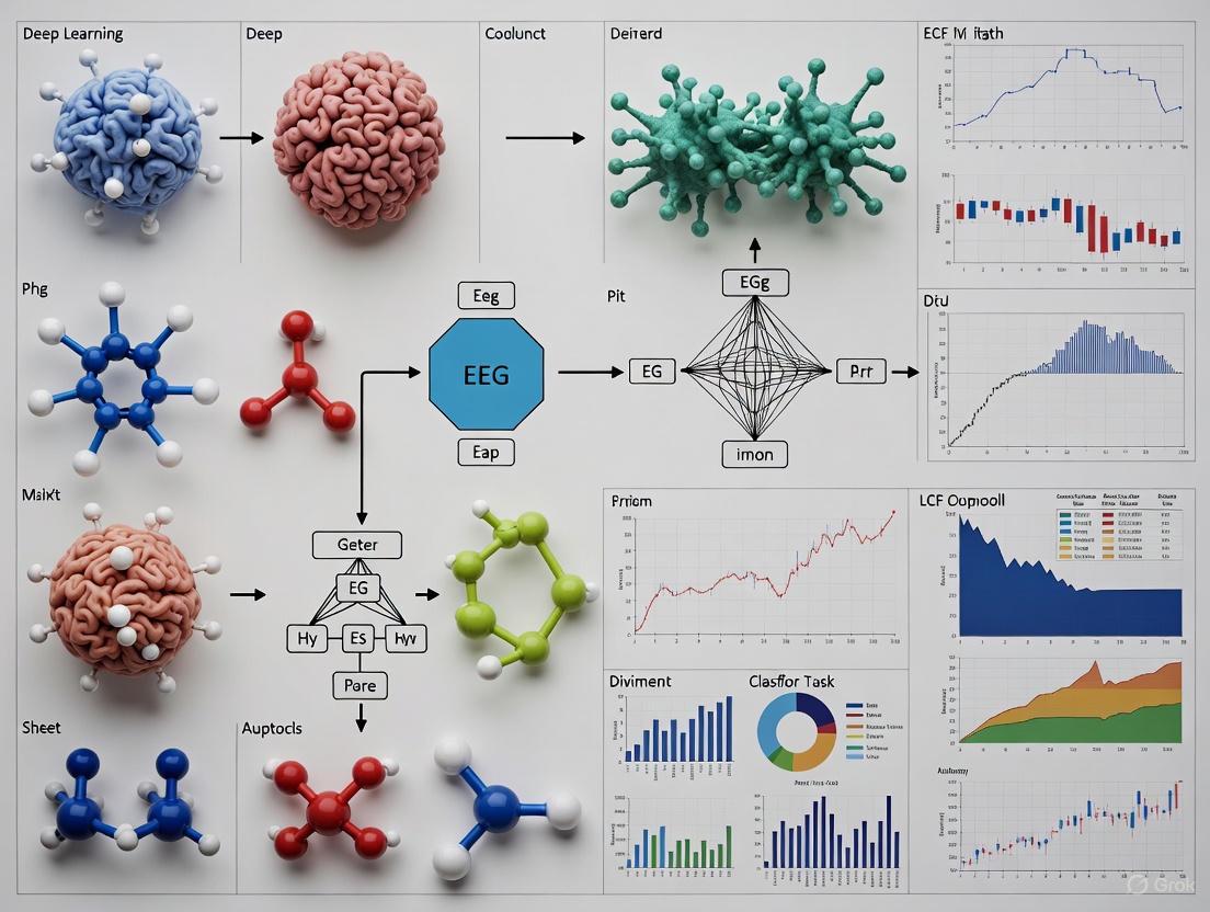
This article provides a comprehensive overview of deep learning methodologies for electroencephalography (EEG) signal classification, tailored for researchers, scientists, and drug development professionals.
Deep Learning in EEG Analysis: A Comprehensive Review of Classification Methods and Clinical Applications
Abstract
This article provides a comprehensive overview of deep learning methodologies for electroencephalography (EEG) signal classification, tailored for researchers, scientists, and drug development professionals. It explores the foundational concepts of EEG analysis, details key deep learning architectures like CNNs, RNNs, and Transformers, and examines their applications in seizure detection, mental task classification, and drug-effect prediction. The review also addresses critical challenges such as data scarcity and model interpretability, offers comparative performance analyses of different models, and discusses the future trajectory of deep learning for enhancing diagnostics and therapeutic development in clinical neuroscience.
Understanding EEG Signals and the Deep Learning Revolution in Neuroscience
Fundamental Principles of Electroencephalography (EEG)
Electroencephalography (EEG) is a non-invasive neurophysiological technique that records the brain's spontaneous electrical activity from the scalp [1]. These signals originate from the summed post-synaptic potentials of large, synchronously firing populations of cortical pyramidal neurons. When excitatory afferent fibers are stimulated, an influx of cations causes post-synaptic membrane depolarization, generating extracellular currents that are detected as voltage fluctuations by electrodes [2]. First recorded in humans by Hans Berger in 1924, EEG has evolved into an indispensable tool for investigating brain function, diagnosing neurological disorders, and advancing neurotechnology [1] [2].
The electrical signals measured by EEG are characterized by their oscillatory patterns, which are categorized into specific frequency bands, each associated with different brain states and functions [3]. The table below summarizes the standard EEG frequency bands and their clinical and functional correlates.
Table 1: Standard EEG Frequency Bands and Their Correlates
| Band | Frequency Range (Hz) | Primary Functional/Clinical Correlates |
|---|---|---|
| Delta (δ) | 0.5 - 4 | Deep sleep, infancy, organic brain disease [4] [3] |
| Theta (θ) | 4 - 8 | Drowsiness, childhood, emotional stress [4] [3] |
| Alpha (α) | 8 - 13 | Relaxed wakefulness, eyes closed, posterior dominant rhythm [1] [3] |
| Beta (β) | 13 - 30 | Active thinking, focus, alertness; can be increased by certain drugs [4] [3] |
| Gamma (γ) | 30 - 150 | High-level information processing, sensory binding [3] |
EEG Signal Acquisition and Preprocessing
The fidelity of an EEG recording is paramount for both clinical interpretation and advanced analytical models. The acquisition process involves several critical components and steps to ensure a high-quality, low-noise signal.
Acquisition Hardware and Electrodes
Modern EEG systems use multiple electrodes placed on the scalp according to standardized systems like the International 10-20 system, which specifies locations based on proportional distances between anatomical landmarks [2]. Electrodes can be invasive (surgically implanted) or, more commonly, non-invasive (placed on the scalp surface) [1].
Table 2: Key Materials and Equipment for EEG Acquisition
| Research Reagent / Equipment | Function and Specification |
|---|---|
| Silver Chloride (Ag/AgCl) Cup Electrodes | High conductivity and low impedance; ideal for high-fidelity signal acquisition [5]. |
| Gold Cup Electrodes | Chemically inert, reducing skin reactions; suitable for long recordings [5]. |
| Conductive Electrolyte Gel/Paste | Establishes a stable, low-impedance electrical connection between the electrode and scalp [5]. |
| High-Impedance Amplifier | Critical for amplifying microvolt-level EEG signals (typically 2-100 µV) without distortion [6]. |
| Digitizer with Anti-aliasing Filter | Converts the analog signal to digital; a suitable filter band must be selected before digitization [5] [6]. |
A proper acquisition protocol requires careful skin preparation to achieve electrode-skin impedance values between 1 kΩ and 10 kΩ [5]. Patients must be instructed to remain still, as movements, blinking, and perspiration can introduce artifacts. Furthermore, the recording environment should be controlled to minimize electromagnetic interference (EMI) from sources like fluorescent lights and cell phones [5].
Preprocessing and Denoising Pipeline
Raw EEG signals are susceptible to various artifacts and noise, making preprocessing a crucial step before analysis or modeling. The primary goal is to isolate the neural signal of interest. The following workflow diagram outlines a standard EEG preprocessing pipeline.
Figure 1: Standard EEG signal preprocessing and denoising workflow.
From Features to Classification: Integration with Deep Learning
Moving from cleaned, preprocessed EEG data to a functional classification model involves feature extraction and the application of sophisticated learning algorithms. This process is central to modern EEG analysis, particularly for Brain-Computer Interfaces (BCIs) and automated diagnosis.
Feature Extraction Methods
Feature extraction transforms the high-dimensional, raw EEG signal into a more manageable set of discriminative descriptors that are informative for the task at hand. The choice of feature is critical for model performance.
Table 3: Common Feature Extraction Methods for EEG Analysis
| Domain | Feature Extraction Method | Description | Suitability for Deep Learning |
|---|---|---|---|
| Frequency | Power Spectral Density (PSD) | Distributes signal power over frequency, often computed via Welch's method [7] [3]. | Good input for fully connected networks. |
| Time-Frequency | Wavelet Transform | Resolves signal in both time and frequency, ideal for non-stationary signals [3]. | Excellent for 2D input to CNNs. |
| Spatial | Common Spatial Patterns (CSP) | Finds spatial filters that maximize variance for one class while minimizing for another [3]. | Preprocessing step for motor imagery tasks. |
| Nonlinear | Higher-Order Spectra, Entropy | Captures complex, dynamic interactions within the signal [1]. | Can be combined with other features. |
Deep Learning Architectures for EEG Classification
Deep learning models can automate feature extraction and classification, often learning complex patterns directly from raw or minimally processed data. The following diagram illustrates a typical deep learning pipeline for EEG classification, highlighting common architectural choices.
Figure 2: Deep learning pipeline for EEG classification tasks.
Different architectures excel in different contexts. Convolutional Neural Networks (CNNs) are highly effective at capturing spatial and temporal patterns [8] [9]. Recurrent Neural Networks (RNNs), particularly Long Short-Term Memory (LSTM) networks, are well-suited for modeling long-range dependencies in time-series data [9]. More recently, Transformer models with customized, sparse attention mechanisms have been developed to process long EEG sequences efficiently while capturing complex temporal relationships [8].
For subject-independent tasks, which are crucial for real-world deployment, one proposed methodology involves using a Deep Neural Network (DNN) fed with precomputed features like Power Spectrum Density (PSD). Principal Component Analysis (PCA) is often applied first to reduce the dimensionality of the PSD features, and the model is trained on data from multiple subjects to learn generalizable patterns [7].
Clinical and Pharmaceutical Applications
EEG's high temporal resolution and non-invasive nature make it a powerful tool in clinical diagnostics and pharmaceutical development.
Disease Diagnosis and Monitoring
EEG is a cornerstone for diagnosing and monitoring a range of neurological and psychiatric conditions. Its applications include:
- Epilepsy: Identification of interictal epileptiform discharges and seizure classification are primary applications [1] [4].
- Sleep Disorders: Analysis of sleep stages and detection of disorders like sleep apnea [1] [8].
- Neuropsychiatric Disorders: Assisting in the diagnosis and study of conditions such as Major Depressive Disorder (MDD), schizophrenia, and attention deficit hyperactivity disorder (ADHD) [8]. Deep learning models have been developed to achieve high accuracy in detecting MDD from EEG signals [8].
Pharmaco-EEG in Drug Development
Pharmaco-electroencephalography (Pharmaco-EEG) is the quantitative analysis of EEG to assess the effects of drugs on the central nervous system (CNS) [4]. It plays a vital role in:
- Early Drug Screening: Identifying compounds with potential therapeutic effects by characterizing their impact on brain activity [8].
- Mechanism Insight: Different drug classes produce distinct, reproducible EEG "fingerprints". For instance, benzodiazepines typically increase beta activity, while many sedatives cause EEG slowing (increased delta/theta) [4] [2].
- Dose Optimization and Toxicity Monitoring: Pharmaco-EEG can help establish the therapeutic window and detect neurotoxic effects, such as excessive background slowing, associated with high drug concentrations [4] [2].
The table below summarizes the EEG responses to selected antiepileptic drugs (AEDs), illustrating how pharmaco-EEG can link drug mechanisms to measurable CNS effects.
Table 4: EEG Frequency Responses to Selected Antiepileptic Drugs (AEDs)
| Drug | Primary Mechanism | Typical EEG Frequency Effect | Clinical/Research Context |
|---|---|---|---|
| Ethosuximide | Blocks T-type Calcium channels | Decrease in Delta, Increase in Alpha [4] | Used for absence seizures; effect on background rhythm. |
| Carbamazepine | Blocks Sodium channels | Increase in Delta and Theta [4] | Slowing can be observed. |
| Benzodiazepines | Potentiates GABA-A receptors | Pronounced increase in Beta activity [4] | Marker of drug engagement and sedative effect. |
| Phenytoin | Blocks Sodium channels | Increase in Beta; Slowing at toxic doses [4] | Can indicate toxicity. |
Electroencephalography (EEG) measures electrical brain activity with high temporal resolution, making it invaluable for neuroscience research and clinical diagnostics. However, its utility is challenged by several inherent signal characteristics. This application note details three fundamental properties of EEG signals—non-stationarity, low signal-to-noise ratio (SNR), and individual variability—that are critical for designing robust deep learning models for EEG classification. We frame these characteristics not merely as obstacles but as informative features that, when properly modeled, can enhance the performance and generalizability of analytical frameworks. The protocols and data summaries provided herein are tailored for researchers, scientists, and drug development professionals engaged in computational analysis of neural data.
Characteristic 1: Non-Stationarity
Definition & Quantitative Profile
Non-stationarity refers to the temporal evolution of the statistical properties (e.g., mean, variance, frequency content) of an EEG signal. Rather than being a continuous, stable process, the EEG is considered a piecewise stationary signal, composed of a sequence of quasi-stable patterns or "metastable" states [10]. The signal's properties can change due to shifts in cognitive task engagement, attention levels, fatigue, and underlying brain state dynamics [11].
Table 1: Quantitative Profile of EEG Non-Stationarity
| Metric | Typical Range/Value | Context & Implications |
|---|---|---|
| Stationary Segment Duration | 0.5 - 4 seconds [12] | Defines the window for reliable statistical estimation; shorter segments challenge traditional analysis. |
| Quasi-Stationary Segment Duration | ~0.25 seconds [11] | Relevant for Brain-Computer Interface (BCI) systems; defines the time scale of stable patterns in dynamic tasks. |
| Age-Related Change in Non-Stationarity | Number of states increases; segment duration decreases with age during adolescence [10] | Indicates brain maturation; analytical models must account for age-dependent dynamical properties. |
Experimental Protocol: Assessing Dynamical Non-Stationarity
This protocol outlines a method for quantifying dynamical non-stationarity in resting-state or task-based EEG data, suitable for investigating developmental trends or clinical group differences [10].
Workflow Overview:
Title: Dynamical Non-Stationarity Assessment Workflow
Step-by-Step Procedures:
Data Acquisition & Preprocessing:
- Acquire EEG data using a standard system (e.g., 128-channel Geodesic Sensor Net).
- Apply standard preprocessing: band-pass filtering (e.g., 0.5-70 Hz), re-referencing to average reference, and artifact correction for blinks and eye movements using Independent Component Analysis (ICA). Remove epochs with persistent artifacts [10].
Segmentation of Time Series:
- Divide the continuous, artifact-free EEG time series into short, possibly overlapping, segments.
- Recommended Segment Length: 0.5 to 2 seconds, based on the expected duration of quasi-stationary states [12].
Modeling and Feature Extraction:
- Fit a model (e.g., an Autoregressive (AR) model) to each segment to approximate the underlying dynamics.
- Extract features from the model (e.g., the coefficients of the AR model) that characterize the signal's properties within that segment [10].
Clustering of Segments:
- Apply a clustering algorithm (e.g., k-means) to the extracted features from all segments.
- Each resulting cluster represents a distinct "stationary state"—a type of brain dynamic that recurs over time.
Quantification of Non-Stationarity:
- Calculate the following key metrics from the clustering results:
- Number of States: The total number of distinct clusters identified. An increase signifies greater dynamical complexity.
- Mean Duration of Stationary Segments: The average time the signal remains in one state before transitioning. A decrease signifies faster switching and higher non-stationarity [10].
- Calculate the following key metrics from the clustering results:
Characteristic 2: Low Signal-to-Noise Ratio
Definition & Noise Source Profile
The EEG signal is notoriously weak, measured in microvolts (millionths of a volt), leading to a low SNR. "Noise" in EEG refers to any recorded signal not originating from the brain activity of interest, significantly complicating data interpretation [13].
Table 2: Profile of Primary Noise Sources in EEG Recordings
| Noise Category | Specific Sources | Characteristics & Impact |
|---|---|---|
| Physiological | Ocular signals (EOG), Cardiac signals (ECG), Muscle contractions (EMG), Swallowing, Irrelevant brain activity [14] | Signals are often 100 times larger than brain-generated EEG; create large-amplitude, stereotypical artifacts that can obscure neural signals [13]. |
| Environmental | AC power lines (50/60 Hz), Room lighting, Electronic equipment (computers, monitors) [14] | Emit electromagnetic fields that are easily detected by sensitive EEG sensors, introducing periodic noise. |
| Motion Artifacts | Unstable electrode-skin contact, Movement of electrode cables [14] | Causes large, low-frequency signal drifts or abrupt signal changes, potentially invalidating data segments. |
Experimental Protocol: Comprehensive SNR Optimization
This protocol provides a multi-stage approach to maximize SNR, encompassing procedures before, during, and after EEG recording.
Workflow Overview:
Title: End-to-End SNR Optimization Pipeline
Step-by-Step Procedures:
Phase 1: Before Recording (Preventive Measures)
- Experimental Design:
- For Event-Related Potential (ERP) studies, design a protocol with sufficient trial repetitions to leverage averaging, which cancels out random noise [13].
- Keep participants focused and comfortable to minimize internal noise and movement artifacts. Provide breaks to allow for blinking and readjustment [13].
- Environmental Control:
- Use a Faraday cage, if available, for electromagnetic isolation [14].
- Remove or turn off non-essential electronic equipment. Replace AC-powered devices with DC alternatives where possible.
- Participant Preparation:
- Ensure the participant is in a comfortable, resting position.
Phase 2: During Recording (Monitoring & Control)
- Electrode Management:
- Quality Control:
- Measure and verify electrode impedances before recording starts. Lower impedance values (typically < 50 kΩ) indicate better contact and signal quality [14].
Phase 3: After Recording (Mathematical Cleaning)
- Manual Inspection: Visually inspect the plotted data to identify obvious artifacts and assess overall data quality [14].
- Algorithmic Cleaning: Apply one or more advanced signal processing techniques:
- Independent Component Analysis (ICA): A blind source separation technique effective for isolating and removing stereotypical artifacts like eye blinks (EOG) and muscle activity (EMG) [14].
- Artifact Subspace Reconstruction (ASR): An online, component-based method for removing large-amplitude, transient artifacts by contrasting data segments to a calibration baseline [14].
- Canonical Correlation Analysis (CCA): Separates signal from noise based on autocorrelation, often outperforming ICA in certain scenarios and usable in real-time [14].
- Sensor Noise Suppression (SNS): Improves SNR by projecting each channel's signal onto the subspace of its neighboring channels, effectively removing unique, non-brain noise [14].
Characteristic 3: Individual Variability
Definition & Quantitative Profile
EEG signals exhibit substantial differences between individuals. This variability is not merely noise but is driven by stable, subject-specific neurophysiological factors. Critically, this subject-driven variability can be more pronounced than the variability induced by task demands [15] [16].
Table 3: Profile of Individual Variability in EEG
| Aspect of Variability | Manifestation | Research Implications |
|---|---|---|
| Across-Subject vs. Across-Block Variation | Across-subject variation in EEG variability and signal strength is much stronger than across-block (task) variation within subjects [15] [16]. | Deep learning models trained on pooled data are prone to learning subject-specific identifiers rather than task-general features, hindering generalization. |
| Relationship to Behavior | Individual differences in behavior (e.g., response times) are better reflected in individual differences in EEG variability, not signal strength [15] [16]. | Signal variability itself is a meaningful biomarker for individual cognitive performance and should be modeled as a feature. |
| Long-Term Stability | Key EEG features (e.g., absolute/relative power in alpha band) show high test-retest reliability over weeks and even years (correlation coefficients ~0.84 over 12-16 weeks) [17]. | Subject-specific signatures are stable over time, validating the use of individual baselines or subject-adaptive models. |
Experimental Protocol: Assessing Subject-Driven Variability
This protocol is designed to systematically quantify and isolate subject-driven variability from task-driven changes in EEG data, which is essential for building generalizable classifiers.
Workflow Overview:
Title: Isolating Subject-Driven Variability Protocol
Step-by-Step Procedures:
Data Collection:
Calculation of Trial-Level Metrics:
- For each trial and participant, calculate two primary types of metrics in overlapping time windows:
Variance Partitioning:
- Perform a systematic analysis to determine the relative sensitivity of the calculated metrics to different sources of variation.
- Compare the magnitude of across-subject variation (differences between people within the same task block) to across-block variation (differences within the same person across different task blocks) [15] [16]. The finding that across-subject variation dominates confirms a strong subject-driven signal.
Linking EEG Metrics to Behavior:
- Correlate individual differences in the EEG metrics (both strength and variability) with individual differences in behavioral performance (e.g., average response time, accuracy).
- Determine which EEG metric (strength or variability) is a stronger predictor of behavior. Research indicates that EEG variability often reflects stable subject identity and is a superior correlate of behavior compared to signal strength [15] [16].
The Scientist's Toolkit
Table 4: Essential Research Reagents & Computational Tools
| Tool/Solution | Primary Function | Application Context |
|---|---|---|
| High-Density EEG System (e.g., 128+ channels) | Captures detailed spatial information of brain electrical activity. | Source localization; high-resolution spatial analysis; Sensor Noise Suppression (SNS). |
| Faraday Cage / Electromagnetically Shielded Room | Blocks environmental electromagnetic interference. | Critical for maximizing SNR in studies not involving movement, especially with sensitive equipment [14]. |
| Wet Electrodes with Conductive Gel | Ensures low impedance and stable electrical contact with the scalp. | The gold standard for high-quality, low-noise recordings; superior to most dry electrodes for SNR [14] [13]. |
| Independent Component Analysis (ICA) | Blind source separation for isolating and removing biological artifacts. | Post-processing cleanup of ocular (EOG) and muscular (EMG) artifacts [14] [10]. |
| Artifact Subspace Reconstruction (ASR) | Statistical, component-based method for removing large, transient artifacts. | Online or offline data cleaning; particularly effective for handling large-amplitude, non-stereotypical noise [14]. |
| Covariate Shift Estimation (e.g., EWMA Model) | Detects changes in the input data distribution of streaming EEG features. | Active adaptation in non-stationary learning for BCIs; triggers model updates when a significant shift is detected [11]. |
| Adaptive Ensemble Learning | Maintains and updates a pool of classifiers to handle changing data distributions. | Used in conjunction with covariate shift detection in BCI systems to maintain performance over long sessions [11]. |
The analysis of Electroencephalography (EEG) signals has undergone a profound transformation, moving from traditional machine learning (ML) methods reliant on handcrafted features to deep learning (DL) approaches that automatically learn hierarchical representations from raw data. This paradigm shift addresses the inherent challenges of EEG signals: their non-stationary nature, low signal-to-noise ratio, and complex spatiotemporal dependencies [3]. Traditional ML pipelines required extensive domain expertise for feature extraction (e.g., using wavelet transform or Fourier analysis) before classification with models like Support Vector Machines (SVM) [18] [3]. In contrast, deep learning models, such as Convolutional Neural Networks (CNNs) and Recurrent Neural Networks (RNNs), directly process raw or minimally preprocessed signals, learning both relevant features and classifiers in an end-to-end manner [18] [19]. This shift has significantly enhanced performance in critical applications ranging from epilepsy seizure detection and motor imagery classification to lie detection, thereby accelerating research in neuroscience, clinical diagnostics, and drug development.
Comparative Analysis: Quantitative Performance
The superiority of deep learning architectures is evidenced by their consistently higher performance metrics across diverse EEG classification tasks compared to traditional machine learning methods. The tables below summarize this performance leap.
Table 1: Performance Comparison of ML vs. DL Models on Specific EEG Tasks
| Task | Traditional ML Model | Accuracy | Deep Learning Model | Accuracy | Reference |
|---|---|---|---|---|---|
| Lie Detection | SVM | Information Missing | CNN | 99.96% | [20] |
| Lie Detection | Linear Discriminant Analysis | 91.67% | Deep Neural Network | Information Missing | [20] |
| Motor Imagery | Various Shallow Models | Information Missing | Fast BiGRU + CNN | 96.9% | [21] |
| Seizure Detection | Models with Handcrafted Features | ~90% (Est.) | CNN, RNN, Transformer | >90% (Common) | [22] |
Table 2: Strengths and Weaknesses of Model Archetypes in EEG Analysis
| Aspect | Traditional Machine Learning | Deep Learning |
|---|---|---|
| Feature Engineering | Manual, requires expert domain knowledge [18] [3] | Automatic, learned from data [18] |
| Computational Cost | Lower | Higher |
| Data Requirements | Lower | Large datasets required |
| Interpretability | Higher (transparent features) | Lower ("black-box" nature) |
| Handling Raw Data | Poor, requires pre-processing | Excellent, can use raw data |
| Spatiotemporal Feature Learning | Limited, often separate | Superior, integrated (e.g., CNN+RNN) [21] |
Detailed Experimental Protocols
Protocol 1: CNN for EEG-Based Lie Detection
This protocol outlines the methodology for achieving state-of-the-art lie detection using a Convolutional Neural Network, as detailed in recent research [20].
- Aim: To classify EEG signals into "truthful" and "deceptive" categories with high accuracy.
- Dataset: A novel dataset acquired from 10 participants using a 14-channel OpenBCI Ultracortex Mark IV EEG headset.
- Experimental Design: A video-based protocol using the Comparison Question Test (CQT) technique. Participants watched clips of a theft crime and were asked to answer questions truthfully in one session and deceptively in another.
- Data Acquisition: EEG signals were recorded from 14 electrodes (FP1, FP2, F4, F8, F7, C4, C3, T8, T7, P8, P4, P3, O1, O2) according to the international 10-20 system, with a sampling frequency of 125 Hz.
- Preprocessing: The raw signals were preprocessed to remove noise and artifacts.
- Model Architecture & Training:
- The preprocessed signals were fed into a CNN model.
- The CNN automatically learned discriminative spatial features from the multi-channel EEG inputs.
- Performance: The model achieved an accuracy of 99.96% on the custom dataset and 99.36% on a public benchmark (Dryad dataset), outperforming traditional ML models like SVM and Multilayer Perceptron (MLP) tested in the same study [20].
Protocol 2: BiGRU-CNN for Motor Imagery Classification
This protocol describes a hybrid deep learning model that captures both spatial and temporal features for classifying imagined movements [21].
- Aim: To decode and classify motor imagery EEG signals into one of four classes: left hand, right hand, both feet, and tongue movement.
- Dataset: BCI Competition IV, Dataset 2a, containing recordings from 22 EEG electrodes.
- Preprocessing: Signals were normalized, and a Fast Fourier Transform (FFT) was applied to obtain frequency components. The data was segmented into small, overlapping time windows.
- Model Architecture & Training:
- A Convolutional Neural Network (CNN) processed the input to extract spatial features from the EEG channels, identifying local wave patterns.
- A Bidirectional Gated Recurrent Unit (BiGRU) analyzed the sequence of features extracted by the CNN to capture long-range temporal dependencies and contextual information in the brain activity.
- The model was trained end-to-end to classify the four motor imagery tasks.
- Performance and Robustness:
- The baseline Fast BiGRU + CNN model achieved 96.9% accuracy.
- Ablation studies confirmed the contribution of both architectural components.
- Data augmentation techniques (Gaussian noise, channel dropout, mixup) were employed to test robustness, revealing that while accuracy on clean data was highest for the baseline, augmented models showed improved resistance to noise [21].
Workflow Visualization
The following diagram illustrates the fundamental shift in the EEG analysis pipeline from a traditional machine learning approach to a deep learning paradigm.
The Scientist's Toolkit: Research Reagent Solutions
For researchers embarking on EEG deep learning projects, the following tools and resources are essential.
Table 3: Essential Tools and Resources for Deep Learning EEG Research
| Tool / Resource | Type | Function in Research |
|---|---|---|
| OpenBCI Ultracortex Mark IV | Hardware | A relatively low-cost, open-source EEG headset for data acquisition; used in lie detection studies with 14-16 channels [20]. |
| EEG-DL Library | Software | A dedicated TensorFlow-based deep learning library providing implementations of latest models (CNN, ResNet, LSTM, Transformer, GCN) for EEG signal classification [19]. |
| BCI Competition IV 2a | Data | A benchmark public dataset for motor imagery classification, containing 22-channel EEG data for 4 classes of movement imagination [21]. |
| Dryad Dataset | Data | A public dataset for lie detection research, employing a standard three-stimuli protocol with image-based stimuli [20]. |
| WebAIM Contrast Checker | Tool | Ensures accessibility and readability of visual results and interface elements in developed tools by verifying color contrast ratios against WCAG guidelines [23]. |
| Transformers & Attention Mechanisms | Algorithm | A class of models gaining attention for seizure detection and iEEG classification, excelling at modeling complex temporal dependencies [22]. |
Electroencephalogram (EEG) analysis plays an indispensable role across contemporary medical applications, encompassing diagnosis, monitoring, drug discovery, and therapeutic assessment [8]. The advent of deep learning has revolutionized EEG analysis by enabling end-to-end decoding directly from raw signals without hand-crafted features, achieving performance that matches or exceeds traditional methods [24]. Deep learning models automatically learn hierarchical representations that capture relevant spectral and spatial patterns in EEG data, making them particularly valuable for analyzing the high-dimensional, multivariate nature of neural signals [8]. This document presents application notes and experimental protocols for five major EEG classification tasks, framed within the context of advanced deep learning approaches for biomedical research and neuropharmacology.
Experimental Protocols & Performance Benchmarks
Table 1: Performance Benchmarks for Major EEG Classification Tasks
| Classification Task | Key Applications | Best-Performing Models | Reported Accuracy | Key EEG Features |
|---|---|---|---|---|
| Medication Classification | Pharmaco-EEG, therapeutic monitoring | Deep CNN (DCNN), Kernel SVM [25] | 72.4-77.8% [25] | Spectral power across frequency bands |
| Motor Imagery | Brain-computer interfaces, neurorehabilitation | CSP with LDA, EEGNet, CTNet [26] [27] | Varies by dataset | Sensorimotor rhythms (mu/beta), ERD/ERS |
| Seizure Detection | Epilepsy monitoring, alert systems | Convolutional Sparse Transformer [8] | Superior to approaches [8] | Spike-wave complexes, rhythmic discharges |
| Sleep Stage Scoring | Sleep disorder diagnosis | Attention-based Deep Learning [26] | Varies by dataset | Delta waves, spindles, K-complexes |
| Pathology Detection | Clinical diagnosis, screening | EEG-CLIP, Deep4 Network [24] | Zero-shot capability [24] | Non-specific aberrant patterns |
Detailed Methodological Protocols
Medication Classification Protocol
Objective: To distinguish between patients taking anticonvulsant medications (Dilantin/phenytoin or Keppra/levetiracetam) versus no medications based solely on EEG signatures [25].
Dataset Preparation:
- Utilize Temple University Hospital EEG Corpus with physician report verification [25]
- Include balanced samples from patients taking Dilantin, Keppra, or no medications
- Preprocess data: bandpass filtering (0.5-70 Hz), artifact removal, segmentation into 5-second epochs
Experimental Procedure:
- Feature-Based Approach:
- Extract spectral features: power spectral density across delta, theta, alpha, beta, gamma bands
- Apply K-best feature selection or Principal Component Analysis (PCA) for dimensionality reduction
- Train Kernel SVM with RBF kernel (C=10-1000, γ=0.1) using 10-fold cross-validation [25]
- Deep Learning Approach:
- Implement Deep Convolutional Neural Network (DCNN) with spatial-temporal layers
- Configure architecture: 4 convolutional blocks with batch normalization and dropout
- Train with Adam optimizer (learning rate=0.001) for 100 epochs with early stopping [25]
Validation:
- Perform 10-fold cross-validation with strict patient-wise splitting
- Compare results against random label baseline using Kruskal-Wallis tests
- Report accuracy, precision, recall, F1-score, and computational efficiency metrics
Motor Imagery Classification Protocol
Objective: To decode imagined movements from sensorimotor rhythms for brain-computer interface applications [27].
Experimental Setup:
- Electrode Selection: Focus on C3, C4, and Cz channels per international 10-20 system [27]
- Time Window: 3-second segments with 0.5-second offset [27]
- Task Paradigm: Randomly cued imagination of right hand vs. left hand movement
Signal Processing Pipeline:
- Preprocessing:
- Apply 8-30 Hz bandpass filter to enhance sensorimotor rhythms
- Perform Common Spatial Pattern (CSP) or Independent Component Analysis (ICA) for source separation [27]
Feature Extraction:
- Calculate log-variance of CSP components
- Extract Renyi entropy for non-linear characterization [27]
Classification:
Multimodal EEG-Text Embedding Protocol (EEG-CLIP)
Objective: To align EEG time-series data with clinical text descriptions in a shared embedding space for versatile zero-shot decoding [24].
Architecture Configuration:
- EEG Encoder: Deep4 CNN (4 convolution-max-pooling blocks with batch normalization) [24]
- Text Encoder: Pretrained BERT model for clinical report processing [24]
- Projection Heads: 3-layer MLP with ReLU activations projecting to 64-dimensional shared space [24]
Training Procedure:
- Data Preparation:
- Contrastive Learning:
- Implement symmetric contrastive loss using cosine similarity
- Train with Adam optimizer (learning rate=5×10⁻³, weight decay=5×10⁻⁴) for 20 epochs [24]
- Batch size: 64 with hard negative mining
Evaluation:
- Zero-shot classification using textual prompts for pathology, age, gender, medication
- Few-shot transfer learning on downstream tasks with limited labeled data
- t-SNE visualization of cross-modal embedding alignment
Visualization of Experimental Workflows
End-to-End EEG Deep Learning Pipeline
EEG-CLIP Multimodal Alignment Architecture
Convolutional Sparse Transformer for EEG Analysis
The Scientist's Toolkit: Research Reagent Solutions
Table 2: Essential Research Tools for EEG Deep Learning
| Tool/Category | Specific Examples | Function/Purpose | Implementation Notes |
|---|---|---|---|
| EEG Datasets | Temple University Hospital EEG Corpus [24] [25] | Large-scale clinical data with medical reports | Contains >25,000 recordings; includes medication metadata |
| Preprocessing Tools | MNE-Python, EEGLAB | Signal cleaning, filtering, artifact removal | Minimal preprocessing preferred for deep learning [28] |
| Deep Learning Architectures | EEGNet, Deep4, Convolutional Sparse Transformer [8] [26] | Task-specific model backbones | EEGNet: compact CNN; Transformer: long-range dependencies |
| Multimodal Frameworks | EEG-CLIP [24] | Contrastive EEG-text alignment | Enables zero-shot classification from textual prompts |
| Specialized Components | Spatial Channel Attention, Common Spatial Patterns | Enhancing spatial relationships in EEG | Critical for capturing brain region interactions [8] [27] |
| Evaluation Metrics | 10-fold cross-validation, Kruskal-Wallis tests | Statistical validation of model performance | Essential for pharmaco-EEG applications [25] |
Advanced Applications and Future Directions
Pharmaco-EEG and Therapeutic Monitoring
The application of deep learning to Pharmaco-EEG represents a paradigm shift in drug development and therapeutic monitoring. The Convolutional Sparse Transformer framework demonstrates remarkable versatility across multiple medical tasks, including disease diagnosis, drug discovery, and treatment effect prediction [8]. By directly processing raw EEG waveforms, this approach captures intricate spatial-temporal patterns that serve as biomarkers for drug efficacy. For anticonvulsant medications, studies show that differential classification between Dilantin and Keppra is achievable with accuracies around 72-74% using Random Forest classifiers, while Deep CNN models achieve 77.8% accuracy when distinguishing medicated patients from controls [25].
Zero-Shot Learning and Cross-Modal Applications
The EEG-CLIP framework pioneers zero-shot classification capabilities by aligning EEG signals with natural language descriptions of clinical findings [24]. This approach enables researchers to query EEG data using textual prompts without task-specific training, opening new possibilities for exploratory analysis and hypothesis testing. The contrastive learning objective brings matching EEG-text pairs closer in the embedding space while pushing non-matching pairs apart, creating a semantically rich representation space that captures fundamental relationships between neural patterns and their clinical interpretations [24].
Methodological Considerations and Preprocessing Impact
Recent evidence suggests that extensive preprocessing pipelines may not always benefit deep learning models, with minimal preprocessing (excluding artifact handling methods) often yielding superior performance [28]. This counterintuitive finding emphasizes the importance of evaluating preprocessing strategies within the context of specific classification tasks and model architectures. Models trained on completely raw data consistently perform poorly, indicating that basic filtering and normalization remain essential, while sophisticated artifact removal algorithms may inadvertently remove task-relevant information [28].
Deep Learning Architectures and Their Transformative Applications in EEG Classification
Deep learning architectures have revolutionized electroencephalography (EEG) analysis by enabling automated feature extraction and enhanced classification of complex brain activity patterns. The selection of an appropriate model is critical for tasks such as motor imagery classification, seizure detection, and emotion recognition [29]. The table below summarizes the core characteristics, advantages, and typical applications of each major architecture in EEG research.
Table 1: Comparison of Core Deep Learning Architectures for EEG Classification
| Architecture | Core Mechanism | Key Advantages for EEG | Primary Limitations | Common EEG Applications |
|---|---|---|---|---|
| Convolutional Neural Network (CNN) [30] | Convolutional and pooling layers for spatial feature extraction [30]. | Excels at identifying spatial patterns and hierarchies from multi-channel electrode data [29]. | Limited innate capacity for modeling temporal dependencies and long-range contexts [29]. | Motor Imagery classification, spatial feature extraction from scalp topographies [31] [32]. |
| RNN / LSTM [30] | Gated units (input, forget, output) to regulate information flow in sequences [30] [33]. | Effectively models temporal dynamics and dependencies in EEG time-series [29]. Handles vanishing gradient problem better than simple RNNs [33]. | Sequential processing limits training parallelization, making it computationally intensive [30] [34]. | Emotion recognition, seizure detection, and other tasks with strong temporal dependencies [29]. |
| Transformer [29] | Self-attention mechanism to weigh the importance of all time points in a sequence [29]. | Superior at capturing long-range dependencies in EEG signals. Enables full parallelization for faster training [29] [33]. | Requires very large datasets; computationally expensive and memory-intensive [30] [29]. | State-of-the-art performance in Motor Imagery, Emotion Recognition, and Seizure Detection [29]. |
Empirical studies demonstrate the performance of these architectures in specific EEG classification tasks. The following table consolidates quantitative results from recent research, providing a benchmark for model selection.
Table 2: Reported Performance of Different Architectures on EEG Classification Tasks
| Model Architecture | EEG Task / Dataset | Reported Performance | Key Experimental Condition |
|---|---|---|---|
| Random Forest (Baseline) [32] | Motor Imagery / PhysioNet | 91.00% Accuracy | Traditional machine learning benchmark with handcrafted features [32]. |
| CNN [32] | Motor Imagery / PhysioNet | 88.18% Accuracy | Used for spatial feature extraction [32]. |
| LSTM [32] | Motor Imagery / PhysioNet | 16.13% Accuracy | Struggled with temporal modeling in this specific setup [32]. |
| CNN-LSTM (Hybrid) [32] | Motor Imagery / PhysioNet | 96.06% Accuracy | Combined spatial (CNN) and temporal (LSTM) feature learning [32]. |
| Proposed Multi-Stage Model [35] | Depression Classification / PRED+CT Dataset | 85.33% Accuracy | Integrated cortical source features, Graph CNN, and adversarial learning [35]. |
| Signal Prediction Method [36] | Motor Imagery / BCI Competition IV 2a | 78.16% Average Accuracy | Used elastic net regression to predict full-channel EEG from a few electrodes [36]. |
Experimental Protocols for Key Architectures
Protocol: CNN-LSTM Hybrid Model for Motor Imagery Classification
This protocol outlines the procedure for implementing a high-performing hybrid CNN-LSTM model, which has demonstrated state-of-the-art accuracy of 96.06% in classifying Motor Imagery tasks [32].
Primary Objective: To accurately classify EEG signals into different motor imagery classes (e.g., left hand vs. right hand movement) by leveraging the spatial feature extraction capability of CNNs and the temporal modeling strength of LSTMs.
Materials and Dataset
- Dataset: PhysioNet EEG Motor Movement/Imagery Dataset [32].
- Software: Python with deep learning libraries (e.g., TensorFlow, PyTorch).
- Pre-processing Tools: Band-pass filters, Independent Component Analysis (ICA) for artifact removal, and normalization utilities.
Experimental Procedure
- Data Preprocessing:
- Apply a band-pass filter (e.g., 4-40 Hz) to isolate relevant frequency bands like Mu and Beta rhythms.
- Remove ocular and muscular artifacts using ICA.
- Segment the continuous EEG data into epochs time-locked to the motor imagery cue.
- Normalize the data per channel to have zero mean and unit variance.
- Model Architecture Configuration:
- CNN Component: Design convolutional layers to process the multi-channel EEG input. Use 2D convolutions to capture spatial patterns across electrodes or 1D convolutions for temporal patterns per channel.
- LSTM Component: Feed the feature sequences extracted by the CNN into an LSTM layer to model temporal dependencies.
- Classification Head: Attach a fully connected layer with a softmax activation function to output class probabilities.
- Model Training:
- Loss Function: Categorical cross-entropy.
- Optimizer: Adam.
- Training Regimen: Train for 30-50 epochs, which is sufficient for the model to converge to peak accuracy in this application [32].
- Performance Validation:
- Evaluate the model on a held-out test set using accuracy as the primary metric.
- Compare performance against traditional machine learning classifiers (e.g., Random Forest) and individual CNN/LSTM models.
- Data Preprocessing:
The workflow for this hybrid approach is summarized in the diagram below.
Protocol: Transformer-based Model for Multi-class EEG Analysis
This protocol describes the application of Transformer architectures, which are increasingly used for their superior ability to handle long-range dependencies in EEG sequences [29].
Primary Objective: To implement a Transformer model for EEG-based classification tasks such as motor imagery, emotion recognition, or seizure detection, leveraging self-attention to capture global context.
Materials and Dataset
- Dataset: Varies by task (e.g., BCI Competition IV 2a for Motor Imagery, DEAP for Emotion recognition) [29] [26].
- Software: Python with PyTorch/TensorFlow and Transformer model libraries.
- Feature Engineering Tools: Optional tools for generating input embeddings (e.g., spectral features, visibility graphs [31]).
Experimental Procedure
- Input Representation and Embedding:
- Represent the multi-channel EEG signal as a sequence of vectors. This can be raw data points, extracted features, or data from individual time points.
- Project the input into a higher-dimensional space using a linear embedding layer.
- Inject positional information into the embeddings using sinusoidal positional encoding, as Transformers themselves are permutation-invariant [29].
- Core Transformer Encoder Configuration:
- Multi-Head Self-Attention: Configure multiple attention heads to allow the model to focus on different aspects of the EEG sequence from different representation subspaces [29].
- Feed-Forward Network: Each encoder layer should contain a position-wise fully connected feed-forward network.
- Residual Connections & Layer Normalization: Employ these around both the self-attention and feed-forward sub-layers to stabilize training [29].
- Task-Specific Head and Training:
- Use the output corresponding to a special classification token ([CLS]) or the mean of the output sequence as a summary representation.
- Pass this representation through a linear classifier to obtain final class labels.
- Train the model with cross-entropy loss and an adaptive optimizer like AdamW.
- Input Representation and Embedding:
Protocol: Subject-Independent Semi-Supervised Learning with SSDA
This protocol addresses the critical challenge of inter-subject variability and limited labeled data by using a Semi-Supervised Deep Architecture (SSDA) [37].
Primary Objective: To train a robust motor imagery classification model that generalizes to new, unseen subjects with minimal labeled data.
Materials and Dataset
- Datasets: BCI Competition IV 2a or PhysioNet Motor Movement/Imagery Dataset [37].
- Software: Python with deep learning frameworks.
Experimental Procedure
- Data Preparation:
- Pool data from multiple subjects, keeping a subset of labels and treating the rest as unlabeled.
- Ensure the test set contains only subjects not present in the training set.
- SSDA Model Construction:
- Unsupervised Component (CST-AE): Build a Columnar Spatiotemporal Auto-Encoder (CST-AE) to learn latent feature representations from all training data (both labeled and unlabeled) by reconstructing the input [37].
- Supervised Component: Train a classifier on the latent features from the labeled data only.
- Joint Training with Center Loss:
- Train the entire network end-to-end, combining the reconstruction loss from the auto-encoder and the classification loss from the classifier.
- Incorporate a center loss term to minimize the distance between embedded features of the same class, enhancing intra-class compactity [37].
- Evaluation:
- Evaluate the final model on the held-out test subjects, reporting classification accuracy.
- Data Preparation:
Table 3: Key Research Reagents and Computational Tools for Deep Learning EEG Analysis
| Item / Resource | Function / Description | Example / Reference |
|---|---|---|
| Public EEG Datasets | Provide standardized, annotated data for model training and benchmarking. | PhysioNet EEG Motor Movement/Imagery Dataset [32]; BCI Competition IV 2a [37]; PRED+CT (Depression) [35]. |
| Pre-processing Tools | Clean raw EEG signals by removing noise and artifacts to improve signal quality. | Band-pass & notch filtering; Independent Component Analysis (ICA); Common Average Reference (CAR). |
| Feature Extraction Methods | Transform raw EEG into discriminative features for model input. | Power Spectral Density (PSD) [31]; Wavelet Transform [32]; Visibility Graph (VG) [31]; Riemannian Geometry [32]. |
| Software & Codebases | Open-source implementations of standard and state-of-the-art models. | EEGNet (Keras/TensorFlow) [26]; Vision Transformer for EEG (PyTorch) [26]. |
| Domain Adaptation Techniques | Improve model generalization across subjects or sessions by mitigating data distribution shifts. | Gradient Reversal Layer (GRL) [35]; Focal Loss for class imbalance [35]. |
Electroencephalography (EEG) analysis has been revolutionized by deep learning (DL), which enables the extraction of complex patterns from neural data for tasks ranging from neurological disorder diagnosis to brain-computer interface development [38] [22]. The performance of these DL models is fundamentally dependent on the quality and formulation of input data. This document provides application notes and detailed protocols for EEG data preprocessing and the creation of effective input formulations specifically for deep learning-based classification research. Within the broader context of a thesis on deep learning EEG analysis, this guide serves as an essential methodological bridge between raw signal acquisition and model development, ensuring that researchers can transform noisy, raw EEG signals into structured inputs that maximize model performance and interpretability for applications in scientific research and drug development.
EEG Data Preprocessing Pipeline
Preprocessing is a critical first step that removes contaminants and enhances the signal-to-noise ratio, ensuring that subsequent analysis reflects neural activity rather than artifacts [39] [38]. The following section outlines a standardized, automated pipeline suitable for most research scenarios.
Core Preprocessing Steps
Table 1: Core EEG Preprocessing Steps and Methodologies
| Processing Step | Description | Common Techniques & Parameters | Outcome |
|---|---|---|---|
| Filtering | Removes unwanted frequency components not relevant to the research question. | - High-pass filter: >0.1 Hz to remove slow drifts [40].- Low-pass filter: <40-80 Hz to suppress muscle noise [40].- Notch filter: 50/60 Hz to eliminate line interference [39]. | A signal focused on the frequency band of interest (e.g., 0.5-40 Hz). |
| Bad Channel Interpolation | Identifies and reconstructs malfunctioning or excessively noisy electrodes. | - Automatic detection: Based on abnormal variance, correlation, or kurtosis.- Interpolation: Using spherical splines or signal averaging from neighboring channels. | A complete channel set with minimal data loss. |
| Artifact Removal | Separates and removes non-neural signals (e.g., from eyes, heart, muscles). | - Independent Component Analysis (ICA): Fitted on filtered data (e.g., 1-40 Hz) to isolate and remove artifact-related components [40].- Automated algorithms: Such as ASR or ICLabel. | A "clean" EEG signal predominantly reflecting cortical origin activity. |
| Epoching | Segments the continuous data into trials time-locked to experimental events. | - Time window: e.g., -0.2 s to +0.8 s around stimulus onset.- Baseline correction: Removes mean DC offset using the pre-stimulus period. | A 3D matrix (epochs × channels × time points) ready for feature extraction. |
| Normalization | Scales the data to a standard range, improving model training stability. | - Z-scoring: Subtracting the mean and dividing by the standard deviation per channel [38].- Robust Scaler: Uses median and interquartile range to mitigate outlier effects. | Data with a mean of zero and a standard deviation of one, or similar bounded range. |
Visualizing the Preprocessing Workflow
The following diagram illustrates the sequential workflow of the standard EEG preprocessing pipeline.
Input Formulations for Deep Learning
Choosing how to represent EEG data is as crucial as the model architecture itself. Deep learning models can ingest EEG data in various formulations, each with distinct advantages for capturing different aspects of the signal.
Table 2: Comparison of Input Formulations for Deep Learning Models
| Input Formulation | Description | Strengths | Weaknesses | Best-Suited Model Architectures |
|---|---|---|---|---|
| Raw Signals | The preprocessed but otherwise unmodified time-series voltage data. | - Preserves complete temporal information.- No feature engineering bias.- Suitable for end-to-end learning. | - High dimensionality.- Susceptible to high-frequency noise.- Requires large datasets. | 1D Convolutional Neural Networks (CNNs), Recurrent Neural Networks (RNNs), Transformers [22]. |
| Spectrograms | A time-frequency representation showing power spectral density (PSD) over time, with power encoded as color [41]. | - Provides a 2D image-like input.- Intuitive visualization of spectral evolution.- Effective for capturing oscillatory patterns. | - Loss of phase information.- Time-frequency resolution trade-off. | 2D Convolutional Neural Networks (CNNs) [41]. |
| Time-Frequency Representations (TFRs) | A group of methods that capture both time and frequency details, such as those generated by wavelet transforms [39] [38]. | - Retains both amplitude and phase information.- Superior resolution for transient events compared to spectrograms. | - Computationally intensive.- Can be high-dimensional. | 2D CNNs, Hybrid CNN-RNNs. |
| Handcrafted Features | Engineered features extracted from the signal (e.g., band power, connectivity metrics, Hjorth parameters). | - Low dimensionality.- Incorporates domain knowledge.- Works with smaller datasets. | - Limited to known phenomena; may miss complex patterns.- Requires expert knowledge. | Support Vector Machines (SVM), k-Nearest Neighbors (k-NN), Fully Connected Neural Networks [39]. |
Generating a Spectrogram Input
Spectrograms are a central tool in quantitative EEG, transforming a 1D signal into a 2D map where time is on the x-axis, frequency on the y-axis, and signal power is represented by color intensity [41]. This makes them ideal for input into standard 2D CNNs.
Protocol 1: Creating an EEG Spectrogram
- Segment the Signal: Divide the continuous preprocessed EEG signal into overlapping time windows (e.g., 2-second segments with 50% overlap). The choice of window length represents a trade-off between temporal and frequency resolution.
- Apply Fourier Transform: For each time window, compute the Short-Time Fourier Transform (STFT). This calculates the power spectral density (PSD) for the frequencies within that window.
- Compute Power: Calculate the magnitude squared of the STFT result to obtain the signal power for each frequency bin.
- Plot the Spectrogram: Display time on the x-axis, frequency on the y-axis, and power as a color gradient. Power is often displayed on a logarithmic scale (decibels) to better visualize both low and high-power components [41].
The diagram below illustrates this process and the resulting data structure.
Advanced Time-Frequency Analysis
For detecting transient events with distinct shapes, such as epileptic spikes, or for precisely localizing activity in both time and frequency, more advanced TFRs are required [39] [41]. The Continuous Wavelet Transform (CWT) is a powerful method for this purpose.
Protocol 2: Implementing Time-Frequency Analysis with Wavelet Transform
- Select a Mother Wavelet: Choose an appropriate wavelet function (e.g., Morlet wavelet) that matches the characteristics of the signal feature of interest.
- Convolve and Transform: Convolve the selected wavelet with the EEG signal at various scales (dilations and translations). Each scale corresponds to a specific frequency band.
- Generate Time-Frequency Map: The output of the CWT is a matrix representing the similarity (coefficient magnitude) between the signal and the wavelet at each time and scale (frequency). This creates a detailed time-frequency map.
- Model Input: The resulting 2D representation (Time × Frequency) of coefficients can be used as input for a 2D CNN, similar to a spectrogram, but often with richer detail for transient features.
The Scientist's Toolkit: Essential Research Reagents & Materials
Table 3: Key Resources for EEG Deep Learning Research
| Item | Function in Research | Example Use Case |
|---|---|---|
| MNE-Python | An open-source Python package for exploring, visualizing, and analyzing human neurophysiological data [42] [40]. | It provides end-to-end functionality, from data I/O and preprocessing (filtering, ICA, epoching) to source localization and statistical analysis. |
| eLORETA | A source localization algorithm used to estimate the cortical origins of scalp-recorded EEG signals [43]. | Estimating the neural sources of a cognitive task or pathological activity (e.g., epileptogenic zone) when individual structural MRIs are unavailable. |
| ICBM 2009c Template & CerebrA Atlas | Standardized anatomical brain templates and atlases [43]. | Used as a shared forward model in source localization pipelines for studies without subject-specific structural data. |
| Independent Component Analysis (ICA) | A blind source separation technique used to isolate and remove artifacts like eye blinks and muscle activity from EEG data [40]. | Cleaning continuous EEG data by identifying and rejecting components correlated with artifacts, preserving neural signals. |
| Support Vector Machine (SVM) | A classical machine learning algorithm effective for classification tasks with high-dimensional data [39]. | A strong baseline model for classifying EEG epochs or extracted features (e.g., PSD) into different cognitive states or conditions. |
| Convolutional Neural Network (CNN) | A class of deep neural networks most commonly applied to analyzing visual imagery, making it suitable for 2D EEG inputs like spectrograms and TFRs [22]. | Automating the detection of seizures from spectrograms or identifying event-related potentials (ERPs) from time-series data. |
| Transformer Architecture | A modern deep learning architecture that uses self-attention mechanisms to weigh the importance of different parts of the input sequence [22]. | Modeling long-range dependencies in raw or segmented EEG time-series for seizure prediction or cognitive state decoding. |
Experimental Protocol: A Sample Classification Workflow
This protocol provides a concrete example of applying the above methodologies to a typical EEG classification problem, such as distinguishing between different cognitive states.
Protocol 3: Experiment on Cognitive State Classification from EEG Spectrograms
- Objective: To classify epochs of EEG data into "Eyes Open" vs. "Eyes Closed" resting states using a 2D CNN.
- Dataset: A publicly available dataset containing resting-state EEG recordings with annotated "Eyes Open" and "Eyes Closed" conditions.
Procedure:
Data Preprocessing:
- Load the continuous EEG data.
- Apply a band-pass filter (e.g., 1-40 Hz) and a 50/60 Hz notch filter.
- Run ICA to remove eye-blink and other ocular artifacts.
- Epoch the data into 4-second segments from both conditions.
- Apply a baseline correction if necessary (though less critical for resting-state analysis compared to ERPs).
Input Formulation:
- For each 4-second epoch and for each EEG channel (or a subset of posterior channels like Pz, O1, Oz, O2), compute the spectrogram using STFT.
- Use a window size of 1 second with 90% overlap to balance resolution.
- Stack the spectrograms from individual channels to create a multi-channel image input (Channels × Frequency × Time). Alternatively, average spectrograms across a channel group.
Model Training & Evaluation:
- Model: Design a 2D CNN with layers for convolution, pooling, dropout (for regularization), and a final softmax output layer.
- Training: Split data into training, validation, and test sets. Train the CNN to minimize cross-entropy loss using an optimizer like Adam.
- Evaluation: Report standard performance metrics on the held-out test set, including accuracy, sensitivity, specificity, and F1-score.
This structured approach to preprocessing and input formulation provides a reproducible foundation for building robust and high-performing deep learning models in EEG research.
Epilepsy is a neurological disorder affecting approximately 65 million people worldwide, with about one-third of patients developing drug-resistant epilepsy (DRE) where anti-seizure medications provide inadequate seizure control [22] [44]. For these patients, surgical intervention remains a potentially curative option, with its success critically dependent on the accurate identification and complete resection of the epileptogenic zone (EZ)—the smallest cortical region whose removal results in seizure freedom [22]. Intracranial EEG (iEEG) monitoring is essential for EZ localization but generates massive datasets that are subject to significant inter-expert variability during visual analysis, creating substantial subjectivity in surgical planning [22] [45]. Deep learning has emerged as a transformative technology for automating seizure detection and EZ localization from iEEG recordings, offering the potential to reduce diagnostic subjectivity, enhance reproducibility, and ultimately improve surgical outcomes in epilepsy care [22] [46].
Key Electrophysiological Biomarkers
iEEG analysis for epilepsy surgery focuses on several key electrophysiological biomarkers that indicate epileptogenic tissue. High-frequency oscillations (HFOs), particularly in the 80-500 Hz range (categorized as ripples [80-250 Hz] and fast ripples [250-500 Hz]), have emerged as crucial biomarkers thought to represent synchronized neuronal firing within the EZ [22]. These oscillations can occur during both interictal and ictal periods, with HFO-rich regions showing significant overlap with the epileptogenic zone [22]. Other important biomarkers include interictal epileptiform discharges (IEDs) and the dynamic spectral changes, connectivity patterns, and temporal signatures that directly reflect seizure activity during ictal periods [22] [45]. Deep learning approaches are increasingly capable of detecting these traditional biomarkers while also identifying subtle, alternative biomarkers that may not be apparent through visual inspection alone [22].
Table 1: Key Electrophysiological Biomarkers in iEEG Analysis
| Biomarker | Frequency Range | Clinical Significance | Detection Challenges |
|---|---|---|---|
| Ripples | 80-250 Hz | Indicate epileptogenic tissue | Distinguishing pathological from physiological HFOs |
| Fast Ripples | 250-500 Hz | Strong correlation with seizure onset zone | Require high-sampling rate iEEG systems |
| Interictal Epileptiform Discharges (IEDs) | Transient spikes/sharp waves | Marker of irritative zone | Can occur independently from seizure onset zone |
| Ictal Patterns | Variable, patient-specific | Direct seizure manifestation | Significant heterogeneity across patients |
Deep Learning Architectures for iEEG Analysis
Various deep learning architectures have been successfully applied to iEEG analysis, each with distinct advantages for capturing spatial and temporal patterns in epileptic activity. Convolutional Neural Networks (CNNs) excel at extracting spatial features from iEEG spectrograms or raw signal patterns [47] [48]. Recurrent Neural Networks (RNNs), particularly those with Long Short-Term Memory (LSTM) and Bidirectional LSTM (BiLSTM) units, effectively model temporal dependencies in sequential iEEG data [22] [48]. More recently, transformer-based architectures with self-attention mechanisms have shown promise for capturing long-range dependencies in iEEG signals [22]. Hybrid models that combine CNNs with RNNs (e.g., CNN-BiLSTM) leverage both spatial feature extraction and temporal sequence modeling, often achieving state-of-the-art performance [47] [48].
Performance Comparison
Recent studies demonstrate the effectiveness of these architectures across various seizure analysis tasks. A hybrid CNN-BiLSTM approach applied to ultra-long-term subcutaneous EEG achieved an area under the receiver operating characteristic curve (AUROC) of 0.98 and area under the precision-recall curve (AUPRC) of 0.50, corresponding to 94% sensitivity with only 1.11 false detections per day [48]. A semi-supervised temporal autoencoder method for iEEG classification achieved AUROC scores of 0.862 ± 0.037 for pathologic vs. normal classification and 0.879 ± 0.042 for artifact detection, demonstrating that semi-supervised approaches can provide acceptable results with minimal expert annotations [45]. Traditional CNN and RNN models frequently exceed 90% accuracy in detecting epileptiform activity, though performance varies significantly based on data quality and preprocessing techniques [22].
Table 2: Performance Comparison of Deep Learning Architectures for Seizure Detection
| Architecture | Application | Key Performance Metrics | Data Type |
|---|---|---|---|
| CNN-BiLSTM [48] | Seizure detection | AUROC: 0.98, Sensitivity: 94%, False detections: 1.11/day | Subscalp EEG |
| Temporal Autoencoder [45] | iEEG classification | AUROC: 0.862 ± 0.037, AUPRC: 0.740 ± 0.740 | Intracranial EEG |
| 1D-CNN with BiLSTM [47] | Multi-class seizure classification | High precision, sensitivity, specificity, F1-score | Scalp EEG |
| Transformer-based [22] | Seizure detection | High accuracy for temporal dependencies | Intracranial EEG |
Experimental Protocols & Methodologies
Protocol 1: CNN-BiLSTM for Seizure Detection
This protocol outlines the methodology for implementing a hybrid CNN-BiLSTM model for seizure detection in long-term EEG monitoring [48].
Data Acquisition & Preprocessing:
- Acquire iEEG data using standard clinical systems with sampling rates ≥ 2000 Hz.
- Apply band-pass filtering (0.5-70 Hz) and notch filtering (50/60 Hz) to remove noise and line interference.
- For subscalp EEG, use two-channel recordings with continuous monitoring over several weeks.
- Segment data into 5-minute epochs with 50% overlap for analysis.
Data Augmentation & Balancing:
- Address class imbalance using K-means Synthetic Minority Oversampling Technique (K-means SMOTE) [47].
- Augment training data with both scalp EEG and iEEG seizures to improve model generalizability [48].
Model Architecture & Training:
- Implement a 9-layer CNN-BiLSTM hybrid architecture.
- Use CNN layers for spatial feature extraction from channel spectrograms.
- Employ BiLSTM layers to capture bidirectional temporal dependencies.
- Train using Truncated Backpropagation Through Time (TBPTT) to reduce computational complexity [47].
- Utilize both softmax (multi-class) and sigmoid (binary) classifiers at the output layer.
Validation & Testing:
- Perform k-fold cross-validation (typically 10-fold) to assess model robustness.
- Benchmark against conventional spectral power classifier algorithms.
- Evaluate using area under ROC curve (AUROC), area under precision-recall curve (AUPRC), sensitivity, and false detection rate.
Protocol 2: Semi-Supervised iEEG Classification
This protocol describes a semi-supervised approach for iEEG classification using temporal autoencoders, ideal for scenarios with limited expert annotations [45].
Data Preparation:
- Collect iEEG recordings from multiple centers with different acquisition systems.
- Have domain experts annotate a small subset of data (≥100 samples per category) representing physiological activity, pathological activity (IEDs, HFOs), muscle artifacts, and power line noise.
- Segment iEEG signals into 3-second windows (15,000 samples at 5 kHz sampling rate).
Temporal Autoencoder Implementation:
- Utilize a temporal autoencoder with self-attention mechanism for dimensionality reduction.
- Train the autoencoder in unsupervised fashion on large-scale unlabeled iEEG datasets.
- Project time series data points into low-dimensional embedding space.
Kernel Density Estimation (KDE) Mapping:
- Apply KDE maps to the embedding space using the limited expert-provided labels.
- Implement an active learning approach where the model suggests samples for expert review to refine class boundaries.
Pseudo-Prospective Validation:
- Test the model on novel patients in a pseudo-prospective framework.
- Use 30-minute resting-state recordings for IED detection as per clinical HFO evaluation protocols.
- Evaluate performance using AUROC and AUPRC metrics on the natural prevalence of IEDs in continuous recordings.
Diagram 1: iEEG Analysis Workflow
The Scientist's Toolkit: Research Reagent Solutions
Table 3: Essential Tools and Software for iEEG Research
| Tool/Category | Specific Examples | Function & Application |
|---|---|---|
| iEEG Acquisition Systems | BrainScope, Neuralynx Cheetah | High-frequency recording (up to 25 kHz) with multi-channel capability |
| Signal Processing Platforms | EEGLAB, MNE-Python, SignalPlant | Preprocessing, filtering, artifact removal, and basic analysis |
| Deep Learning Frameworks | TensorFlow, PyTorch, Keras | Implementing CNN, RNN, transformer architectures for iEEG |
| Data Annotation Tools | SignalPlant, Custom MATLAB GUIs | Expert manual labeling of epileptiform events and artifacts |
| Public iEEG Datasets | FNUSA Dataset, Mayo Clinic Dataset | Benchmarking and validation of novel algorithms |
| Specialized Analysis Packages | Temporal Autoencoder implementations | Semi-supervised learning with limited labeled data |
Signaling Pathways and Computational Workflows
The computational framework for iEEG analysis transforms raw neural signals into clinically actionable insights through a multi-stage processing pipeline. The pathway begins with raw iEEG acquisition using stereotactic depth electrodes or subdural grids with sampling rates sufficient to capture HFOs (typically ≥2000 Hz) [45]. Signal preprocessing then removes artifacts and normalizes data, followed by feature extraction through deep learning architectures that automatically detect spatiotemporal patterns associated with epileptogenicity [22]. The model outputs are then translated into clinical决策支持 through epileptogenicity indices and EZ probability maps that inform surgical planning [22].
Diagram 2: Deep Learning Architecture
Challenges and Future Directions
Despite significant advances, several challenges remain in the clinical implementation of deep learning for iEEG analysis. Data scarcity and heterogeneity in iEEG acquisition protocols across centers creates significant obstacles to model generalizability [22]. The "black box" nature of deep learning models raises concerns about interpretability in clinical settings where surgical decisions have profound consequences [22]. There is also a critical need for standardized validation frameworks and prospective clinical trials to establish the efficacy of these approaches in improving surgical outcomes [22] [49].
Future research directions include the development of explainable AI techniques to enhance model interpretability, transfer learning approaches to adapt models across different recording systems and patient populations, and neuromorphic computing implementations for real-time, low-power seizure detection in implantable devices [22] [49]. The integration of multimodal data (combining iEEG with structural/functional MRI and clinical metadata) represents another promising avenue for improving localization accuracy [22]. As these technologies mature, they hold significant potential to transform epilepsy surgery from a subjective art to a data-driven science, ultimately improving outcomes for patients with drug-resistant epilepsy.
Subject-Independent Mental Task Classification for Brain-Computer Interfaces
Subject-independent mental task classification represents a significant paradigm shift in brain-computer interface (BCI) research, addressing the critical challenge of variability in brain signals across different individuals. Traditional BCI systems require extensive calibration for each user, limiting their practical deployment and scalability. Subject-independent classification aims to create generalized models that perform effectively on new users without subject-specific training data, leveraging advanced deep learning architectures and transfer learning strategies to overcome individual neurophysiological differences [50] [51].
The fundamental challenge in subject-independent BCI systems stems from the substantial variability in electroencephalography (EEG) patterns across individuals. These differences arise from factors including skull thickness, brain anatomy, cognitive strategies, and mental states, creating what is known as the "cross-subject domain shift" problem [50]. This variability means that a model trained on one subject's data often performs poorly when applied to another subject, a phenomenon referred to as negative transfer [50]. Recent advancements in deep learning and transfer learning have enabled researchers to develop techniques that learn invariant features across subjects, paving the way for more robust and practical BCI systems.
Within the broader context of deep learning EEG analysis classification research, subject-independent classification represents a crucial step toward real-world BCI applications. By reducing or eliminating the need for individual calibration, these systems can significantly decrease setup time and cognitive fatigue for users while improving the overall usability of BCI technology [50]. This approach is particularly valuable for clinical applications, where patients with severe motor disabilities may struggle with lengthy calibration procedures.
Key Methodological Approaches
Transfer Learning and Domain Adaptation
Transfer learning has emerged as a powerful framework for addressing cross-subject variability in EEG classification. The core principle involves leveraging knowledge gained from multiple source subjects to improve performance on target subjects with limited or no training data. Two primary approaches have dominated this space: task adaptation, where a model is fine-tuned for specific tasks, and domain adaptation, where input data is adjusted to create more consistent representations across users [50].
Euclidean Alignment (EA) has gained significant traction as an effective domain adaptation technique due to its computational efficiency and compatibility with deep learning models. EA operates by reducing differences between the data distributions of different subjects through covariance-based transformations. Specifically, it adjusts the mean and covariance of each subject's EEG data to resemble a standard form, effectively aligning the statistical properties of EEG signals across individuals [50]. This alignment process enables deep learning models to learn more generalized features that transfer better to new subjects.
Experimental evaluations demonstrate that EA substantially improves subject-independent classification performance. When applied to shared models trained on data from multiple subjects, EA improved decoding accuracy for target subjects by 4.33% while reducing model convergence time by over 70% [50]. These improvements highlight the practical value of EA in developing efficient and accurate subject-independent BCI systems.
Advanced Deep Learning Architectures
Recent research has explored sophisticated deep learning architectures specifically designed for subject-independent EEG classification. The Composite Improved Attention Convolutional Network (CIACNet) represents one such advanced architecture that combines multiple complementary components for robust feature extraction [52]. CIACNet integrates a dual-branch convolutional neural network (CNN) to extract rich temporal features, an improved convolutional block attention module (CBAM) to enhance feature selection, a temporal convolutional network (TCN) to capture advanced temporal dependencies, and multi-level feature concatenation for comprehensive feature representation [52].
The attention mechanism within CIACNet plays a crucial role in subject-independent classification by dynamically weighting the importance of different EEG features. This allows the model to focus on neurophysiologically relevant patterns that generalize across subjects while ignoring subject-specific artifacts or noise [52]. Empirical results demonstrate CIACNet's strong performance on standard benchmark datasets, achieving accuracies of 85.15% on the BCI IV-2a dataset and 90.05% on the BCI IV-2b dataset [52].
Another significant architectural advancement comes from foundation models pre-trained using self-supervised learning on large-scale EEG datasets. Inspired by the HuBERT framework originally developed for speech processing, these models learn generalized representations of EEG signals that capture diverse electrophysiological features [53]. Once pre-trained, these foundation models can be efficiently adapted to various BCI tasks, including subject-independent classification, with minimal fine-tuning. This approach is particularly valuable for real-world applications where data from target subjects is limited [53].
Table 1: Performance Comparison of Subject-Independent Classification Methods
| Method | Architecture | Dataset | Accuracy | Key Advantages |
|---|---|---|---|---|
| Euclidean Alignment with Shared Models [50] | Deep Learning with Domain Adaptation | Multiple Public Datasets | +4.33% improvement | 70% faster convergence, simple implementation |
| CIACNet [52] | Dual-branch CNN + Attention + TCN | BCI IV-2a | 85.15% | Comprehensive feature representation, temporal modeling |
| CIACNet [52] | Dual-branch CNN + Attention + TCN | BCI IV-2b | 90.05% | Attention mechanism, multi-level features |
| Ensemble of Individual Models with EA [50] | Multiple Individual Models | Multiple Public Datasets | +3.7% improvement with 3-model ensemble | Reduces individual variability |
| Foundation Models with Self-Supervised Learning [53] | Transformer-based | Multiple Tasks | State-of-the-art on several benchmarks | Leverages large unlabeled datasets, strong generalization |
Experimental Protocols and Validation
Data Preparation and Preprocessing
Standardized data preparation is essential for reproducible subject-independent EEG classification research. The process typically begins with collecting EEG data from multiple subjects performing specific mental tasks. For motor imagery classification, common tasks include imagining movements of the left hand, right hand, feet, or tongue [50] [52]. EEG signals are recorded using multi-channel systems, with the data represented as matrices containing channels and time steps.
A critical preprocessing step for subject-independent classification is Euclidean Alignment, which transforms each subject's data to reduce inter-subject variability. The alignment process involves:
- Calculating the mean covariance matrix for each subject's trials
- Applying transformations based on these covariance matrices to align each subject's EEG data to a standard reference
- Ensuring the aligned data maintains task-relevant information while minimizing subject-specific characteristics [50]
Additional standard preprocessing steps include bandpass filtering to isolate frequency bands of interest (e.g., mu, beta, or gamma rhythms), resampling to a consistent sampling rate, and artifact removal to minimize the impact of eye movements, muscle activity, and other sources of noise [50] [52].
Model Training and Evaluation Strategies
Robust evaluation methodologies are crucial for validating subject-independent classification approaches. The leave-one-subject-out cross-validation strategy is widely employed, where data from all but one subject is used for training, and the left-out subject's data is used for testing [50]. This approach provides a realistic assessment of how well the model will generalize to completely new subjects.
Researchers typically compare two main training paradigms: shared models trained on data from multiple subjects and individual models tailored for each subject. Shared models create a single classification network using data from all available subjects, while individual models are trained separately for each subject [50]. Ensemble methods that combine predictions from multiple individual models have also shown promise for improving classification accuracy and robustness [50].
Fine-tuning strategies play an important role in adapting pre-trained models to new subjects. Linear probing, where only the final classification layer is retrained while keeping earlier layers fixed, has proven effective for subject adaptation without requiring extensive computational resources or large amounts of subject-specific data [50].
Table 2: Standard Experimental Protocols for Subject-Independent BCI Research
| Protocol Component | Standard Implementation | Purpose in Subject-Independent Classification |
|---|---|---|
| Data Partitioning | Leave-one-subject-out cross-validation | Realistic generalization assessment to new subjects |
| Baseline Models | Shared vs. Individual models | Performance comparison and ablation studies |
| Evaluation Metrics | Classification accuracy, Kappa score, Information Transfer Rate | Comprehensive performance assessment |
| Alignment Methods | Euclidean Alignment, Riemannian Alignment | Reduction of inter-subject variability |
| Statistical Analysis | Repeated-measures ANOVA with Bonferroni correction | Determination of statistical significance |
Implementation Workflow
The following diagram illustrates the complete workflow for subject-independent mental task classification, integrating data processing, model training, and deployment phases:
Subject-Independent Mental Task Classification Workflow
This workflow encompasses the major stages involved in developing and deploying subject-independent classification systems, from initial data collection through final deployment on new subjects. The deep learning architecture options highlighted in blue represent the key model designs that have demonstrated effectiveness for this challenging problem.
The Scientist's Toolkit
Research Reagent Solutions
Table 3: Essential Materials and Tools for Subject-Independent BCI Research
| Tool/Resource | Type | Function in Research | Example Implementations |
|---|---|---|---|
| EEG Acquisition Systems | Hardware | Records raw brain signals with multi-electrode setups | OpenBCI [54], medical-grade EEG systems |
| Signal Processing Toolboxes | Software | Preprocessing, filtering, and artifact removal | EEGLAB, MNE-Python, BCILAB |
| Deep Learning Frameworks | Software | Implementation and training of neural network models | TensorFlow, PyTorch, Keras |
| Public EEG Datasets | Data Resource | Benchmarking and validation of algorithms | BCI Competition IV-2a & 2b [52], OpenNeuro |
| Euclidean Alignment Code | Algorithm | Domain adaptation for cross-subject generalization | Custom implementations based on [50] |
| Model Evaluation Suites | Software | Standardized performance assessment and statistical testing | scikit-learn, custom evaluation scripts |
Subject-independent mental task classification represents a pivotal advancement in BCI research, directly addressing the critical challenge of cross-subject variability that has long hindered practical deployment of these systems. Through the integration of domain adaptation techniques like Euclidean Alignment, sophisticated deep learning architectures such as CIACNet, and innovative training paradigms including foundation models and ensemble methods, researchers have demonstrated substantial improvements in classification accuracy and generalization capability.
The experimental protocols and implementation workflows detailed in this application note provide a robust foundation for further research and development in this domain. As these methodologies continue to mature, subject-independent classification approaches will play an increasingly important role in translating BCI technology from laboratory environments to real-world applications, particularly in clinical settings where rapid setup and minimal user calibration are essential for practical implementation. Future research directions likely include more advanced self-supervised learning approaches, hybrid architectures that combine the strengths of multiple methodologies, and larger-scale validation across diverse populations and task paradigms.
Within the broader scope of deep learning electroencephalography (EEG) analysis classification research, predicting drug-target interactions (DTIs) and mechanisms of action (MoA) represents a transformative application. Pharmaco-EEG, the quantitative analysis of drug-induced changes in brain electrical activity, provides a functional readout of a compound's effect on the central nervous system (CNS) [55]. The core premise is that psychotropic drugs, by binding to molecular targets, modify the electrical behavior of neurons, producing specific and analyzable changes in EEG signals [55]. Deep learning models, particularly Convolutional Neural Networks (CNNs), are exceptionally suited to decode these complex, multidimensional EEG patterns and link them to specific biological mechanisms, thereby accelerating CNS-active drug discovery and reducing late-stage failure rates [55] [56].
Key Research and Quantitative Evidence
Recent studies demonstrate the viability of deep learning models for DTI and MoA prediction using pharmaco-EEG data. The following table summarizes key quantitative findings from seminal research in this domain.
Table 1: Quantitative Performance of Deep Learning Models in Pharmaco-EEG Analysis
| Study / Model | Primary Objective | Key Performance Metrics | Noteworthy Findings |
|---|---|---|---|
| ANN4EEG (CNN) [55] [56] | Drug-target interaction prediction from intracranial EEG (i-EEG). | N/A (Methodology-focused) | Establishes a transdisciplinary approach using i-EEG, LFP, MUA, and SUA signals for DTI prediction and CNS drug discovery. |
| mAChR Index (Elastic Net) [57] | Classification of muscarinic acetylcholine receptor antagonism (scopolamine) from EEG. | Accuracy: 90 ± 2%Sensitivity: 92 ± 4%Specificity: 88 ± 4% | An integrated index of 14 EEG biomarkers outperformed any single biomarker (e.g., relative delta power, accuracy 79%). Demonstrated high test-retest stability (r=0.64). |
| Antidepressant Response Prediction (Random Forest) [58] | Prediction of antidepressant treatment response at week 12 using baseline and 1-week EEG/clinical data. | Accuracy: 88%(Model with all features) | A combination of eLORETA features, scalp EEG power, and clinical data (e.g., "concentration difficulty" scores) yielded the highest prediction accuracy. |
Experimental Protocols
Protocol: Developing an Integrated EEG Biomarker Index for MoA Classification
This protocol is adapted from studies that successfully created a robust biomarker index for classifying cholinergic antagonism, a methodology that can be generalized to other MoAs [57].
A. Data Acquisition and Preprocessing
- Equipment: Use a low-noise EEG system with appropriate electrode caps (e.g., 64-channel) according to the 10-20 international system.
- Recording Parameters: Record resting-state EEG from subjects (e.g., healthy volunteers or animal models) under both baseline and post-drug administration conditions. Sampling rate should be ≥ 500 Hz.
- Preprocessing: Apply standard preprocessing pipelines: band-pass filtering (e.g., 0.5-70 Hz), notch filtering (e.g., 50/60 Hz), artifact removal (e.g., ocular, muscle), and bad channel interpolation. Segment data into non-overlapping epochs.
B. Multi-Dimensional Feature Extraction For each epoch, extract a comprehensive set of biomarkers that characterize the spectral and temporal dynamics of neuronal oscillations. These form the initial feature vector.
- Spectral Features: Calculate relative and absolute power in standard frequency bands (delta, theta, alpha, beta, gamma).
- Temporal Dynamics Features:
- Oscillation-Burst Lifetime: Quantify the short-time scale temporal structure of narrow-band oscillations by extracting the amplitude envelope and identifying bursts.
- Detrended Fluctuation Analysis (DFA): Quantify long-range temporal correlations and scale-invariant properties of the EEG signal.
C. Machine Learning Model Training and Index Construction
- Feature Selection & Weighting: Use a regularized classifier like Elastic Net on the baseline vs. peak drug effect data. This algorithm performs feature selection by assigning zero weight to non-informative biomarkers.
- Index Optimization: Sort the selected biomarkers by their absolute weight. Incrementally add biomarkers in order of decreasing weight to a classifier and plot performance metrics (accuracy, AUC) against the number of features. Apply the "elbow method" to identify the optimal, minimal number of biomarkers for the final index.
- Validation: Perform cross-validation (e.g., 100 iterations) to obtain a robust estimate of performance. Test the final index on an independent cohort without retraining to assess generalizability.
Protocol: CNN-Based DTI Prediction from Intracranial EEG
This protocol outlines a deep learning approach for predicting drug-target interactions directly from intracranial EEG recordings, as exemplified by the ANN4EEG project [55] [56].
A. Advanced Data Collection
- Recording Modalities: Record multi-level neural activity from animal models following drug administration. This includes:
- Intracranial EEG (i-EEG)/Electrocorticogram (ECoG): Mesoscale network activity.
- Local Field Potential (LFP): Population-level activity within a brain region.
- Multi-Unit Activity (MUA) & Single-Unit Activity (SUA): Action potentials from small neuronal populations or individual neurons.
- Patch Clamp: Detailed electrophysiological properties of individual neurons.
- Dataset Curation: Assemble a training dataset of compounds with known mechanisms of action and therapeutic value.
B. CNN Model Design and Training
- Input Preparation: Preprocess and segment the neural signals. Convert time-series data into a suitable input format, potentially as 2D spectrograms or 1D arrays.
- Network Architecture: Implement a Convolutional Neural Network (CNN). The architecture should include:
- Convolutional Layers: To extract local, translation-invariant features from the input signals.
- Pooling Layers: For dimensionality reduction and to introduce spatial hierarchy.
- Fully Connected Layers: To integrate features for the final classification.
- Training: Train the model in a supervised manner to classify the input neural data into predefined categories of drug targets or mechanisms of action.
C. Prediction and Validation
- Mechanism Identification: Use the trained CNN to predict the MoA of novel compounds based on their elicited neural signal profile. The model can identify similarities in the mechanisms of action by clustering the outputs of the network's final layer.
- Experimental Validation: Crucially, validate the model's predictions using classical pharmacological methods (e.g., binding assays, behavioral tests) to confirm the predicted effects and targets.
Signaling Pathways and Experimental Workflow
The following diagram illustrates the logical workflow and computational pipeline for deep learning-based DTI prediction from electrophysiological data.
The Scientist's Toolkit: Research Reagent Solutions
This table details essential materials, tools, and software required for conducting research in pharmaco-EEG-based DTI and MoA prediction.
Table 2: Essential Research Tools for Pharmaco-EEG and DTI Prediction
| Item / Reagent | Function / Application | Examples / Specifications |
|---|---|---|
| Low-Noise EEG System | Recording of scalp-level brain electrical activity from human subjects. | Systems from Brain Products, Biosemi, Neuroscan. |
| Intracranial Microelectrodes & Data Acquisition | Recording of i-EEG, LFP, MUA, and SUA from animal models. | Microprobes (e.g., NeuroNexus), Multi-Electrode Arrays (MEA), acquisition systems (e.g., Biopac) [55]. |
| Patch Clamp Setup | Detailed electrophysiological characterization of drug effects on individual neurons. | Standard patch clamp rig with micromanipulators and amplifier. |
| Programmable Pulse Generator | Precise delivery of electrical stimuli in neurophysiological experiments. | A.M.P.I. pulse generators [55]. |
| Computational Resources | Training and running complex deep learning models on large electrophysiological datasets. | High-performance computing (HPC) clusters or workstations with powerful GPUs (e.g., 32 TFLOPS supercomputer) [55]. |
| Deep Learning Frameworks | Building, training, and validating custom neural network architectures. | TensorFlow, PyTorch, Keras (typically implemented in Python). |
| AlphaFold 3 | Predicting 3D structures of protein targets and their interactions with drug molecules, providing structural context for MoAs [59]. | AlphaFold 3 for protein-ligand interaction prediction. |
| Public Datasets & Databases | Access to gene expression, cell viability, and drug interaction data for model training and validation. | LINCS L1000, CTD, STITCH, SIDER, Protein Data Bank [60] [59] [61]. |
Overcoming Challenges: Data, Generalization, and Model Optimization Strategies
Addressing Data Scarcity and Heterogeneity in EEG Acquisition
Electroencephalography (EEG) is a fundamental neuroimaging technique in neuroscience and clinical diagnostics, valued for its non-invasive nature, high temporal resolution, and safety profile [1]. The application of deep learning to EEG analysis promises transformative advances in detecting neurological disorders, enabling brain-computer interfaces (BCIs), and quantifying drug efficacy. However, this potential is critically constrained by two interconnected challenges: data scarcity and data heterogeneity [22] [62].
Data scarcity arises from the difficulty and cost of collecting large, well-annotated EEG datasets, particularly in clinical populations. Deep learning models, being data-hungry, often overfit on small datasets, leading to poor generalization [62]. Data heterogeneity manifests as significant variations in data characteristics across different recording sessions, subjects, and experimental setups. Key sources of heterogeneity include the use of different EEG acquisition equipment with varying electrode numbers (e.g., from 14 to 64 channels) and layouts, inconsistent experimental protocols, and inherent biological variability between subjects [63] [62]. This heterogeneity creates domain shifts that degrade model performance when applied to new data sources.
Framed within deep learning EEG classification research, addressing these challenges is not merely a preprocessing step but a prerequisite for developing robust, generalizable models that can be reliably deployed in both research and clinical settings, including pharmaceutical development where consistent biomarkers are essential.
Experimental Protocols for Managing Heterogeneous Data
Protocol 1: Standardized Data Acquisition and Preprocessing
A consistent acquisition and preprocessing pipeline is vital to mitigate heterogeneity and ensure data quality from the outset.
2.1.1 Materials and Equipment
- EEG Acquisition System: Select a system (e.g., NeuroScan SynAmps 2, Emotiv EPOC X) based on required channel count, sampling rate, and portability needs [63].
- Electrode Caps: Use caps following the international 10-20 system or other standardized layouts for consistent electrode placement.
- Preprocessing Software: Utilize tools in MATLAB, Python (MNE, PyEEG), or other environments for signal denoising and feature extraction [64] [1].
2.1.2 Procedure
- Equipment Setup and Configuration:
Data Acquisition:
Preprocessing:
- Denoising: Apply techniques such as Independent Component Analysis (ICA) to remove artifacts from eye blinks, muscle movement, and line noise [65] [1].
- Re-referencing: Re-reference signals to a common average or mastoid reference.
- Filtering: Apply notch filters (e.g., 50/60 Hz) and band-pass filters to isolate frequencies of interest (e.g., Delta: 1-4 Hz, Theta: 4-8 Hz, Alpha: 8-13 Hz, Beta: 13-30 Hz, Gamma: >31 Hz) [64].
- Epoching: Segment data into trials time-locked to experimental events.
Feature Engineering (Optional for Deep Learning):
Table 1: Standardized Parameters for EEG Data Acquisition
| Parameter | Recommended Setting | Purpose |
|---|---|---|
| Sampling Rate | ≥ 250 Hz (min), 1000-4000 Hz (high-res) | Avoids aliasing, captures high-frequency components |
| Filtering (Acquisition) | Band-pass 0.5-70 Hz | Removes very low and high-frequency drifts/noise |
| Reference Electrode | Common Average, Linked Mastoids | Standardizes electrical reference point |
| Electrode Layout | International 10-20 System | Ensures consistency and anatomical correspondence |
| Event Synchronization | High-precision markers (wired preferred) | Accurately aligns stimuli/response with EEG data |
Protocol 2: Transfer Learning for Knowledge Aggregation
Transfer learning leverages knowledge from a data-rich source domain to improve performance on a data-scarce target domain, directly addressing data scarcity and cross-dataset heterogeneity.
2.2.1 Materials
- Source Datasets: Large-scale public EEG corpora (e.g., TUH EEG Corpus) or aggregated data from multiple internal studies [66].
- Computational Framework: Machine learning frameworks (e.g., TensorFlow, PyTorch) with support for Graph Neural Networks (GNNs) or transformers [62] [66].
2.2.2 Procedure
- Source Domain Pre-training:
- Pre-train a model on the large source dataset. This can be done in a supervised manner on a related task or via self-supervised learning, where the model learns to reconstruct masked segments of the EEG signal [66].
Model Adaptation:
- Architecture Selection: For datasets with different electrode configurations, use a GNN-based framework. GNNs can model the functional brain network by representing electrodes as nodes and their spatial or functional relationships as edges, making them inherently adaptable to various graph structures [62].
- Domain Alignment: Incorporate a latent alignment block that projects features from different domains (subjects or datasets) into a shared feature space, minimizing domain shift [62].
Target Domain Fine-tuning:
- Replace and retrain the final classification layer of the pre-trained model using the limited target dataset.
- Optionally, perform further fine-tuning of a subset of the model's layers with a low learning rate to adapt feature representations to the target domain [66].
Diagram 1: Transfer Learning Workflow
Application Notes and Technical Solutions
Data Augmentation to Alleviate Scarcity
Data augmentation artificially expands training datasets by creating modified copies of existing EEG signals, improving model robustness.
- Synthetic Data Generation: Generative Adversarial Networks (GANs) can create synthetic EEG traces that preserve the statistical properties of real data, effectively enlarging the training set [1].
- Signal Transformations: Apply simple, label-preserving transformations in the time or frequency domain, including:
- Gaussian Noise: Adding small random noise.
- Time Warping: Slightly speeding up or slowing down the signal.
- Magnitude Warping: Multiplying the signal by a smooth curve to vary amplitude.
Advanced Architectures for Heterogeneous Inputs
Standard convolutional and recurrent neural networks struggle with variable input dimensions. The following architectures are better suited for heterogeneous EEG.
- Graph Neural Networks (GNNs): GNNs are a natural fit for EEG data. Electrodes are treated as nodes in a graph, with edges representing spatial proximity or functional connectivity. This structure allows the model to handle different electrode layouts and numbers natively, as the graph structure can be defined per dataset or subject [62].
- Transformer-Based Foundation Models: Models like Neuro-GPT are pre-trained on massive, heterogeneous EEG datasets (e.g., TUH EEG Corpus) using self-supervised objectives. The pre-trained model provides a powerful, generic feature extractor that can be efficiently fine-tuned on small, downstream tasks (e.g., motor imagery classification) with minimal data, demonstrating strong generalizability [66].
Diagram 2: GNN for Heterogeneous Layouts
Feature Selection and Dimensionality Reduction
High-dimensional feature vectors can exacerbate the curse of dimensionality in small datasets.
- Genetic Algorithms (GA) for Feature Selection: GAs provide a robust, data-driven method for selecting an optimal subset of features from a large pool of time, frequency, and time-frequency domain features. The GA uses a fitness function (e.g., classification accuracy) to evolve and select features that maximize performance while reducing redundancy and the risk of overfitting [64].
- Standardization of Feature Vectors: After selection, normalize features (e.g., Z-scoring) per subject to mitigate subject-specific variations in signal amplitude and baseline [64].
Table 2: Computational Solutions for Data Scarcity and Heterogeneity
| Method | Principle | Application Context |
|---|---|---|
| Transfer Learning | Leverages knowledge from a related source domain | Adapting a model pre-trained on a large public dataset to a small in-house clinical dataset |
| Graph Neural Networks (GNNs) | Models data as graphs to handle variable electrode layouts | Integrating multiple EEG datasets with different channel numbers and positions [62] |
| Self-Supervised Learning (SSL) | Pre-trains models using unlabeled data via pretext tasks (e.g., masked signal reconstruction) | Creating powerful foundation models (e.g., Neuro-GPT) from vast, unlabeled EEG corpora [66] |
| Genetic Algorithm (GA) Feature Selection | Uses evolutionary optimization to find an optimal, non-redundant feature subset | Reducing dimensionality and improving model generalization on small, high-dimensional datasets [64] |
The Scientist's Toolkit
Table 3: Essential Research Reagents and Computational Tools
| Item / Resource | Function / Purpose | Example(s) / Notes |
|---|---|---|
| High-Density EEG Systems | Precise acquisition of brain electrical activity with high spatial resolution. | NeuroScan SynAmps 2 (64+ channels), Brain Products systems. Ideal for rigorous clinical research [63]. |
| Portable/Wearable EEG Systems | Enables data collection in naturalistic settings and large-scale studies. | Emotiv EPOC X (14 channels), InteraXon Muse. Useful for consumer-grade BCI and ecological momentary assessment [63]. |
| Field-Programmable Gate Array (FPGA) | Allows for scalable, high-throughput, on-chip EEG acquisition and real-time processing. | Custom systems for building low-power, high-speed BCI applications with customizable electrode scalability [63]. |
| Standardized EEG Datasets | Provides benchmark data for model development and testing. | TUH EEG Corpus (for pre-training), BCI Competition IV Dataset 2a (for motor imagery tasks) [66]. |
| Graph Neural Network (GNN) Framework | Deep learning architecture for handling heterogeneous electrode configurations and modeling functional connectivity. | PyTorch Geometric; capable of learning from multiple datasets with different sensor layouts [62]. |
| Genetic Algorithm (GA) Library | Provides an optimization engine for automated feature selection from high-dimensional EEG features. | DEAP (Python); used to evolve feature subsets that maximize classifier performance [64]. |
In deep learning for electroencephalography (EEG) analysis, data augmentation serves as a critical regularization technique to combat overfitting and enhance model generalization, particularly given the frequent scarcity and high noise levels in biomedical datasets. This document provides detailed application notes and experimental protocols for three potent data augmentation strategies—Mixup, Window Shifting, and Masking—specifically contextualized within EEG classification research. These techniques artificially expand training datasets by manipulating the temporal, spatial, and feature characteristics of EEG signals, leading to more robust and accurate brain-computer interface (BCI) systems. We summarize quantitative performance comparisons, delineate step-by-step implementation methodologies, and visualize experimental workflows to serve researchers and scientists in the field.
The application of deep learning to EEG analysis faces significant challenges, including limited dataset sizes, pronounced class imbalances, and the non-stationary, low signal-to-noise ratio nature of neural signals [67]. Data augmentation artificially increases the diversity and volume of training data by creating modified copies of existing data, which is a proven strategy to mitigate overfitting and improve the generalization of deep learning models [68] [69]. Within the domain of EEG analysis, effective augmentation must preserve the underlying spatiotemporal and physiological characteristics of the brain's electrical activity [67].
This document focuses on three advanced augmentation techniques highly relevant to EEG time-series data:
- Mixup: A spatial-feature interpolation technique that encourages smoother decision boundaries.
- Window Shifting: A temporal augmentation method that builds invariance to the timing of event-related potentials.
- Masking: A regularization-oriented approach that forces the model to learn from incomplete data, improving robustness.
Their efficacy is demonstrated by their impact on classification accuracy in deep learning models for EEG, such as Convolutional Neural Networks (CNNs), Long Short-Term Memory (LSTM) networks, and their hybrids [32].
Data augmentation techniques have been quantitatively shown to enhance the performance of EEG classification models. The table below summarizes the improvements attributed to various augmentation strategies and model architectures on benchmark datasets.
Table 1: Quantitative Impact of Data Augmentation on EEG Classification Models
| Model / Technique | Dataset | Key Augmentation | Reported Accuracy | Notes | Source |
|---|---|---|---|---|---|
| Hybrid CNN-LSTM | PhysioNet EEG Motor Movement/Imagery | GAN-based synthetic data | 96.06% | Highest accuracy; combines spatial (CNN) and temporal (LSTM) feature extraction. | [32] |
| ResNet-based CNN with Attention | MIT-BIH Arrhythmia | Time-domain concatenation & Focal Loss | 99.78% | Manages class imbalance; robust across ECG/EEG. | [70] |
| ResNet-based CNN with Attention | UCI Seizure EEG | Time-domain concatenation & Focal Loss | 99.96% | Novel augmentation increases signal complexity. | [70] |
| Random Forest (Traditional ML) | PhysioNet EEG Motor Movement/Imagery | Not Specified | 91.00% | Baseline for comparison without deep learning-specific augmentation. | [32] |
| GMM-Based Augmentation + Classifier | BCI Competition IV 2a | Gaussian Mixture Model feature reconstruction | +29.84% (Improvement) | Retains spatiotemporal characteristics; improves upon non-augmented baseline. | [67] |
Detailed Experimental Protocols
Protocol 1: Mixup for EEG Spatial-Feature Interpolation
Principle: Mixup generates virtual training samples by performing a linear interpolation between two random input data points and their corresponding labels. This technique regularizes the model to favor simple linear behavior between training examples and reduces overfitting [71].
Materials:
- Raw EEG data (
X_train) of shape(n_samples, n_channels, n_timesteps) - One-hot encoded labels (
y_train) of shape(n_samples, n_classes)
Methodology:
- Parameter Setting: Define the mixing coefficient
λ(lambda). Typically,λis drawn from a symmetric Beta distribution,Beta(α, α), whereαis a hyperparameter (e.g., 0.4). - Sample Selection: For each sample
iin a mini-batch, randomly select another samplejfrom the same batch. - Mixing Coefficient Sampling: Sample
λfromBeta(α, α). - Data Mixing: Create a mixed data sample:
x_mixed = λ * x_i + (1 - λ) * x_j. - Label Mixing: Create a mixed label:
y_mixed = λ * y_i + (1 - λ) * y_j. - Model Training: Use the pair
(x_mixed, y_mixed)for model training instead of, or in addition to, the original samples.
Considerations for EEG:
- Apply Mixup to pre-processed and standardized EEG signals to ensure meaningful interpolation.
- The choice of
αcontrols the interpolation strength. A smallerαproducesλnear 0 or 1, resulting in less mixing, while a largerαyields more blended samples.
EEG Mixup Augmentation Workflow
Protocol 2: Window Shifting for Temporal Augmentation
Principle: The Window Shifting technique artificially expands the dataset by creating multiple, slightly offset time windows from the original signal. This helps the model become invariant to the precise temporal location of features, which is crucial for generalizing across different trials and subjects [72].
Materials:
- A continuous or long-segmented EEG signal.
- Defined window length (
L) for model input (e.g., 2 seconds). - Defined shift step (
S), which is smaller thanL.
Methodology:
- Parameter Definition: Set the fixed window length
L(e.g., 512 data points) and the shift stepS(e.g., 64 data points, corresponding to ~87.5% overlap). - Signal Segmentation: Apply a sliding window of length
Lto the original EEG signal. - Window Progression: Advance the window by
Sdata points for each new segment until the entire signal is traversed. - Label Assignment: Assign the original label of the signal to each generated window segment.
- Dataset Expansion: The resulting dataset will contain
floor((N - L) / S) + 1samples per original signal, whereNis the total signal length.
Considerations for EEG:
- This method is particularly effective for tasks like Motor Imagery classification, where the exact onset of mental execution may vary [32].
- Overlapping windows dramatically increase dataset size. Computational resources must be considered when choosing the shift step
S.
Window Shifting Augmentation Workflow
Protocol 3: Masking for Improved Feature Robustness
Principle: Masking involves randomly occluding portions of the input data, forcing the model to not rely on any single feature or time point and to learn more robust representations. This is analogous to Cutout or Random Erasing in computer vision [69].
Materials:
- Pre-processed EEG data.
Methodology:
- Mask Parameter Definition: Set the mask parameters:
mask_ratio: The fraction of the input to be masked (e.g., 10-20%).mask_type: The pattern of the mask (e.g., 'random', 'temporalblock', 'channelblock').
- Mask Generation: For each sample in a mini-batch, generate a binary mask
Mof the same dimensions as the input EEG sample.- For a random mask, set a random
mask_ratioof elements inMto 0. - For a temporal block mask, set a contiguous block of time steps across all channels to 0.
- For a channel block mask, set all time steps for a random subset of channels to 0.
- For a random mask, set a random
- Data Application: Apply the mask to the original data:
x_masked = x * M. - Model Training: Train the model using the masked data
x_maskedwith the original labely.
Considerations for EEG:
- Temporal block masking simulates short-term signal loss or artifacts.
- Channel block masking forces the model to be robust to the failure of specific electrodes, a common issue in real-world BCI applications.
- The mask ratio should be carefully tuned to avoid destroying critical information necessary for learning.
Masking Augmentation Workflow
The Scientist's Toolkit: Research Reagent Solutions
The successful implementation of the aforementioned protocols relies on a suite of software tools and datasets. The table below lists essential "research reagents" for EEG data augmentation.
Table 2: Essential Research Reagents for EEG Data Augmentation
| Tool/Resource | Type | Primary Function in Augmentation | Relevance to Protocol |
|---|---|---|---|
| PyTorch / TensorFlow | Deep Learning Framework | Provides flexible environment for implementing custom augmentation logic (e.g., Mixup, Masking) and training complex models (CNN, LSTM). | Essential for Protocols 1, 2, 3. |
| BCI Competition IV 2a | Public Dataset | Benchmark EEG dataset for Motor Imagery; used for validating and comparing augmentation method performance. | Validation for all protocols. [67] |
| PhysioNet EEG Motor Movement/Imagery Dataset | Public Dataset | Large, publicly available dataset containing both actual and imagined movements; ideal for training and testing data-hungry models like hybrids and GANs. | Validation for all protocols. [32] |
| Gaussian Mixture Model (GMM) | Statistical Model | Used for model-based augmentation by decomposing and reconstructing EEG features while preserving data distribution. | Related to advanced masking/feature reconstruction. [67] |
| Generative Adversarial Network (GAN) | Generative Model | Generates highly realistic, synthetic EEG data to balance classes and expand training sets, addressing data scarcity directly. | Related to synthetic data generation for training. [32] |
| Short-Time Fourier Transform (STFT) | Signal Processing Tool | Converts 1D time-series signals into 2D time-frequency representations (spectrograms), enabling image-based augmentations. | Can be a preprocessing step before augmentation. [72] |
Electroencephalography (EEG) analysis is fundamental to advancements in neuroscience, brain-computer interfaces (BCIs), and neuropharmacology. The inherent characteristics of EEG signals—including their non-stationarity, low signal-to-noise ratio, and high-dimensional nature—make feature engineering and dimensionality reduction critical preprocessing steps for effective deep learning model training. This document provides detailed application notes and experimental protocols for key techniques in this domain: Power Spectral Density (PSD), Principal Component Analysis (PCA), and automated feature learning. Framed within a broader thesis on deep learning for EEG classification, this guide equips researchers and drug development professionals with practical methodologies to enhance their analytical pipelines, ensuring robust and interpretable results in both clinical and research settings.
Power Spectral Density (PSD) for Frequency-Domain Feature Extraction
Theoretical Foundations and Application Notes
Power Spectral Density (PSD) is a fundamental feature extraction method that characterizes the power distribution of EEG signals across different frequency bands. It is particularly effective for identifying event-related synchronization (ERS) and desynchronization (ERD), which are crucial for detecting cognitive states and the efficacy of psychoactive compounds [73]. EEG signals are characterized by weak intensity, low signal-to-noise ratio, and non-stationary, non-linear, time-frequency-spatial properties, making PSD an adaptive and robust feature that reflects time, frequency, and spatial characteristics [74] [73].
The WDPSD (Weighted Difference of Power Spectral Density) method is an advanced PSD-based technique designed for 2-class motor imagery-based BCIs. Its key innovation lies in extracting features from the weighted difference of PSD matrices from an optimal channel couple, thereby enhancing class separability and robustness to non-stationarity [74] [73]. Furthermore, PSD features can be integrated with graph-based methods, such as Visibility Graphs (VG), which convert time series into complex networks to capture non-linear temporal dynamics, providing a complementary approach to standard frequency-domain analysis [31].
Experimental Protocol: WDPSD for Motor Imagery BCI
Objective: To extract discriminative features for classifying left-hand vs. right-hand motor imagery using the WDPSD method.
Materials and Dataset:
- EEG System: A minimum of 64-channel EEG amplifier with active electrodes.
- Dataset: BCI Competition IV Dataset 2a or 2b.
- Software: MATLAB or Python with MNE-Python and Scikit-learn.
Procedure:
- Data Preprocessing:
- Bandpass filter raw EEG signals to 0.5-35 Hz.
- Segment data into epochs related to the motor imagery cue (e.g., 0-4 seconds post-cue).
- Apply artifact removal techniques, such as Independent Component Analysis (ICA), to remove ocular and muscle artifacts.
PSD Calculation:
- Compute the PSD for all channels and trials. Use either Short-Time Fourier Transform (STFT) with a 512-sample Hamming window and 500-sample overlap, or Continuous Wavelet Transform (CWT) with a Morlet wavelet for better time-frequency resolution [73].
- The output is a 3D matrix:
Trials × Channels × Frequency_Power.
Optimal Channel Couple Selection:
- For all possible pairs of channels, calculate the PSD difference matrix.
- Evaluate each pair based on its non-stationarity (e.g., using statistical tests like the Kruskal-Wallis test across trials) and class separability (e.g., Fisher's discriminant ratio).
- Select the channel pair that maximizes class separability while minimizing non-stationarity.
Weight Matrix Calculation and Feature Extraction:
- For the selected channel couple, compute a weight matrix that reflects the trial-to-trial stability (non-stationarity) of the PSD difference.
- Extract the final feature vector by multiplying the PSD difference matrix by the weight matrix and vectorizing the result.
Validation:
- Validate the features using a subject-independent cross-validation scheme.
- Classify using a Linear Discriminant Analysis (LDA) or Support Vector Machine (SVM) classifier and report accuracy, precision, and recall.
Table 1: Performance of WDPSD on BCI Competition IV Dataset 2a
| Subject | Classification Accuracy (%) | Frequency Band (Hz) | Optimal Channels |
|---|---|---|---|
| S1 | 88.5 | μ (8-13) | C3, C4 |
| S2 | 79.2 | β (13-30) | C3, CPz |
| S3 | 92.1 | μ (8-13) | C3, C4 |
| S4 | 84.7 | β (13-30) | C3, C4 |
| Average | 86.1 | - | - |
Dimensionality Reduction with Principal Component Analysis (PCA)
Theoretical Foundations and Caveats
Principal Component Analysis (PCA) is a linear dimensionality reduction technique that projects high-dimensional data into a lower-dimensional subspace defined by orthogonal principal components (PCs) that capture the maximum variance. In EEG analysis, it is often used to reduce the computational load and mitigate the curse of dimensionality before classification [75].
However, a critical application note is that PCA rank reduction can be detrimental when used as a preprocessing step for Independent Component Analysis (ICA). Research has demonstrated that reducing data rank by PCA to retain even 99% of the original variance adversely affects the number of physiologically plausible "dipolar" independent components recovered and reduces their stability across bootstrap replications [76] [77]. For instance, decomposing a principal subspace retaining 95% of data variance reduced the mean number of recovered dipolar ICs from 30 to 10 per dataset and reduced median IC stability from 90% to 76% [76]. Therefore, it is recommended to avoid PCA rank reduction before ICA decomposition to preserve source localization accuracy and component reliability.
Experimental Protocol: PCA for Emotion Recognition from EEG
Objective: To apply PCA for dimensionality reduction in an EEG-based emotion recognition task and evaluate its impact on classifier performance.
Materials and Dataset:
- EEG System: A low-cost, consumer-grade EEG headset (e.g., 4-channel: TP9, AF7, AF8, TP10).
- Dataset: EEG Brainwave Dataset: Feeling Emotions (publicly available on Kaggle).
- Software: Python with Pandas, Scikit-learn, and MNE-Python.
Procedure:
- Data Preprocessing and Feature Engineering:
- Load the pre-processed EEG dataset, which contains 12 minutes of data per subject across multiple emotional states.
- Extract features from the pre-processed data. Standard features include:
- Band Power: Mean power in delta, theta, alpha, beta, and gamma bands.
- Statistical Features: Mean, variance, skewness, and kurtosis of the signal in each epoch.
- Hjorth Parameters: Activity, mobility, and complexity.
- This creates a high-dimensional feature vector per epoch.
Dimensionality Reduction with PCA:
- Standardize the feature matrix (zero mean and unit variance).
- Apply PCA to the standardized feature matrix.
- Determine the number of components to retain by analyzing the scree plot (plot of explained variance) and selecting the number that captures >95% of the cumulative variance. Typically, this drastically reduces the feature dimension.
Classification and Evaluation:
- Split the reduced dataset into training and testing sets (80/20 split).
- Train multiple classifiers (e.g., Linear Regression, K-Nearest Neighbors (KNN), Naive Bayes) on the PCA-reduced training set.
- Evaluate classifiers on the test set using metrics such as Accuracy and Area Under the Curve (AUC).
Comparative Analysis:
- Compare the performance and computational time of classifiers trained on the full feature set versus the PCA-reduced set.
Table 2: Impact of PCA on Classifier Performance for Emotion Recognition
| Classifier | AUC (Full Feature Set) | AUC (After PCA) | Computational Time Reduction (%) |
|---|---|---|---|
| Linear Regression | 50.0 | 99.5 | ~65% |
| KNN | 87.7 | 98.1 | ~70% |
| Naive Bayes | 67.5 | 85.6 | ~60% |
| MLP | 67.8 | 99.3 | ~75% |
| SVM | 76.3 | 99.1 | ~80% |
Automated Feature Learning with Deep Learning
Multi-Domain Feature Fusion and Domain Generalization
Automated feature learning through deep learning bypasses manual feature engineering, allowing models to learn optimal representations directly from raw or minimally processed data. A leading trend is multi-domain feature fusion, which integrates temporal, spectral, and spatial information to create a comprehensive feature set [78]. For instance, one framework uses Discrete Wavelet Transform (DWT) for time-frequency features and extracts spatial features from this denoised information, followed by a two-step dimension reduction strategy to select the most discriminative features [78].
A significant challenge in subject-independent models is domain shift caused by inter-subject variability. Domain Generalization (DG) techniques address this by learning domain-invariant representations. Promising DG methods integrated with deep learning architectures include:
- Deep CORAL: Aligns second-order statistics (covariances) of feature representations across source domains [79].
- Variance Risk Extrapolation (VREx): Penalizes the variance of risks across domains to encourage uniform performance [79].
- Domain Adversarial Neural Networks (DANN): Uses an adversarial discriminator to learn features that are indistinguishable across domains [79].
Experimental Protocol: Multi-Domain Feature Fusion for Pathology Detection
Objective: To implement a feature-based framework for automatic EEG pathology detection (normal vs. abnormal) using multi-domain feature fusion and two-step dimension reduction.
Materials and Dataset:
- EEG System: Clinical-grade, high-density (>= 32 channels) EEG system.
- Dataset: Temple University Hospital Abnormal EEG Corpus (TUAB).
- Software: Python with MNE-Python, PyWavelets, and Scikit-learn.
Procedure:
- Time-Frequency Feature Extraction:
- Apply a multi-resolution decomposition technique like Discrete Wavelet Transform (DWT) to decompose each channel's signal into frequency sub-bands (e.g., delta, theta, alpha, beta).
- From each sub-band, extract a set of statistical features: mean, standard deviation, skewness, kurtosis, and energy. This constructs a rich time-frequency feature vector.
Spatial Feature Extraction:
- Instead of using raw signals, compute spatial features from the denoised time-frequency information. One approach is to calculate the correlation or coherence between channels for each frequency band.
- Alternatively, extract Hjorth parameters across the spatial dimension (e.g., across channels over the sensorimotor cortex).
Feature Fusion and Two-Step Dimension Reduction:
- Step 1 (Multi-view Aggregation): Concatenate the time-frequency and spatial features into a high-dimensional fused feature vector. Apply a lightweight aggregation (e.g., feature selection based on mutual information) to the time-frequency component before fusion.
- Step 2 (Statistical Significance Analysis): Use a non-parametric test (e.g., Mann-Whitney U test) on the fused feature set to select only those features that show a statistically significant difference (p-value < 0.05) between normal and pathological EEG classes.
Classification and Evaluation:
- Feed the final optimal feature set into an ensemble classifier such as Light Gradient Boosting Machine (LightGBM) or eXtreme Gradient Boosting (XGBoost).
- Evaluate the model using a strict subject-independent cross-validation protocol, reporting accuracy, sensitivity, specificity, and F1-score.
The following workflow diagram illustrates the complete process from raw EEG data to classification.
The Scientist's Toolkit: Research Reagent Solutions
Table 3: Essential Tools and Datasets for EEG Feature Engineering Research
| Item Name | Specifications / Source | Primary Function in Research |
|---|---|---|
| Neurofax EEG-1200C | Nihon Kohden | Clinical-grade EEG acquisition; provides high-fidelity, multi-channel data for building and validating analysis pipelines. |
| BCI Competition IV Datasets | https://www.bbci.de/competition/iv/ | Benchmark datasets (e.g., Dataset 2a) for developing and testing motor imagery BCI algorithms, enabling direct comparison with state-of-the-art. |
| TUAB Corpus | Temple University Hospital | Large publicly available database of abnormal EEGs; essential for training and testing automated pathology detection models in a clinical context. |
| MNE-Python | https://mne.tools/ | Open-source Python package for exploring, visualizing, and analyzing human neurophysiological data; core tool for data preprocessing and feature extraction. |
| FastICA Algorithm | Scikit-learn / MNE-Python | Standard algorithm for performing Independent Component Analysis; critical for artifact removal and blind source separation. |
| Visibility Graph (VG) Code | https://github.com/asmab89/VisibilityGraphs.git | Implements the conversion of EEG time series into complex networks, enabling the analysis of non-linear temporal dynamics and graph-theoretical feature extraction. |
This document has outlined critical protocols for feature engineering and dimensionality reduction in EEG analysis, spanning traditional methods like PSD and PCA to advanced automated feature learning and domain generalization. The experimental protocols provide a concrete starting point for researchers to implement these techniques. As the field evolves, the integration of multi-domain features and the development of models robust to domain shift will be paramount for creating reliable, subject-independent EEG classification systems. These advancements are particularly crucial for drug development, where objective, EEG-based biomarkers can significantly enhance the assessment of neurological and psychiatric treatments.
In deep learning for Electroencephalogram (EEG) analysis, model architecture alone does not guarantee success. The optimization strategies employed during training are equally critical for achieving high performance in classification tasks such as seizure detection, emotion recognition, and cognitive load assessment. Multi-stage training and adaptive learning rates have emerged as powerful techniques to enhance model robustness, improve convergence, and achieve state-of-the-art results across diverse EEG applications. This document provides a comprehensive technical overview of these strategies, complete with experimental protocols, performance comparisons, and practical implementation guidelines tailored for research scientists and drug development professionals working at the intersection of neuroscience and artificial intelligence.
Core Concepts and Mechanisms
Multi-stage Training
Multi-stage training involves dividing the learning process into distinct phases, each with specific optimization objectives and training configurations. This approach has demonstrated significant performance improvements in EEG classification by allowing models to first learn general features before fine-tuning on more specific patterns.
In recent comparative analyses of deep learning architectures for harmful brain activity detection, multi-stage training strategies proved equally important as architectural choices for achieving optimal performance [80]. This approach typically begins with a pre-training phase on a related task or dataset, followed by systematic fine-tuning on the target task. Studies have shown that training strategies, data preprocessing, and augmentation techniques are as critical to model success as architecture choice, with multi-stage approaches demonstrating superior performance in EEG classification tasks [80].
The multi-stage paradigm is particularly valuable for addressing the high variability in EEG signals across individuals and recording sessions. By exposing models to diverse data distributions in a structured manner, these strategies enhance generalization capabilities—a crucial requirement for clinical applications.
Adaptive Learning Rates
Adaptive learning rate algorithms dynamically adjust the step size during optimization based on gradient behavior, enabling more efficient convergence and improved performance on complex EEG datasets. These methods automatically tune the learning rate for each parameter, overcoming challenges associated with fixed learning rates that often lead to slow convergence or oscillation around minima.
While the specific adaptive algorithms used (Adam, AdamW, etc.) were not detailed in the search results, their importance is implied through the documented performance improvements in EEG classification tasks. The integration of these optimizers with multi-stage frameworks has enabled researchers to achieve more stable training and higher accuracy across various EEG classification benchmarks [80] [81].
Performance Analysis and Comparative Results
Quantitative Performance of Multi-stage Training
Table 1: Performance Comparison of Training Strategies for EEG Classification
| Model Architecture | Training Strategy | Dataset | Accuracy (%) | Sensitivity (%) | Specificity (%) | Improvement Over Baseline |
|---|---|---|---|---|---|---|
| TinyViT + EfficientNet | Multi-stage training | HMS-HBAC [80] | Not Specified | Not Specified | Not Specified | Superior performance vs. single-stage |
| AMS-PAFN [81] | Standard single-stage | CHB-MIT | 97.39 | Not Specified | 92.55 | Baseline |
| AMS-PAFN [81] | With DFS module | CHB-MIT | Not Specified | Not Specified | +6.87 (absolute) | +6.87% Specificity |
| AMS-PAFN [81] | With MCPA module | CHB-MIT | Not Specified | Not Specified | +5.54 (absolute) | +5.54% Specificity |
| Multi-domain EEG [82] | Orthogonal constraints | CL-Drive/CLARE | SOTA performance | Not Specified | Not Specified | Outperformed single-domain |
Multi-stage training strategies have demonstrated consistent improvements across various EEG classification tasks. In a comprehensive comparison of deep learning approaches for harmful brain activity detection, models employing multi-stage training—particularly TinyViT and EfficientNet architectures—achieved superior performance compared to single-stage training approaches [80].
Specialized modules that incorporate adaptive mechanisms have shown particularly impressive results. The Dynamic Frequency Selection (DFS) module in the AMS-PAFN architecture improved specificity by 6.87% in seizure recognition tasks, while the Multi-Scale Phase-Aware Fusion (MCPA) module enhanced cross-scale synchronization by 5.54% [81]. These findings underscore the value of adaptive, multi-phase approaches for optimizing specific performance metrics in EEG analysis.
Benefits of Multi-stage and Adaptive Approaches
Table 2: Impact of Advanced Training Strategies on EEG Classification Tasks
| Strategy | Key Advantages | EEG Applications | Observed Effects |
|---|---|---|---|
| Multi-stage Training | Improved generalization, Better convergence, Robustness to noise | Seizure detection [80], Cognitive load classification [82] | Enhanced performance on clinical datasets, Reduced overfitting |
| Adaptive Learning Mechanisms | Dynamic feature emphasis, Automatic parameter tuning, Stable optimization | Emotion recognition [83], Mental health monitoring [84] | Higher accuracy, Improved specificity/sensitivity balance |
| Orthogonal Constraints [82] | Increased inter-class separation, Improved intra-class clustering | Cognitive load classification | Better discrimination between cognitive states |
| Multi-domain Attention [82] | Enhanced inter-domain relationships, Complementary feature utilization | Cognitive load classification | Superior performance vs. single-domain |
The implementation of multi-stage training and adaptive learning strategies provides multiple advantages for EEG classification tasks. These approaches demonstrate particular strength in improving model generalization across diverse populations and recording conditions—a persistent challenge in EEG analysis due to significant inter-subject variability [84].
Additionally, adaptive mechanisms enable more efficient handling of the non-stationary characteristics of EEG signals. By dynamically adjusting to signal properties, these strategies enhance feature extraction and representation learning, ultimately leading to more accurate and robust classification performance [81] [82].
Experimental Protocols and Methodologies
Multi-stage Training Protocol for EEG Classification
Stage 1: Initial Pre-training
- Objective: Learn general EEG feature representations from a large-scale dataset
- Duration: 50-100 epochs, depending on dataset size and complexity
- Data Configuration: Utilize mixed EEG datasets including various conditions and subjects
- Learning Rate: Start with higher initial rate (e.g., 1e-3) with cosine decay scheduler
- Regularization: Apply strong data augmentation (XY Masking, Mixup, Window Shifting) [80]
- Validation: Monitor both training and validation loss for signs of overfitting
Stage 2: Domain-Specific Fine-tuning
- Objective: Adapt general features to specific EEG classification task
- Duration: 30-50 epochs with early stopping based on validation performance
- Data Configuration: Task-specific EEG datasets (e.g., seizure, emotion, cognitive load)
- Learning Rate: Reduced initial rate (e.g., 1e-4) with gradual decay
- Regularization: Lighter augmentation focused on task-relevant variations
- Validation: Track task-specific metrics (accuracy, sensitivity, specificity)
Stage 3: Specialized Component Integration
- Objective: Optimize performance of specialized adaptive modules
- Duration: 20-30 epochs with frozen base layers
- Data Configuration: Balanced subsets addressing specific challenges
- Learning Rate: Lower rates (e.g., 1e-5) for sensitive component tuning
- Modules: Integrate DFS [81], orthogonal constraints [82], or attention mechanisms
- Validation: Comprehensive evaluation on held-out test sets
Protocol for Adaptive Multi-scale Phase-Aware Fusion Network
The AMS-PAFN framework provides a sophisticated example of adaptive learning mechanisms for EEG seizure recognition [81]:
Phase 1: Dynamic Frequency Selection Implementation
- Deploy the DFS module using Gumbel-SoftMax for adaptive spectral filtering
- Initialize frequency importance scoring network with He normal initialization
- Apply reparameterization with relaxation variable τ for differentiable frequency selection
- Train with temperature annealing (τ=5→1) over initial 20 epochs
- Utilize FFT-transformed EEG signals X ∈ R^(B×L) as input
Phase 2: Multi-scale Feature Extraction
- Implement hierarchical downsampling at multiple temporal resolutions
- Capture macro-rhythmic fluctuations (0.5-4 Hz) through 4× downsampling
- Extract micro-transient spikes (8-30 Hz) with 2× downsampling
- Apply temperature-controlled multi-head attention across scales
- Employ feature pyramid network for cross-scale integration
Phase 3: Phase-Aware Fusion
- Implement Multi-Scale Phase-Aware (MCPA) fusion module
- Compute phase coherence across different temporal scales
- Apply phase-sensitive weighting to enhance synchronized components
- Utilize learnable fusion coefficients initialized at 0.5
- Employ residual connections to preserve original feature integrity
Multi-domain EEG Representation Learning Protocol
For cognitive load classification, the multi-domain approach with orthogonal mapping provides another adaptive framework [82]:
Stream A: Time-Domain Processing
- Input raw EEG signals through 1D convolutional encoder
- Extract temporal features with kernel sizes [3, 5, 7] for multi-scale analysis
- Apply batch normalization and ELU activation functions
- Utilize temporal attention with 8 heads for feature weighting
Stream B: Frequency-Domain Processing
- Compute Power Spectral Density for 5 standard EEG bands
- Generate multi-spectral topography maps as 2D representations
- Process through 2D CNN encoder with ResNet-34 backbone
- Extract spectral-spatial features with global average pooling
Multi-Domain Fusion
- Employ attention-based fusion with orthogonal constraints
- Project time and frequency embeddings to shared space
- Apply orthogonality loss to maximize inter-class separation
- Utilize multi-head cross-domain attention (4 heads)
- Balance domain contributions with learnable weighting (α=0.6)
Implementation Framework and Visualization
Multi-stage Training Workflow
Figure 1: Multi-stage Training Workflow for EEG Classification
Adaptive Learning Rate Strategy
Figure 2: Adaptive Learning Rate Strategy Across Training Stages
Research Reagents and Computational Tools
Table 3: Essential Research Reagents and Computational Tools for EEG Training Optimization
| Tool/Resource | Type | Function | Example Applications |
|---|---|---|---|
| CL-Drive Dataset [82] | Data Resource | Cognitive load classification with EEG | Multi-domain representation learning |
| CLARE Dataset [82] | Data Resource | Cognitive load assessment benchmarks | Model validation and comparison |
| CHB-MIT Dataset [81] | Data Resource | Scalp EEG recordings for seizure detection | Epilepsy recognition systems |
| PRED+CT Dataset [35] | Data Resource | Depression classification with EEG | Mental health monitoring |
| sLORETA Algorithm [35] | Software Tool | Cortical source reconstruction | Feature extraction for depression classification |
| Gumbel-SoftMax [81] | Algorithm | Differentiable discrete distribution sampling | Dynamic frequency selection in AMS-PAFN |
| Orthogonal Constraints [82] | Mathematical Method | Enforcing orthogonality in feature spaces | Multi-domain EEG representation learning |
| Multi-head Attention [82] | Neural Mechanism | Capturing dependencies across dimensions | Time-frequency feature fusion |
| Continuous Wavelet Transform [80] | Signal Processing | Time-frequency representation | Spectrogram generation for EEG analysis |
| Double Banana Montage [80] | EEG Configuration | Standard electrode placement | Brain region-specific analysis |
Multi-stage training and adaptive learning rates represent foundational strategies for advancing deep learning applications in EEG analysis. The experimental evidence and protocols presented demonstrate significant performance improvements across diverse classification tasks including seizure detection, cognitive load assessment, and mental health monitoring. As the field progresses, several emerging trends warrant particular attention: the development of more sophisticated adaptive mechanisms that dynamically adjust training strategies based on real-time performance feedback; the integration of multi-modal data streams to provide complementary information; and the creation of standardized benchmarking frameworks to enable fair comparison across methodologies. These advances will further solidify the role of optimized training strategies in developing robust, clinically applicable EEG classification systems that can withstand the challenges of real-world variability and complexity.
Tackling Model Interpretability and Computational Efficiency for Clinical Deployment
Application Note: The Dual Challenge in Clinical EEG Analysis
The integration of deep learning for electroencephalography (EEG) analysis into clinical practice is fundamentally constrained by two interconnected barriers: the "black box" nature of complex models and the computational burden of real-time processing. Overcoming these limitations is essential for developing trustworthy, accessible, and effective clinical decision-support systems, particularly in domains such as epilepsy monitoring, neonatal care, and neuropsychiatric diagnosis [22] [85]. This document outlines standardized protocols and evaluation frameworks to advance model interpretability and computational efficiency, enabling robust clinical deployment.
Table 1: Performance and Computational Characteristics of Representative EEG Deep Learning Models
| Model Architecture | Application Context | Reported Accuracy/ AUC | Key Strengths | Computational & Interpretability Notes |
|---|---|---|---|---|
| Convolutional Neural Network (CNN) [86] | EEG Emotion Classification | 95.21% (Arousal) | Amenable to visualization techniques (Grad-CAM) for spatial localization. | Moderate computational cost; interpretability requires additional modules. |
| Transformer with Time-Series Imaging [87] | Epileptic Seizure Prediction | 98.7% (CHB-MIT) | High accuracy on public benchmarks; captures complex spatio-temporal features. | High computational demand; attention maps can provide some interpretability. |
| Fully Convolutional Network [88] | Neonatal Seizure Detection | High AUC (vs. SVM baselines) | Independent of input length; preserves temporal relationships for localization. | More efficient than dense networks; features are learned, not engineered. |
| Enhanced ConvNet (Latest Advances) [88] | Neonatal Seizure Detection | Outperformed baseline model | Achieved greater performance gains from architectural advances than from data alone. | Optimized architecture improves performance without drastically increasing cost. |
| RBF Neural Network (PSO optimized) [89] | Dynamic EEG Reconstruction | NRMSE: 0.0671 ± 0.0074 | High signal reconstruction accuracy; fixed-point analysis offers potential biomarkers. | Computationally efficient; model states are interpretable as system dynamics. |
Table 2: Comparison of Interpretability Techniques for EEG Deep Learning Models
| Technique | Underlying Principle | Model Compatibility | Clinical Output | Limitations |
|---|---|---|---|---|
| Gradient-weighted Class Activation Mapping (Grad-CAM) [86] | Uses gradients flowing into the final convolutional layer to produce a coarse localization map. | CNN-based architectures | Highlights brain regions (electrodes) most relevant to the classification. | Low-resolution heatmaps; requires specific model layers. |
| Attention Mechanisms [22] [87] | Weights the importance of different input sequence parts (time points, channels). | Transformer, RNNs, Hybrid Models | Identifies critical temporal segments and spatial channels contributing to the decision. | Can be complex to visualize for high-dimensional data; may not reveal feature interactions. |
| Fixed-Point Analysis (RBF Networks) [89] | Analyzes the stable states of the dynamic system modeled by the neural network. | RBF and other dynamic models | Provides quantitative markers (e.g., for brain aging or pathology) from system dynamics. | Specific to dynamic models; clinical meaning of fixed points requires validation. |
| Channel Contribution Scoring [22] | Simulates epileptogenicity indices by scoring the contribution of individual iEEG channels. | CNN, RNN, Transformers | Directly informs surgical planning by suggesting EZ/SOZ margins for resection. | Dependent on high-quality, localized iEEG recordings. |
Experimental Protocols for Model Evaluation
Protocol for Validating Interpretability Methods
Aim: To quantitatively and qualitatively assess the validity and clinical utility of model interpretability outputs. Materials: A curated EEG dataset with expert-annotated labels (e.g., seizure onset zones, epileptiform discharges). A trained deep learning model (e.g., CNN, Transformer). Methodology:
- Model Inference and Saliency Map Generation: For a given input EEG epoch, run the model to obtain a prediction. Generate the saliency map (e.g., using Grad-CAM, attention weights) [86].
- Quantitative Overlap Analysis: Compare the saliency map against expert annotations. Calculate metrics such as:
- Intersection over Union (IoU): Measures the overlap between the highlighted region in the saliency map and the expert-annotated region.
- Pointing Game Accuracy: Checks if the point of maximum saliency falls within an annotated region.
- Qualitative Expert Review: Present the EEG signal, model prediction, and corresponding saliency map to a clinical neurophysiologist blinded to the model's prediction.
- Ablation Study: Systematically remove or perturb the input features (e.g., specific time segments or channels) identified as important by the saliency map. A significant drop in model performance confirms the importance of these features.
Protocol for Benchmarking Computational Efficiency
Aim: To evaluate the feasibility of deploying a model in resource-constrained or real-time clinical environments. Materials: A trained model, a standardized hardware setup (e.g., a single GPU and a CPU-only system), and a representative EEG dataset. Methodology:
- Inference Speed: Measure the average time taken to process one minute of multi-channel EEG data. Conduct this test on both GPU and CPU to simulate high-performance and edge computing scenarios.
- Model Size: Record the number of parameters (in millions or billions) and the disk space (in MB) required to store the model.
- Resource Consumption: Monitor peak memory usage (RAM and VRAM) and power consumption during inference.
- Performance-Efficiency Trade-off: Plot the model's accuracy (or AUC) against its inference time and model size. This visualization helps in selecting the optimal model for a given clinical constraint [88] [85].
Visualization of Workflows and System Architectures
Clinical EEG Model Evaluation Workflow
Efficient and Interpretable Model Design
The Scientist's Toolkit: Key Research Reagents & Materials
Table 3: Essential Resources for Developing Clinical EEG Deep Learning Systems
| Category / Item | Specification / Example | Primary Function in Research & Development |
|---|---|---|
| Public EEG Datasets | CHB-MIT Scalp EEG Database [87], DEAP Dataset for Emotion Analysis [86] | Serves as benchmark data for training, validating, and comparing model performance across different labs. |
| Preprocessing & Feature Extraction Tools | Independent Component Analysis (ICA) [89], Bandpass Filtering (1–35 Hz) [89], Wavelet Transforms [87] | Removes artifacts (e.g., ocular, muscle) and extracts clinically relevant signal components from raw EEG. |
| Deep Learning Frameworks | PyTorch, TensorFlow [88] | Provides the software infrastructure for building, training, and testing complex neural network models. |
| Interpretability Libraries | Grad-CAM implementations, Attention Visualization tools [86] | Generates saliency maps and other explanations to decipher the model's decision-making process. |
| Hardware for Deployment | GPU clusters (for training), Low-power CPUs or Edge devices (for deployment) [85] | Provides the computational power for model development and enables feasible real-time clinical application. |
| Model Optimization Tools | Pruning, Quantization, Knowledge Distillation [88] | Reduces model size and computational requirements, facilitating deployment on resource-constrained hardware. |
Benchmarking Performance: A Comparative Analysis of Models and Validation Frameworks
Electroencephalography (EEG) analysis has been transformed by deep learning, offering powerful tools for decoding neural signals in brain-computer interfaces (BCIs), neurological diagnosis, and cognitive monitoring. This application note provides a structured benchmark and detailed experimental protocols for four pivotal deep learning architectures—CNNs, RNNs, Transformers, and the specialized EEGNet—within the context of EEG classification research. The content is framed to support a broader thesis on deep learning for EEG analysis, offering scientists and drug development professionals a practical guide for model selection and implementation. We synthesize performance metrics from recent studies, deliver step-by-step methodological protocols, and outline essential computational tools to accelerate research in this domain.
Model Architectures and Performance Benchmarking
2.1 Core Architectural Principles: Each model family possesses distinct inductive biases that shape its applicability for EEG signal processing. Convolutional Neural Networks (CNNs) employ hierarchical filters to extract spatially local patterns, making them adept at identifying features from EEG electrode arrays [30]. Recurrent Neural Networks (RNNs), particularly Long Short-Term Memory (LSTM) networks, incorporate gating mechanisms to model temporal dependencies and long-range sequences, which is crucial for capturing the dynamic nature of brain signals [30] [91]. Transformer-based models utilize self-attention mechanisms to dynamically weigh the importance of different time points and/or channels, effectively capturing complex, global dependencies in the data [92] [93]. EEGNet is a compact convolutional architecture specifically engineered for EEG, utilizing depthwise and separable convolutions to efficiently extract robust spatial and temporal features while mitigating overfitting [92] [94].
2.2 Quantitative Performance Comparison: The table below summarizes the classification performance of these architectures across various EEG tasks, as reported in recent literature. Accuracy and F1-score are primary metrics for comparison.
Table 1: Performance Benchmark of Deep Learning Models on EEG Classification Tasks
| Model Architecture | Specific Model | EEG Task (Dataset) | Key Performance Metrics | Reported Advantages |
|---|---|---|---|---|
| Transformer | Spectro-temporal Transformer | Inner Speech (8-word) [92] | Accuracy: 82.4%, Macro-F1: 0.70 | Superior discriminative power; effective with wavelet time-frequency features & attention. |
| CNN (Specialized) | EEGNet | Inner Speech (8-word) [92] | Accuracy: <82.4%, Macro-F1: <0.70 | Lightweight, efficient design suitable for compact models. |
| Hybrid (CNN-RNN) | CNN-LSTM | Parkinson's Disease Diagnosis [95] | Best performing DL architecture | Captures long-range temporal dependencies effectively. |
| RNN | Stacked Bidirectional RNN | Imagined Digits (MindBigData) [91] | Accuracy: 96.18% (MUSE), 71.60% (EPOC) | Excellent for high-temporal-resolution signals; exploits past/future context. |
| Transformer-CNN Hybrid | Trans-EEGNet | HIE Severity Grading [94] | Outperforms previous methods in computation time & feature extraction | Combines EEGNet's spatial feature extraction with Transformer's strength in long-term dependencies. |
| Transformer | EEGformer | SSVEP, Emotion, Depression [93] | Best classification performance across three diverse EEG datasets | Unifies learning of temporal, regional, and synchronous EEG characteristics. |
2.3 Model Selection Guidelines: The choice of model is dictated by the specific characteristics of the EEG task and data. Transformers are increasingly setting new benchmarks in complex cognitive tasks like inner speech decoding and multi-class brain activity analysis, particularly due to their ability to model global context [92] [93]. The CNN-LSTM hybrid presents a powerful alternative for tasks where capturing long-range temporal dynamics is critical, as evidenced in disease diagnosis [95]. EEGNet remains a strong, parameter-efficient baseline for general EEG classification, especially with limited computational resources or data [92]. Bidirectional RNNs are exceptionally well-suited for imagined speech classification where high temporal resolution is paramount [91].
Experimental Protocols for EEG Model Benchmarking
This section provides a detailed, replicable protocol for benchmarking deep learning models on an inner speech EEG classification task, based on a recent comparative study [92].
3.1 Data Acquisition and Preprocessing
- Dataset: Utilize a publicly available bimodal EEG-fMRI dataset, such as the "Inner speech EEG-fMRI dataset" (OpenNeuro accession ds003626) [92].
- Participants: Data from 4 healthy, right-handed participants performing structured inner speech tasks involving 8 target words (e.g., 'child', 'four') is used. One participant was excluded due to excessive EEG artifacts [92].
- EEG Recording: Record using a 73-channel BioSemi Active Two system. Sampling rate and other parameters should follow the original dataset specifications [92].
- Preprocessing with MNE-Python:
- Bandpass Filtering: Apply a 0.1–50 Hz finite impulse response (FIR) filter to remove slow drifts and high-frequency noise.
- Epoching: Segment the continuous data into trials (epochs) around the stimulus onset (e.g., -200 ms to 800 ms).
- Artifact Rejection: Automatically reject epochs with amplitudes exceeding ±300 μV and interpolate bad channels.
3.2 Model Training and Evaluation Configuration
- Validation Strategy: Implement Leave-One-Subject-Out (LOSO) cross-validation to rigorously test model generalizability across individuals. In each fold, data from three participants are used for training, and the remaining one for testing [92].
- Core Performance Metrics: Calculate Accuracy, Macro-F1 score, Precision, and Recall to comprehensively evaluate model performance.
- Comparative Models:
- EEGNet: Implement the standard lightweight CNN architecture.
- Spectro-temporal Transformer: Implement a model that uses wavelet decomposition for time-frequency analysis and a self-attention mechanism.
- (Optional) Hybrid CNN-LSTM: A suitable baseline for temporal dependency modeling [95].
The following workflow diagram illustrates the key stages of this experimental protocol:
The Scientist's Toolkit: Key Research Reagents and Computational Solutions
This section catalogs essential software, data, and model resources required for establishing a robust EEG deep learning research pipeline.
Table 2: Essential Research Reagents and Computational Solutions for EEG Deep Learning
| Tool/Solution Name | Type | Primary Function in Research | Key Features / Rationale for Use |
|---|---|---|---|
| MNE-Python | Software Library | EEG Preprocessing & Analysis | Industry-standard for EEG data manipulation, filtering, epoching, and visualization [92]. |
| Inner speech EEG-fMRI dataset (ds003626) | Reference Dataset | Model Benchmarking | Publicly available on OpenNeuro; provides high-quality, bimodal data for covert speech decoding [92]. |
| EEGNet | Pre-defined Model Architecture | Efficient EEG-Specific Baseline | A compact CNN designed for EEG, providing a strong, efficient baseline for classification tasks [92] [94]. |
| Spectro-temporal Transformer | Advanced Model Architecture | State-of-the-Art Cognitive Decoding | Leverages self-attention and wavelet transforms for superior performance on complex tasks like inner speech [92]. |
| 1D Convolutional Neural Network (1D-CNN) | Model Architecture | Raw Temporal Signal Processing | Effective for extracting features directly from raw or minimally processed EEG time series [96] [93]. |
| Bidirectional LSTM (Bi-LSTM) | Model Architecture | Temporal Dependency Modeling | Captures contextual information from both past and future time points in a sequence, ideal for sequence labeling [91]. |
Advanced Architectural Diagram: Trans-EEGNet Hybrid Model
The integration of convolutional and attention mechanisms represents a cutting-edge approach in EEG analysis. The Trans-EEGNet model, which combines the strengths of EEGNet and Transformer, has demonstrated state-of-the-art performance in tasks such as Hypoxic-Ischemic Encephalopathy (HIE) severity grading [94]. The architecture diagram below delineates its core components and data flow.
This application note establishes a structured framework for benchmarking deep learning models in EEG classification, underscoring the ascendancy of attention-based models like Transformers and sophisticated hybrids like Trans-EEGNet for complex decoding tasks. The provided protocols and benchmarks offer a foundational toolkit for researchers embarking on thesis work in this domain. The field is rapidly evolving, with future progress contingent upon expanding vocabulary sizes in inner speech paradigms, enhancing cross-subject generalization, and validating models in real-time, clinical BCI applications [92]. The integration of multimodal data (e.g., EEG-fMRI) and the development of more parameter-efficient attention mechanisms present promising avenues for future research, pushing the boundaries of what is achievable in neural decoding and its applications in therapeutics and drug development.
Performance Metrics and Cross-Validation in EEG Classification Studies
Electroencephalography (EEG) remains a cornerstone technique in brain-computer interface (BCI) and cognitive neuroscience research due to its non-invasive nature, high temporal resolution, and relative affordability [97]. The integration of deep learning methodologies into EEG analysis has revolutionized the classification of neural signals, enabling more sophisticated decoding of cognitive states, motor imagery, and responses to visual stimuli [98] [99]. However, the reliability and reproducibility of findings in this domain are critically dependent on two fundamental aspects: the choice of performance metrics and the implementation of rigorous cross-validation schemes. Recent evidence indicates that the selection of cross-validation procedures can significantly bias reported classification accuracies, potentially inflating metrics by up to 30.4% in some cases [100]. This application note details standardized protocols and metrics to enhance the validity and comparability of deep learning-based EEG classification research, framed within the broader context of advancing reproducible neuroinformatics.
Performance Metrics for EEG Classification
A comprehensive evaluation of EEG classification models extends beyond simple accuracy to include multiple complementary metrics that provide a holistic view of model performance, particularly important given the typically unbalanced nature of neural datasets.
Table 1: Key Performance Metrics for EEG Classification
| Metric | Formula | Interpretation | Use Case |
|---|---|---|---|
| Accuracy | (TP+TN)/(TP+TN+FP+FN) | Overall correctness | General model assessment |
| Precision | TP/(TP+FP) | Reliability of positive predictions | Critical when false positives are costly |
| Recall (Sensitivity) | TP/(TP+FN) | Ability to detect true positives | Critical when false negatives are costly |
| F1-Score | 2×(Precision×Recall)/(Precision+Recall) | Harmonic mean of precision and recall | Balanced measure for uneven class distributions |
| Cohen's Kappa | (Po−Pe)/(1−Pe) | Agreement accounting for chance | Inter-rater reliability in classification |
| Matthews Correlation Coefficient (MCC) | (TP×TN−FP×FN)/√((TP+FP)(TP+FN)(TN+FP)(TN+FN)) | Balanced measure for binary classification | Robust for all class imbalance scenarios |
| Area Under Curve (AUC) | Area under ROC curve | Discrimination ability across thresholds | Overall diagnostic power |
Exemplifying rigorous metric reporting, one study achieved an impressive AUC average of 0.9998, Cohen's Kappa of 0.9552, and Matthews correlation coefficient of 0.9819 for multiclass motor movement classification, demonstrating the value of comprehensive reporting [97]. Similarly, in lie detection research, models have been evaluated using accuracy, F1 score, recall, and precision, with Convolutional Neural Networks (CNNs) reaching 99.96% accuracy on novel datasets [20].
Cross-Validation Schemes in EEG Research
Cross-validation represents a critical methodological choice that significantly impacts the validity and reported performance of EEG classification models. The fundamental challenge stems from temporal dependencies and non-stationarities inherent in EEG data, which can lead to artificially inflated performance metrics when improperly addressed [100].
Table 2: Cross-Validation Schemes in EEG Classification
| Scheme | Procedure | Advantages | Limitations | Reported Impact |
|---|---|---|---|---|
| Leave-One-Sample-Out | Each sample tested once; all others train | Maximizes training data | High variance; vulnerable to temporal dependencies | Inflation up to 43% vs. independent tests [100] |
| K-Fold (Non-Blocked) | Data split randomly into k folds | Reduced variance vs. leave-one-out | May leak temporal information between folds | Classifier performance variations up to 30.4% [100] |
| Blocked/Structured K-Fold | Respects experimental block structure | Realistic generalization estimate | Requires careful experimental design | Essential for valid results in block-designed paradigms |
| Leave-One-Subject-Out | All data from one subject as test set | Measures cross-subject generalization | May underestimate within-subject performance | Crucial for clinical translation |
The critical importance of cross-validation selection is demonstrated by research showing that classification accuracies for Riemannian Minimum Distance (RMDM) classifiers can differ by up to 12.7%, while Filter Bank Common Spatial Pattern (FBCSP) based Linear Discriminant Analysis (LDA) may differ by up to 30.4% depending solely on cross-validation implementation [100]. These differences directly impact research conclusions and reproducibility.
Experimental Protocols for EEG Classification
Standardized EEG Processing Workflow
Figure 1: Standard EEG classification workflow with iterative refinement.
Protocol 1: Visual Stimuli Classification Using Hybrid Neural Networks
Objective: To classify raw EEG signals evoked by visual stimuli using an end-to-end deep learning approach without handcrafted features [98].
Experimental Design:
- Participants: Dataset-dependent; large-scale validation across multiple public datasets recommended
- EEG Acquisition: Standard international 10-20 system electrode placement; sampling rate ≥128 Hz
- Stimuli Presentation: Serial visual presentation paradigm with randomized stimulus order
Procedure:
- Data Acquisition: Record raw EEG signals during visual stimulus presentation
- Preprocessing:
- Bandpass filtering (0.5-45 Hz)
- Artifact removal (ocular, muscular)
- Re-referencing to common average
- Model Architecture:
- Reweight module for adaptive channel weighting
- Local-temporal module (4-layer 1D CNN with residual connections)
- Spatial-integration module (1D spatial convolution)
- Global-temporal module (Transformer block)
- Training:
- Optimizer: Adam (learning rate: 0.001)
- Batch size: 32-64 depending on dataset size
- Regularization: Dropout (rate: 0.3-0.5)
- Validation: Structured k-fold cross-validation respecting block design
Key Findings: This hybrid local-global neural network achieved state-of-the-art results on multiple datasets, demonstrating that raw signals can outperform handcrafted frequency-domain features when processed with appropriate architectures [98].
Protocol 2: Lie Detection System Using Novel Acquisition Protocol
Objective: To automatically detect deceptive states from EEG signals using deep learning classifiers [20].
Experimental Design:
- Participants: 10 subjects (age 18-23), no psychological disorders
- EEG Acquisition: OpenBCI Ultracortex Mark IV headset (14 channels), 125 Hz sampling
- Stimuli: Custom video-based protocol simulating theft crime scenarios
Procedure:
- Protocol Design:
- Comparison Question Test (CQT) technology
- Three short crime video clips with questioning
- Two sessions: truth-telling vs. deceptive responses
- Data Acquisition:
- Electrodes: FP1, FP2, F4, F8, F7, C4, C3, T8, T7, P8, P4, P3, O1, O2
- Reference: Linked ear lobes
- Sampling: 125 Hz
- Preprocessing:
- Bandpass filtering (0.5-30 Hz)
- Epoch extraction around stimulus presentation
- Baseline correction
- Classifier Comparison:
- Multilayer Perceptron (MLP)
- Long Short-Term Memory (LSTM)
- Convolutional Neural Network (CNN)
- Validation: Subject-wise cross-validation with separate test set
Key Findings: CNN achieved superior performance with 99.96% accuracy on the novel dataset and 99.36% on the benchmark Dryad dataset, demonstrating protocol effectiveness [20].
The Scientist's Toolkit: Essential Research Reagents and Materials
Table 3: Essential Resources for Deep Learning EEG Research
| Category | Item | Specification | Function | Example/Reference |
|---|---|---|---|---|
| Hardware | EEG Acquisition System | Medical-grade for clinical; research-grade (OpenBCI) for prototyping | Signal recording with minimal noise | OpenBCI Ultracortex Mark IV [20] |
| Software | Deep Learning Frameworks | TensorFlow, PyTorch with GPU support | Model development and training | Hybrid Local-Global NN [98] |
| Data | Public Benchmark Datasets | EEGmmidb, OpenMIIR, Dryad | Method validation and comparison | Dryad Dataset for lie detection [20] |
| Preprocessing Tools | EEG Processing Pipelines | MNE-Python, EEGLAB, FieldTrip | Signal cleaning, filtering, epoching | Automated artifact removal |
| Validation Frameworks | Custom Cross-Validation Code | Structured k-fold, leave-one-subject-out | Bias-free performance estimation | Block-structure respecting CV [100] |
Methodological Considerations and Recommendations
Addressing Temporal Dependencies in EEG
Temporal dependencies in EEG signals represent a critical challenge that can artificially inflate performance metrics if not properly addressed during cross-validation. These dependencies arise from multiple sources including:
- Neural Sources: Intrinsic autocorrelation in neural time-series [100]
- Experimental Factors: Block-designed paradigms creating condition-specific dynamics
- Physiological Confounds: Gradual changes in arousal, drowsiness, or adaptation [100]
Recommendation: Implement structured cross-validation that strictly respects the temporal block structure of experimental designs. Training and test splits should not contain samples from the same experimental block, ensuring that classification relies on genuine cognitive state differences rather than temporal artifacts.
Reporting Standards for Transparent Research
Comprehensive reporting of methodology is essential for reproducibility and accurate interpretation of results. Based on systematic reviews, only 25% of studies provide sufficient details regarding their data-splitting procedures despite 93% reporting the cross-validation method used [100].
Minimum Reporting Requirements:
- Detailed description of cross-validation scheme including split rationale
- Explicit statement of whether temporal dependencies were considered
- Complete performance metrics beyond accuracy (F1, MCC, Kappa)
- Dataset characteristics including subject count and trial numbers
- Preprocessing parameters and quality control measures
Robust performance metrics and rigorous cross-validation methodologies form the foundation of valid and reproducible deep learning applications in EEG classification. The protocols and guidelines presented in this document provide a framework for conducting methodologically sound research that accurately represents model capabilities and generalizability. As the field advances toward real-world applications and clinical translation, adherence to these standards will ensure that reported performances reflect true neurophysiological decoding rather than methodological artifacts. Future directions should focus on developing consensus standards for cross-validation in EEG research and creating more sophisticated validation frameworks that account for the complex multivariate temporal dependencies inherent in neural signals.
Electroencephalography (EEG) provides a non-invasive window into brain activity, making it a cornerstone for brain-computer interface (BCI) systems, cognitive monitoring, and neurological disorder diagnostics. A fundamental challenge in EEG-based deep learning is designing models that can generalize across the vast physiological variability between individuals. This analysis directly compares two foundational paradigms: subject-dependent and subject-independent models. Subject-dependent models are trained and tested on data from the same individual, while subject-independent models are trained on a cohort of subjects and tested on entirely unseen individuals [101] [102]. The choice between these approaches involves a critical trade-off between personalization and generalization, with profound implications for the clinical applicability and scalability of EEG technologies. This document provides a detailed comparison of their performance and outlines standardized protocols for their implementation, tailored for researchers and drug development professionals working at the intersection of computational neuroscience and biomedicine.
The performance disparity between subject-dependent and subject-independent models is consistent across various EEG tasks, as shown in the quantitative summary below.
Table 1: Comparative Performance Across EEG Classification Tasks
| EEG Task Classification | Subject-Dependent Accuracy (%) | Subject-Independent Accuracy (%) | Key Algorithm(s) |
|---|---|---|---|
| Inner Speech Decoding [101] | 46.60 | 32.00 | BruteExtraTree, ShallowFBCSPNet |
| Finger Movement Imagery [103] | 59.17 | 39.30 | Support Vector Machine (SVM) |
| Motor Imagery Decoding [104] | 82.93 | 68.52 | Time-Frequency-Spatial-Graph (TFSG) Features |
| Imagined Speech Detection [102] | 81.70 | 69.40 (Strict LOSO) | MRF-EEGNet with LSTM |
| 78.10 (with 10% Calibration) |
The data consistently shows that subject-dependent models achieve superior accuracy by leveraging individual-specific neural patterns [101] [102]. However, subject-independent models offer the crucial advantage of not requiring calibration data from new users, which is essential for scalable, plug-and-play BCI systems [79]. Strategies such as lightweight subject calibration, where a model is pre-trained on a group and then fine-tuned with a small amount of data from a new subject (e.g., 10%), can significantly bridge this performance gap, achieving an accuracy of 78.1% in imagined speech detection [102].
Experimental Protocols
To ensure reproducible and comparable results in EEG deep learning research, adhering to standardized experimental protocols for both subject-dependent and subject-independent paradigms is essential.
Subject-Dependent Protocol
This protocol is designed to maximize model performance for a single individual.
- Objective: To train and validate a model on data from a single subject to achieve optimal personalized accuracy.
- Dataset Partitioning: Data from one subject is split into training, validation, and test sets using a chronological or k-fold cross-validation strategy. A typical split is 70% for training, 15% for validation, and 15% for testing. It is critical to ensure trials are shuffled to prevent the model from learning temporal biases.
- Feature Engineering: Extract features that capture subject-specific patterns.
- Spatial Features: Use Common Spatial Patterns (CSP) to enhance discriminability between mental task classes [104] [105].
- Spectral Features: Calculate Power Spectral Density (PSD) in standard frequency bands (e.g., Delta, Theta, Alpha, Beta, Gamma) [31] [7].
- Temporal Dynamics: Employ Visibility Graph (VG) features to convert time-series signals into complex networks, capturing non-linear temporal dynamics [31].
- Model Training: Train a classifier such as a Deep Neural Network (DNN) or Support Vector Machine (SVM) using the extracted features. The validation set should be used for hyperparameter tuning and early stopping.
- Performance Validation: Report accuracy, precision, recall, and F1-score on the held-out test set, which contains completely unseen data from the same subject.
Subject-Independent Protocol
This protocol evaluates a model's ability to generalize to completely new, unseen individuals.
- Objective: To train a model on data from multiple subjects and evaluate its performance on one or more subjects that were excluded from the training set.
- Dataset Partitioning: The Leave-One-Subject-Out (LOSO) cross-validation is the gold standard.
- Iteratively, data from N-1 subjects form the training pool, and the data from the one left-out subject is used for testing.
- This process is repeated until every subject has served as the test subject once.
- The final performance is the average across all test subjects [102].
- Feature Engineering: Focus on extracting domain-invariant features.
- Use Time-Frequency-Spatial-Graph (TFSG) multi-domain features to create a comprehensive and robust feature space that can accommodate inter-subject variability [104].
- Apply Domain Generalization (DG) techniques during model training, such as:
- Model Training: Train deep learning models like EEGNet or TSception integrated with the chosen DG method on the pooled data from the N-1 training subjects [79].
- Performance Validation: The model is evaluated directly on the left-out subject's data without any fine-tuning. Performance metrics are aggregated over all left-out subjects to report the final subject-independent accuracy.
The following workflow diagram illustrates the logical relationship and procedural differences between these two experimental pathways.
The Scientist's Toolkit
Successful implementation of the aforementioned protocols relies on a suite of computational and data resources. The following table details key reagents and tools for EEG deep learning research.
Table 2: Essential Research Reagents and Tools for EEG Deep Learning
| Tool / Reagent | Type | Primary Function | Example Use Case |
|---|---|---|---|
| BCI Competition IV Dataset 2a [104] [105] | Benchmark Data | Provides standardized MI EEG data for model training and benchmarking. | Evaluating motor imagery classification algorithms. |
| "Thinking Out Loud" Dataset [101] | Benchmark Data | Contains inner speech EEG recordings; used for decoding silent thoughts. | Research on imagined speech BCIs for communication. |
| Common Spatial Patterns (CSP) [104] [105] | Algorithm | Spatial filter for feature extraction; maximizes variance between two classes. | Enhancing discriminability of left-hand vs. right-hand motor imagery. |
| Visibility Graph (VG) [31] | Algorithm | Converts time-series into graph structures to model complex temporal dynamics. | Capturing non-linear, time-dependent properties of EEG signals. |
| Time-Frequency-Spatial-Graph (TFSG) [104] | Feature Vector | A fused multi-domain feature providing a comprehensive signal characterization. | Creating a robust feature space for subject-independent decoding. |
| Domain Generalization (DG) [79] | Training Strategy | Techniques like VREx and Deep CORAL that learn subject-invariant features. | Improving model performance on unseen subjects (LOSO validation). |
| Lightweight Calibration [102] | Adaptation Strategy | Fine-tuning a pre-trained model with minimal data from a new user. | Rapidly personalizing a subject-independent model for a new subject. |
The integration of deep learning (DL) for intracranial electroencephalogram (iEEG) analysis represents a paradigm shift in the surgical management of drug-resistant epilepsy (DRE). Accurate localization of the epileptogenic zone (EZ) is the cornerstone of successful epilepsy surgery, yet traditional dependence on visual iEEG inspection is marked by significant inter-expert variability and subjectivity [22]. Deep learning models, particularly those leveraging multi-branch architectures and complex feature extraction, have demonstrated superior performance in identifying epileptogenic signals, thus offering a pathway to enhanced surgical precision and improved patient outcomes [106] [22]. This document outlines application notes and experimental protocols for the clinical validation and integration of these DL model outputs into surgical planning workflows.
Quantitative Performance of Deep Learning Models for iEEG Analysis
The validation of any deep learning model for clinical use requires rigorous benchmarking against established standards and datasets. The table below summarizes the reported performance metrics of various DL architectures in iEEG analysis for EZ localization and seizure detection.
Table 1: Performance Metrics of Deep Learning Models in iEEG Analysis
| Model Architecture | Database/Context | Sensitivity (%) | Accuracy (%) | Specificity (%) | Notes |
|---|---|---|---|---|---|
| Multi-Branch Deep Learning Fusion Model (Bi-LSTM-AM + 1D-CNN) [106] | Bern-Barcelona iEEG Database | 97.78 | 97.60 | 97.42 | Identifies epileptogenic signals from the brain's epileptogenic area. |
| Multi-Branch Deep Learning Fusion Model (Bi-LSTM-AM + 1D-CNN) [106] | Clinical Stereo-EEG Database | - | 92.53 (Intra-subject) | - | Demonstrates robustness on a large-scale private clinical dataset. |
| Multi-Branch Deep Learning Fusion Model (Bi-LSTM-AM + 1D-CNN) [106] | Clinical Stereo-EEG Database | - | 88.03 (Cross-subject) | - | Highlights the challenge of generalizability across subjects. |
| Traditional CNN/RNN/LSTM Models [22] | Various iEEG Seizure Detection | >90 | >90 | >90 | Established baseline performance for seizure and epileptiform activity identification. |
Experimental Protocols for Model Development and Validation
Protocol 1: Multi-Branch Deep Learning Fusion for Epileptogenic Signal Identification
This protocol is adapted from a study that achieved state-of-the-art performance on public and clinical iEEG databases [106].
1. Objective: To develop and validate a model that fuses multi-domain handcrafted features and deep features for robust identification of epileptogenic signals from iEEG data.
2. Materials and Input Data:
- Data: Intracranial EEG (iEEG) or stereo-EEG (sEEG) recordings.
- Data Source: Publicly available benchmark databases (e.g., Bern-Barcelona) and/or institutional clinical iEEG databases.
- Preprocessing: Standard preprocessing including band-pass filtering and notch filtering to remove line noise.
3. Methodology:
- Feature Extraction Branch 1 (Handcrafted Features):
- Extract multi-domain features (e.g., temporal, spectral, nonlinear) from the raw iEEG signals to construct a time-series feature sequence.
- Input this sequence into a Bi-directional Long Short-Term Memory Attention Machine (Bi-LSTM-AM) classifier. The attention mechanism helps the model focus on clinically relevant segments.
- Feature Extraction Branch 2 (Deep Features):
- Use the raw time-series iEEG signals as input to a one-dimensional Convolutional Neural Network (1D-CNN).
- The 1D-CNN performs end-to-end deep feature extraction and classification.
- Fusion and Classification:
- Integrate the abstracted features from both branches (Bi-LSTM-AM and 1D-CNN) to obtain a deep fusion feature set.
- Use a final classification layer (e.g., fully connected layer with softmax) to generate the output (epileptogenic vs. non-epileptogenic).
- Handling Class Imbalance: Employ resampling techniques (e.g., SMOTE, random over/under-sampling) to split imbalanced sample sets into balanced subsets for model training.
4. Validation:
- Perform k-fold cross-validation on public databases to benchmark against state-of-the-art methods.
- Conduct both intra-subject and cross-subject validation on clinical datasets to assess model robustness and generalizability.
Protocol 2: An End-to-End Framework for EEG Classification with Visibility Graphs
This protocol explores an alternative feature extraction method that converts EEG time series into complex networks to capture temporal dynamics [31].
1. Objective: To create an end-to-end EEG classification framework that integrates Power Spectral Density (PSD) and Visibility Graph (VG) features with deep learning architectures.
2. Materials and Input Data:
- Data: Scalp or intracranial EEG signals.
- Feature Sets: Power Spectral Density (PSD) and Visibility Graph (VG) features.
3. Methodology:
- Feature Extraction:
- PSD Features: Calculate the power spectral density to capture frequency-domain characteristics of the EEG signals.
- Visibility Graph (VG) Features: Transform the EEG time series into a graph network. Extract graph-theoretical measures (e.g., clustering coefficient, path length) to quantify the temporal structure and connectivity of the signal.
- Model Architectures: Evaluate and compare the following DL architectures:
- MLP (Multi-Layer Perceptron): A baseline feedforward network.
- LSTM (Long Short-Term Memory): For modeling temporal dependencies.
- InceptionTime: A CNN-based architecture for efficient capture of hierarchical temporal patterns.
- ChronoNet: An architecture designed to seamlessly integrate temporal and frequency-domain features.
- Training and Evaluation: Train models using the combined PSD and VG features and evaluate based on accuracy, precision, recall, and F1-score.
Integration into Surgical Planning and Clinical Workflow
The ultimate test of a DL model is its seamless integration into the clinical pathway to inform surgical decisions.
Workflow Integration:
- Data Acquisition & Preprocessing: iEEG is recorded from implanted electrodes, followed by noise removal and downsampling [22].
- Model Inference: The validated DL model processes the preprocessed iEEG data.
- Output Generation: The model generates an EZ localization map or a seizure onset zone (SOZ) probability heatmap, scoring the contribution of each electrode channel to the epileptogenic network [22].
- Clinical Interpretation & Decision-Making: The model's output is overlaid with structural (MRI) and functional neuroimaging data. Clinicians use this integrated information to define resection margins, aiming to maximize seizure freedom while minimizing damage to eloquent brain areas [22].
The Scientist's Toolkit: Research Reagent Solutions
Table 2: Essential Tools and Resources for Deep Learning-based EEG Analysis
| Item/Resource | Function/Description | Example/Reference |
|---|---|---|
| Public iEEG Databases | Benchmark datasets for model training and validation. | Bern-Barcelona iEEG database [106] |
| Deep Learning Architectures | Core computational models for feature extraction and classification. | 1D-CNN, Bi-LSTM-AM [106], InceptionTime, ChronoNet [31], Transformers [22] |
| Feature Extraction Methods | Techniques to convert raw EEG into discriminative features. | Multi-domain features (Spectral, Temporal) [106], Visibility Graphs (VG) [31], Power Spectral Density (PSD) [31] |
| Class Imbalance Algorithms | Computational techniques to handle uneven class distributions in medical data. | Resampling methods (e.g., SMOTE) [106] |
| Multimodal Fusion Platforms | Software/hardware for integrating DL outputs with other data for surgical planning. | Co-registration of iEEG output with structural (MRI) and functional neuroimaging [22] |
Visualization of a Multi-Branch Deep Learning Model Architecture
The following diagram illustrates the architecture of a high-performance multi-branch fusion model, as described in Protocol 1.
Conclusion
Deep learning has undeniably transformed EEG analysis, moving beyond traditional methods to achieve robust classification across a spectrum of neurological applications. The synthesis of findings reveals that while architectures like CNNs, RNNs, and Transformers are powerful, success is equally dependent on sophisticated data preprocessing, augmentation, and training strategies. Key challenges remain, including the need for larger, standardized datasets and improved model interpretability for clinical trust. Future directions point towards the development of more generalized, subject-independent models, the integration of multimodal neuroimaging data, and the rise of real-time, low-power neuromorphic computing systems. For biomedical research and drug development, these advancements pave the way for more precise diagnostics, personalized therapeutic strategies, and accelerated discovery of central nervous system-active drugs, ultimately promising significant improvements in patient care.
References
- 1. Electroencephalography Signal Processing: A Comprehensive Review... [https://pmc.ncbi.nlm.nih.gov/articles/PMC10385593/]
- 2. Electroencephalogram-based pharmacodynamic measures: a review - PMC [https://pmc.ncbi.nlm.nih.gov/articles/PMC3637024/]
- 3. Neural Decoding of EEG Signals with Machine Learning: A Systematic Review - PMC [https://pmc.ncbi.nlm.nih.gov/articles/PMC8615531/]
- 4. Quantitative Pharmaco-Electroencephalography in Antiepileptic Drug Research - PMC [https://pmc.ncbi.nlm.nih.gov/articles/PMC6153969/]
- 5. and Improvement: Materials... - LifeSync Store EEG Signal Acquisition [https://lifesync.com/blogs/blog/eeg-signal-acquisition-and-improvement-materials-and-methodology]
- 6. (PDF) eeg signal acquisition [https://www.academia.edu/28843430/EEG_SIGNAL_ACQUISITION]
- 7. Deep Neural Network for EEG Signal-Based Subject-Independent Imaginary Mental Task Classification - PMC [https://pmc.ncbi.nlm.nih.gov/articles/PMC9955721/]
- 8. Unified Convolutional Sparse Transformer for Disease Diagnosis, Monitoring, Drug Development, and Therapeutic Effect Prediction from EEG Raw Data - PMC [https://pmc.ncbi.nlm.nih.gov/articles/PMC11048286/]
- 9. [2203.10009] Analyzing EEG Data with Machine and Deep Learning: A Benchmark [https://arxiv.org/abs/2203.10009]
- 10. Exploring Age-Related Changes in Dynamical Non - Stationarity in... [https://journals.plos.org/plosone/article?id=10.1371/journal.pone.0057217]
- 11. Covariate shift estimation based adaptive ensemble learning for... [https://pmc.ncbi.nlm.nih.gov/articles/PMC7086459/]
- 12. - Non in stationarities . Methods of segmentations of the EEG ... EEG [http://brain.bio.msu.ru/papers/chp2000/3.htm]
- 13. Signal to Noise in EEG - NMSBA [https://www.nmsba.com/neuromarketing-companies/neuromarketing-2021/signal-to-noise-in-eeg]
- 14. How To Reduce Noise In EEG Recordings [11 Solutions] | Mentalab [https://mentalab.com/reduce-noise-in-eeg-recordings/]
- 15. EEG variability: Task-driven or subject-driven signal of interest? - PubMed [https://pubmed.ncbi.nlm.nih.gov/35240300/]
- 16. EEG variability: Task-driven or subject-driven signal of interest? - ScienceDirect [https://www.sciencedirect.com/science/article/pii/S105381192200163X]
- 17. Long-term intra-individual variability of the background EEG in normals - ScienceDirect [https://www.sciencedirect.com/science/article/pii/S1388245799001224]
- 18. Frontiers | A systematic comparison of deep for... learning methods [https://www.frontiersin.org/journals/neuroinformatics/articles/10.3389/fninf.2023.1067095/full]
- 19. GitHub - SuperBruceJia/EEG-DL: A Deep Learning library for EEG Tasks (Signals) Classification, based on TensorFlow. [https://github.com/SuperBruceJia/EEG-DL]
- 20. Signals Classification Using Novel Acquisition EEG for... | IIETA Protocol [https://iieta.org/journals/isi/paper/10.18280/isi.300113]
- 21. Motor Imagery EEG using Classification Deep Learning [https://www.linkedin.com/pulse/eeg-motor-imagery-classification-using-deep-learning-adeeba-rafi-jnrif]
- 22. Frontiers | Deep in intracranial learning for seizure detection... EEG [https://www.frontiersin.org/journals/neuroscience/articles/10.3389/fnins.2025.1677898/full]
- 23. Color Contrast Guidelines for UI Accessibility - Galactic Design Subscription - Shukr Design [https://shukrdesign.com/seobot/color-contrast-guidelines-for-ui-accessibility]
- 24. Frontiers | EEG -CLIP: learning representations from natural... EEG [https://www.frontiersin.org/journals/robotics-and-ai/articles/10.3389/frobt.2025.1625731/full]
- 25. Deep learning and feature based medication classifications from EEG in a large clinical data set | Scientific Reports [https://www.nature.com/articles/s41598-020-70569-y]
- 26. - eeg · GitHub Topics · GitHub classification [https://github.com/topics/eeg-classification]
- 27. (PDF) A Review on the Components of EEG -based... - Academia.edu [https://www.academia.edu/72767923/A_Review_on_the_Components_of_EEG_based_Motor_Imagery_Classification_with_Quantitative_Comparison]
- 28. The More, the Better? Evaluating the Role of EEG Preprocessing for... [https://pubmed.ncbi.nlm.nih.gov/40031716/]
- 29. Transformers in EEG Analysis: A Review of Architectures and Applications in Motor Imagery, Seizure, and Emotion Classification - PMC [https://pmc.ncbi.nlm.nih.gov/articles/PMC11902326/]
- 30. CNN vs. RNN vs. LSTM vs. Transformer : A Comprehensive... | Medium [https://medium.com/@smith.emily2584/cnn-vs-rnn-vs-lstm-vs-transformer-a-comprehensive-comparison-b0eb9fdad4ce]
- 31. Frontiers | Enhanced visibility graph for EEG classification [https://www.frontiersin.org/journals/neuroscience/articles/10.3389/fnins.2025.1541062/full]
- 32. Enhanced EEG signal classification in brain computer interfaces using hybrid deep learning models | Scientific Reports [https://www.nature.com/articles/s41598-025-07427-2]
- 33. vs RNN vs GRU vs LSTM - GeeksforGeeks Transformers [https://www.geeksforgeeks.org/deep-learning/rnn-vs-lstm-vs-gru-vs-transformers/]
- 34. Sequence Models Compared : RNNs, LSTMs, GRUs, and Transformers [https://aiml.com/compare-the-different-sequence-models-rnn-lstm-gru-and-transformers/]
- 35. Integrating Cortical Source Reconstruction and Adversarial Learning for EEG Classification - PMC [https://pmc.ncbi.nlm.nih.gov/articles/PMC12389973/]
- 36. EEG Signal Prediction for Motor Imagery Classification in Brain–Computer Interfaces [https://www.mdpi.com/1424-8220/25/7/2259]
- 37. New Method for Motor Imagery Classification Using EEG [https://scisimple.com/en/articles/2025-08-27-new-method-for-motor-imagery-classification-using-eeg--akyxo55]
- 38. Unlocking Brain Secrets with EEG Analysis [https://www.numberanalytics.com/blog/eeg-analysis-computational-neuroscience-guide]
- 39. Guide to Electroencephalography: From EEG to Classification Data [https://www.neuroelectrics.com/blog/guide-to-electroencephalography-from-eeg-data-to-classification]
- 40. Overview of MEG/EEG analysis with MNE-Python — MNE 1.10.2 documentation [https://mne.tools/stable/auto_tutorials/intro/10_overview.html]
- 41. A Primer on EEG - PMC Spectrograms [https://pmc.ncbi.nlm.nih.gov/articles/PMC8901534/]
- 42. Data Visualization [https://neuro.inf.unibe.ch/AlgorithmsNeuroscience/Tutorial_files/DataVisualization.html]
- 43. Frontiers | Evaluation of EEG - pre and source localization... processing [https://www.frontiersin.org/journals/neuroimaging/articles/10.3389/fnimg.2025.1479569/full]
- 44. International Epilepsy Day 2025: Looking Back at Progress for Cell and Gene Therapy | CGTlive® [https://www.cgtlive.com/view/international-epilepsy-day-2025-looking-back-progress-cell-gene-therapy]
- 45. Utilization of temporal autoencoder for semi-supervised intracranial ... [https://www.nature.com/articles/s41598-023-27978-6?error=cookies_not_supported&code=e10c1a78-a4ee-40f1-bdf8-3c152fdb5e49]
- 46. AI-based localization of the epileptogenic using zone intracranial EEG [https://pubmed.ncbi.nlm.nih.gov/40853284/]
- 47. A Hybrid Deep Learning Approach for Epileptic Seizure Detection in EEG signals - PubMed [https://pubmed.ncbi.nlm.nih.gov/37037252/]
- 48. using ultra-long-term subcutaneous... Seizure detection [https://pubmed.ncbi.nlm.nih.gov/41056137/]
- 49. Machine and deep learning methods for epileptic seizure recognition using EEG data: A systematic review - PubMed [https://pubmed.ncbi.nlm.nih.gov/40562194/]
- 50. Advancements in EEG and Deep for Learning - Simple Science BCI [https://scisimple.com/en/articles/2025-08-28-advancements-in-eeg-and-deep-learning-for-bci--a30en1m]
- 51. Frontiers | Current Status, Challenges, and Possible Solutions of... [https://www.frontiersin.org/journals/neurorobotics/articles/10.3389/fnbot.2020.00025/full]
- 52. Frontiers | A composite improved attention convolutional network for... [https://www.frontiersin.org/journals/neuroscience/articles/10.3389/fnins.2025.1543508/full]
- 53. [2506.01867] EEG Foundation Models for BCI Learn Diverse Features... [https://arxiv.org/abs/2506.01867]
- 54. OBM Neurobiology | Interactive and Deep -Powered... Learning [https://www.lidsen.com/journals/neurobiology/neurobiology-09-03-302]
- 55. ANN4 EEG – Deep -based i- learning classification with... EEG [https://cmi.to/ann4eeg/]
- 56. GitHub - neuroeeg/ANN4 EEG : ANN4 EEG is a convolutional neural... [https://github.com/neuroeeg/ANN4EEG]
- 57. EEG machine learning for accurate detection of cholinergic intervention and Alzheimer’s disease | Scientific Reports [https://www.nature.com/articles/s41598-017-06165-4]
- 58. Frontiers | Leveraging Machine Learning Approaches for Predicting Antidepressant Treatment Response Using Electroencephalography (EEG) and Clinical Data [https://www.frontiersin.org/journals/psychiatry/articles/10.3389/fpsyt.2018.00768/full]
- 59. Neuropsychopharmacology in the era of artificial intelligence and biomolecule prediction software - PMC [https://pmc.ncbi.nlm.nih.gov/articles/PMC12208904/]
- 60. Frontiers | Prediction of adverse drug reactions based on... [https://www.frontiersin.org/journals/pharmacology/articles/10.3389/fphar.2025.1448106/full]
- 61. GitHub - oleksandrsirenko/ mechanisms - of - action -moa- prediction ... [https://github.com/oleksandrsirenko/mechanisms-of-action-moa-prediction]
- 62. EEG Decoding for Datasets with Heterogenous Electrode Configurations using Transfer Learning Graph Neural Networks [https://arxiv.org/html/2306.13109]
- 63. Design and implementation of a scalable and high-throughput EEG acquisition and analysis system | Moore and More [https://link.springer.com/article/10.1007/s44275-024-00017-w]
- 64. Genetic algorithm for feature selection of EEG heterogeneous data - ScienceDirect [https://www.sciencedirect.com/science/article/abs/pii/S0957417422025076]
- 65. Managing EEG studies: How to prepare and what to do once data collection has begun - PMC [https://pmc.ncbi.nlm.nih.gov/articles/PMC11276027/]
- 66. Neuro-GPT: Developing A Foundation Model for EEG -Bohrium [https://www.bohrium.com/paper-details/neuro-gpt-developing-a-foundation-model-for-eeg/929076021410398423-108614]
- 67. EEG Data Augmentation Method Based on the Gaussian Mixture Model [https://www.mdpi.com/2227-7390/13/5/729]
- 68. Data Augmentation with Masks — mxnet documentation [https://mxnet.apache.org/versions/1.2.1/tutorials/python/data_augmentation_with_masks.html]
- 69. Enhancing Image Classification with Augmentation: Data Augmentation Techniques for Improved Image Classification [https://arxiv.org/html/2502.18691v1]
- 70. [2507.12645] A Novel Data Augmentation Strategy for Robust Deep Learning Classification of Biomedical Time-Series Data: Application to ECG and EEG Analysis [https://arxiv.org/abs/2507.12645]
- 71. Cutout, Mixup , and Cutmix: Implementing Modern Image... | Medium [https://medium.com/data-science/cutout-mixup-and-cutmix-implementing-modern-image-augmentations-in-pytorch-a9d7db3074ad]
- 72. Improved Fault Detection Using Shifting ... Window Data Augmentation [https://www.mdpi.com/1996-1073/17/16/3956]
- 73. An effective feature method by extraction of... power spectral density [https://link.springer.com/article/10.1007/s11517-017-1761-4]
- 74. An effective feature method by extraction of... power spectral density [https://pubmed.ncbi.nlm.nih.gov/29497931/]
- 75. Comparative analysis of dimensionality techniques for... reduction [https://pmc.ncbi.nlm.nih.gov/articles/PMC11578865/]
- 76. Applying dimension reduction to EEG data by Principal Component Analysis reduces the quality of its subsequent Independent Component decomposition - PubMed [https://pubmed.ncbi.nlm.nih.gov/29526744/]
- 77. Applying dimension reduction to EEG data by Principal Component Analysis reduces the quality of its subsequent Independent Component decomposition - ScienceDirect [https://www.sciencedirect.com/science/article/abs/pii/S1053811918302143]
- 78. diagnostics of electroencephalography... | PLOS One Automatic [https://journals.plos.org/plosone/article?id=10.1371/journal.pone.0310348]
- 79. Domain-generalized Deep Learning for Improved Subject-independent Emotion Recognition Based on Electroencephalography - PMC [https://pmc.ncbi.nlm.nih.gov/articles/PMC12235040/]
- 80. Comparative Analysis of Deep Learning Approaches for Harmful Brain Activity Detection Using EEG [https://arxiv.org/html/2412.07878v1]
- 81. Frontiers | Adaptive multi-scale phase-aware fusion network for EEG ... [https://www.frontiersin.org/journals/neurology/articles/10.3389/fneur.2025.1631064/full]
- 82. Multi-Domain EEG Representation Learning with Orthogonal Mapping... [https://arxiv.org/html/2511.12394v1]
- 83. From Neural Networks to Emotional Networks: A Systematic Review of... [https://pmc.ncbi.nlm.nih.gov/articles/PMC11940461/]
- 84. Frontiers | Leveraging deep learning for robust EEG analysis in mental health monitoring [https://www.frontiersin.org/journals/neuroinformatics/articles/10.3389/fninf.2024.1494970/full]
- 85. Advancements and Challenges of Artificial Intelligence-Assisted Electroencephalography in Epilepsy Management - PMC [https://pmc.ncbi.nlm.nih.gov/articles/PMC12194755/]
- 86. Interpretable EEG Emotion Classification via CNN Model and... [https://pubmed.ncbi.nlm.nih.gov/40867216/]
- 87. Dual-Modality Transformer with Time Series Imaging for Robust Epileptic Seizure Prediction [https://www.mdpi.com/2076-3417/15/3/1538]
- 88. Analysis of the impact of deep learning know-how and data in modelling neonatal EEG | Scientific Reports [https://www.nature.com/articles/s41598-024-78979-y]
- 89. Frontiers | Dynamic reconstruction of electroencephalogram data using RBF neural networks [https://www.frontiersin.org/journals/neuroscience/articles/10.3389/fnins.2025.1557763/full]
- 90. Recent advances in clinical electroencephalography - PubMed [https://pubmed.ncbi.nlm.nih.gov/38230652/]
- 91. -based EEG of imagined digits using a classification ... recurrent neural [https://pubmed.ncbi.nlm.nih.gov/37001511/]
- 92. Frontiers | Deep Learning for Inner Speech Recognition: A Pilot... [https://www.frontiersin.org/journals/human-neuroscience/articles/10.3389/fnhum.2025.1668935/abstract]
- 93. Frontiers | EEGformer: A transformer–based brain activity classification method using EEG signal [https://www.frontiersin.org/journals/neuroscience/articles/10.3389/fnins.2023.1148855/full]
- 94. Advanced Trans-EEGNet Deep Learning Model for Hypoxic-Ischemic Encephalopathy Severity Grading [https://www.mdpi.com/2227-7390/12/24/3915]
- 95. [2507.13716] Benchmarking of EEG Analysis Techniques for... [https://arxiv.org/abs/2507.13716]
- 96. and Convolutional Recurrent in Neural of... Networks Classification [https://arxiv.org/html/2405.16901v2]
- 97. Evaluation of Machine Learning Algorithms for Classification of EEG Signals [https://www.mdpi.com/2227-7080/10/4/79]
- 98. A hybrid local-global neural network for visual classification using raw EEG signals | Scientific Reports [https://www.nature.com/articles/s41598-024-77923-4]
- 99. Automatic classification of eeg signals, based on image interpretation of spatio-temporal information | medRxiv [https://www.medrxiv.org/content/10.1101/2025.02.10.25322019v2.full]
- 100. Frontiers | The impact of cross - validation choices on pBCI... [https://www.frontiersin.org/journals/neuroergonomics/articles/10.3389/fnrgo.2025.1582724/full]
- 101. Frontiers | Stochasticity as a solution for overfitting—A new model and... [https://www.frontiersin.org/journals/human-neuroscience/articles/10.3389/fnhum.2025.1484470/full]
- 102. -Independent Imagined Speech Detection via Cross- Subject ... Subject [https://arxiv.org/html/2511.13739v1]
- 103. Frontiers | Classification of finger movements through optimal EEG ... [https://www.frontiersin.org/journals/human-neuroscience/articles/10.3389/fnhum.2025.1633910/full]
- 104. Graph-based feature learning methods for subject - dependent and... [https://link.springer.com/article/10.1007/s11571-025-10291-5]
- 105. Motor imagery EEG signal classification using novel deep learning algorithm | Scientific Reports [https://www.nature.com/articles/s41598-025-00824-7]
- 106. Computer-Aided Intracranial EEG Signal Identification Method Based... [https://pubmed.ncbi.nlm.nih.gov/34064889/]